Muscular dystrophy is a class of inherited disorders characterised by muscle weakness and wasting. Over 40 forms of muscular dystrophy have been identified, based on underlying genetic and molecular etiology, clinical manifestation and prognosis (Ref. Reference Emery1). The most severe form, Duchenne muscular dystrophy (DMD, MIM 310200), and the milder Becker muscular dystrophy (BMD) are the most common lethal genetic disorders of children, with DMD affecting ~1 in 3500 newborn males (Ref. Reference Emery and Muntoni2). The second and third most common muscular dystrophies are myotonic dystrophy (DM1) and facioscapulohumeral muscular dystrophy (FSHD) (Refs Reference Wheeler3, Reference Tawil4).
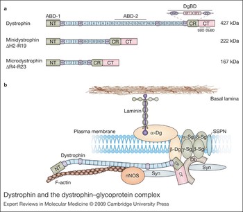
The molecular pathology of various muscular dystrophies is diverse because of the heterogeneity of the defective proteins involved. Understanding the role of these proteins and their interactions will be a crucial aspect of the search for therapeutic targets. Many muscular dystrophies result from defects in muscle-membrane-associated proteins that help maintain the structural integrity of muscle fibres (Refs Reference Bansal and Campbell5, Reference Davies and Nowak6). DMD is caused by mutations in the dystrophin gene (official symbol, DMD for human and Dmd for mouse), most of which result in translational frameshifts and failure to express a functional protein (Ref. Reference Monaco7). The four major domains of dystrophin (N-terminal, central rod, cysteine-rich and C-terminal) mediate a link between the subsarcolemmal cytoskeleton and a complex series of protein–protein interactions at the sarcolemma (reviewed in Ref. Reference Abmayr, Chamberlain and Winder8) (Fig. 1). The N-terminal and central rod domains interact with filamentous actin. The rod domain contains 24 repeats that are homologous to those in spectrin, providing a flexible and elastic region connecting the end domains that are crucial for dystrophin function. The third and fourth are the cysteine-rich and C-terminal domains, which contain many of the protein-interaction domains essential for signalling and assembly of the dystrophin–glycoprotein complex (DGC) (Fig. 1). The most critical binding site in dystrophin is the dystroglycan-binding domain (DgBD), which is made up of a WW domain (Ref. Reference Huang9) at the end of the rod domain and the adjacent cysteine-rich domain. Inactivation of the DgBD renders dystrophin nonfunctional (reviewed in Ref. Reference Abmayr, Chamberlain and Winder8).
Dystrophin and the dystrophin–glycoprotein complex. (Legend; See previous page for figure) (a) Comparison of the structural domains in full-length dystrophin with those of highly functional miniaturised versions, which have been engineered in response to the limited packaging capacity of delivery vectors for gene therapy. Full-length dystrophin contains four major domains: an N-terminal (NT) cytoskeletal-binding domain, a rod domain composed of 24 spectrin-like repeats (1–24) and four hinge regions (H1–H4), a cysteine-rich (CR) and a C-terminal (CT) domain. The two known actin-binding domains (ABD-1 and ABD-2) are within the N-terminal and rod domains, respectively. The dystroglycan-binding domain (DgBD) is composed of a WW motif (in hinge 4) connected to two EF-hand-like motifs and a ZZ domain (the two EF-hand and ZZ regions comprise the so-called cysteine-rich ‘CR’ domain). Towards the C-terminus is a syntrophin-binding domain (SBD) and a dystrobrevin-binding domain (DbBD). Minidystrophin and microdystrophin retain most of the necessary regions for the signalling and structural roles of dystrophin, as well as the ability to assemble members of the dystrophin–glycoprotein complex (DGC) at the plasma membrane. (b) Important interactions among members of the DGC, many of which are defective in various muscular dystrophies. Within striated muscle fibres, dystrophin binds to cytoskeletal proteins such as filamentous actin (F-actin) at its N-terminal domain. The rod domain encodes a second actin-binding domain in spectrin-like repeats 11–17, and repeats 16–17 also participate in binding to neuronal nitric oxide synthase (nNOS). Although localisation of nNOS has been found to require the presence of both repeats 16–17 and syntrophin, the precise three-dimensional structure of the dystrophin–nNOS complex is speculative (Ref. Reference Lai78). The DgBD anchors β-dystroglycan to dystrophin, and might help to assemble other proteins. The CT domain binds to and localises members of the syntrophin (Syn) and dystrobrevin (Db) protein families. Studies suggest that up to four syntrophins could attach to the DGC at any one time, two to dystrophin and two to dystrobrevin (Ref. Reference Newey180). At present it is unclear how many of these syntrophins are attached to nNOS, as it has been shown that syntrophin can also bind to other proteins, such as sodium channels and aquaporin-4 (Refs Reference Adams, Mueller and Froehner181, Reference Gee182). In addition to dystrophin, dystrobrevin is known to bind to another member of the DGC, which has yet to be identified. Abbreviations: Dg, dystroglycan; Sg, sarcoglycan; SSPN, sarcospan.

Dystrophin is thought to have a primarily structural role, linking the cytoskeleton to the extracellular matrix via the DGC (Fig. 1b) (Refs Reference Yoshida and Ozawa10, Reference Ervasti and Campbell11). This linkage transduces the forces of contraction to the extracellular matrix to protect myofibres from contraction-induced injury (Ref. Reference Petrof12). The absence of dystrophin results in membrane instability and repeated tears in the sarcolemma with calcium entry into the muscle cell (Refs Reference Turner13, Reference Batchelor and Winder14). Stretch-activated calcium channels might have a role in this process, because their blockade reduces membrane permeability and loss of force in dystrophic muscle following eccentric contractions (Ref. Reference Whitehead15). The resulting cascade of events forces muscle fibres to undergo cycles of degeneration and regeneration until repair capacity is no longer sufficient, and muscle fibres are replaced by adipose and fibrous connective tissue (Ref. Reference Emery and Muntoni2). In BMD, mutations typically maintain the mRNA reading frame but lead to reduced expression, or expression of truncated forms of dystrophin in striated muscle (Refs Reference Monaco7, Reference Baumbach16). Mutations in other components of the DGC result in a number of other muscular dystrophies. For example, sarcoglycanopathy results in several limb-girdle muscular dystrophies, and integrin or laminin deficiencies result in congenital muscular dystrophy (Refs Reference Ozawa17, Reference Campbell18).
Despite tremendous effort and major advances in our understanding of the molecular basis for the muscular dystrophies, no cure has been found. Symptom management and prolonging mobility is therefore the primary focus of clinical interventions (Refs Reference Manzur19, Reference Chamberlain and Rando20). Many hopes rest on recent advances in gene and cell therapies to prevent muscle degeneration and potentially reverse dystrophy-related damage. The goal of gene therapy is to deliver a functional copy of the gene, or repair the damaged gene, such that it produces sufficient product to halt the dystrophic phenotype. Methods of gene delivery include both viral and nonviral vectors. Current cell therapies involve transplantation of stem or progenitor cells with skeletal myogenic potential that can fuse with existing myofibres or form new ones. To avoid the host-versus-graft immune response, a patient's own cells that have been corrected for the genetic defect could be used. Upon transplantation, these cells would ideally engraft and regenerate the muscle, as well as repopulate the muscle stem cell (satellite cell) niche.
Mammalian preclinical testing often uses the X-linked muscular dystrophy Dmdmdx mouse model of DMD (hereafter mdx), or the canine cxmd model (Ref. Reference Banks, Chamberlain and Krauss21). In this review, many of the therapies have been developed and will be discussed with respect to models of DMD; however, these approaches are applicable to a wide range of muscle and genetic disorders.
Gene therapy for muscular dystrophies
Approaches
Gene therapy approaches for treatment of DMD must either develop methods to deliver dystrophin, or repair the locus within a patient's genome (Fig. 2). As dystrophin encodes a very large 14 kb mRNA (the gene itself spans about 2.1 Mb), therapeutic delivery is a significant challenge (Refs Reference Chamberlain, Caskey and Appel22, Reference Chamberlain23). However, the observation of large genomic deletions in some very mildly affected BMD patients (Refs Reference England24, Reference Matsumura25) prompted construction of highly functional ‘mini’ and ‘micro’ versions of dystrophin to facilitate gene transfer using viral vectors such as adeno-associated virus, which have a limited carrying capacity (Refs Reference Harper26, Reference Phelps27, Reference Sakamoto28, Reference Wang, Li and Xiao29). Internally deleted dystrophins, illustrated in Fig. 1a, retain their N-terminal actin-binding and C-terminal dystroglycan-binding domains, which are thought to contain most of the necessary regions for dystrophin's role in signalling, structural support and assembly of dystrophin-associated proteins at the cell membrane (Refs Reference Harper26, Reference Ishikawa-Sakurai30) (Fig. 1). Although inclusion of actin- and dystroglycan-binding domains is crucial, meticulous design of the deletions in the rod domain is also essential for maintaining functionality and rescuing the dystrophic phenotype (Ref. Reference Harper26).
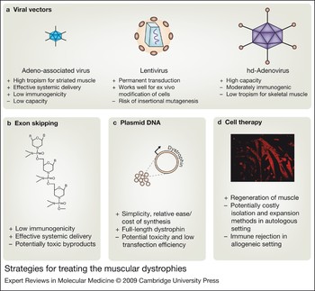
Strategies for treating the muscular dystrophies. Major advantages and limitations of viral vectors, exon skipping, plasmid DNA and cell therapies. Each virus-based delivery system (a) has particular strengths, such as high striated muscle tropism (adeno-associated virus), ability to integrate into the host genome (lentivirus), and packaging capacity [helper-dependent (hd)-adenovirus]. However, viral systems must carefully address safety concerns such as insertional mutagenesis, where provirus integration into the genome might alter the structure or expression of nearby genes, and immunogenicity of capsid proteins. Exon skipping (b) is a promising new approach in which mutations can be bypassed using antisense oligonucleotides of various chemistries that modify splicing of pre-mRNAs. Shown is the phosphorodiamidate morpholino oligomer (PMO), which incorporates morpholine rings linked by phosphorodiamidate groups instead of the ribose rings linked by phosphodiester groups found in RNA. R, cell-penetrating moiety; B, RNA nucleobases. (c) Plasmid DNA is a straightforward approach and can potentially deliver full-length dystrophin. Current studies are focused on lowering toxicity of transfection reagents as well as improving delivery efficiency. (d) Cell-based therapies might be able to regenerate muscle by replacing muscle fibres lost during the course of progressive muscle-wasting conditions such as Duchenne muscular dystrophy. Transplanted cells derived from the patient (autologous setting) require genetic modification to include a functional copy of the defective gene, whereas donor cells (allogeneic setting) are at risk for immune rejection. Image shows differentiating muscle cells in culture, with immunofluorescent staining for myosin heavy chain (red). Most therapies must be adapted for systemic delivery and targeted specifically to striated muscle using muscle-specific promoters to regulate expression of the delivered transgene.

It has long been considered in the field of gene therapy that expression of a delivered transgene not normally expressed in a host could incite an immune response. Evidence from immunological studies indicates that dystrophin could act as a neoantigen in this manner (Refs Reference Huard31, Reference Ohtsuka32, Reference Gilchrist33). If the dystrophin neoantigen is released from degenerating muscles and absorbed by antigen-presenting cells, this could stimulate both cytotoxic and humoral immune responses, presenting a significant challenge for gene transfer in DMD (reviewed in Ref. Reference Wells, Ferrer and Wells34).
A possible alternative is to use utrophin, a highly similar protein to dystrophin in both structure and properties (Ref. Reference Tinsley35). Utrophin has been suggested to have a comparable functional role in muscle, with the potential to compensate for dystrophin (Refs Reference Matsumura36, Reference Winder37). Although it is primarily expressed at the neuromuscular junctions in adult muscle (Refs Reference Nguyen38, Reference Khurana39), it is speculated that elevated expression of utrophin in some DMD patients partially compensates for lack of dystrophin (Refs Reference Mizuno40, Reference Weir, Morgan and Davies41). Delivering and upregulating endogenous utrophin are therefore potential therapies for DMD. Full-length utrophin transferred via adenoviral vector ameliorates the dystrophic phenotype in the limb muscles of mdx mice (Ref. Reference Deol42). In addition, mini-utrophins significantly improve the pathophysiology of dystrophic mdx and dystrophin–/– and utrophin–/– (mdx:utrn –/–) double-knockout transgenic animals, as well as dystrophic dogs (Refs Reference Tinsley43, Reference Deconinck44, Reference Cerletti45). Recently, microutrophins have been shown to alleviate a wide range of histopathological features of the mdx:utrn –/– model when delivered systemically using recombinant adeno-associated viral vectors (Ref. Reference Odom46). Assessment of muscle morphology, mass, fibre size, and contractile properties suggests that microutrophin might work similarly to microdystrophin. These results raise the possibility of delivering micro-utrophins to dystrophic muscle as a way of ameliorating pathology, without eliciting a cellular immune response against exogenous dystrophin.
Adeno-associated virus
Recombinant vectors derived from adeno-associated virus (rAAV) are one of the most promising methods for delivery of genes to striated muscle (Fig. 2a). The wild-type virus is a nonpathogenic single-stranded DNA parvovirus that requires a helper virus to replicate (Refs Reference Atchison, Casto and Hammon47, Reference Muzyczka, Berns, Knipe and Howley48). The recombinant form carrying a desired transgene can be produced at high titres in the absence of helper virus, and it can infect both dividing and nondividing cells (Ref. Reference Podsakoff, Wong and Chatterjee49). Although its small size and range of target tissues facilitates dissemination, the limited packaging capacity (less than 5 kb) precludes delivery of full-length dystrophin. However rAAV has been used to successfully deliver microdystrophin systemically to all striated muscle (Ref. Reference Gregorevic50).
At least nine AAV serotypes have been identified in primates, referred to as AAV1–AAV9 (Ref. Reference Gao51). The different serotypes display various tropisms in vivo (Refs Reference Chao52, Reference Duan53, Reference Grimm54, Reference Halbert, Allen and Miller55). rAAV genomes persist as nonintegrated episomes following infection of cells (Refs Reference Duan56, Reference Schnepp57), except at very high doses, where low levels of integration have been found in cultured cells, liver and muscle (Refs Reference Chamberlain58, Reference Inagaki59). Stable gene expression following rAAV injection into muscle has been reported for up to 2 years in mice and more than 7 years in dogs and rhesus monkeys (Refs Reference Herzog60, Reference Manno61). However, the mostly episomal DNA transferred by rAAV in muscle will almost certainly be lost over an extended period of time because of natural muscle turnover during exercise.
AAV vectors have been widely used for gene therapy studies because they appeared unable to elicit a host cellular immune response in numerous studies. A Phase I human trial for haemophilia b showed that rAAV2 was safe, with no adverse events reported among patients who received intramuscular injections of up to 1014 vector genomes (Ref. Reference Manno61). The authors also reported that pre-existing antibodies to AAV2 in patients had no effect on myofibre transduction. However, in some cases, such as in dystrophic muscles from the mdx mouse or the sarcoglycan-deficient hamster, an inflammatory response has been observed following delivery of rAAV vectors that express an immunogenic protein, such as E. coli β-galactosidase, under the control of a ubiquitously active promoter, such as CMV (Refs Reference Gregorevic50, Reference Yuasa62). Use of a muscle-specific promoter can often block this response (Refs Reference Gregorevic50, Reference Hartigan-O'Connor63). Other proteins delivered to the mdx mouse, such as human dystrophin, have not elicited a cellular immune response (Refs Reference Harper26, Reference Wang, Li and Xiao29, Reference Gregorevic50). Nonetheless, as investigators have expanded studies to larger animals and started to use higher doses, it is becoming clear that most, if not all, AAV serotypes can elicit a cellular immune response. Recent studies have found such responses to both rAAV2 and rAAV6 in dogs (Ref. Reference Wang64), and similar observations have been made in humans (Refs Reference Manno65, Reference Mingozzi and High66, Reference Mingozzi67). Nonetheless, the immune response is often mild, and studies in dystrophic dogs have shown that it can be blocked with brief immunosuppression (Ref. Reference Wang64). Transient immune suppression has also been applied with success in nonhuman primates (Ref. Reference Jiang68).
Systemic delivery of rAAV will be a critical aspect of administration, given the need to target all striated muscle, including the heart and diaphragm, for an effective DMD therapy. Targeting skeletal muscle alone will strain the dystrophic heart and aggravate cardiomyopathy (Ref. Reference Townsend69). A major breakthrough for muscular dystrophy gene therapy came with the discovery that several serotypes of AAV could transduce muscle tissue body-wide following intravascular delivery. Initial studies of systemic delivery used vectors derived from AAV6 (Ref. Reference Gregorevic50), but subsequent studies have also had success with AAV1, AAV8 and AAV9 (Refs Reference Mah70, Reference Wang71, Reference Inagaki72). Feasibility of the systemic approach coupled with early intervention is validated by successful transduction of all skeletal muscle in newborn dogs in the absence of immune suppression (Ref. Reference Yue73).
The intimate association between myofibres and capillaries allows extravasated vectors to come into close contact with the surface of muscle cells. Thus, AAV serotypes readily transduce muscle following intravascular delivery. The mechanisms responsible for vector extravasation remain unclear, and few cell surface receptors have been identified for AAV1, AAV6, AAV8 or AAV9. Also, little is known of the intracellular events following vector uptake by myofibres that enable vector decapsidation and gene expression (reviewed in Ref. Reference Schultz and Chamberlain74). Identification of these mechanisms could well lead to modified delivery protocols to enhance body-wide muscle transduction using lower vector doses.
Split-vector approaches for full-length dystrophin delivery by rAAVs
Restoration of the major functions of dystrophin is key to alleviating deficits in DMD, and internally truncated dystrophins lose some functionality (Refs Reference Harper26, Reference Banks75). For example, although most mini- and microdystrophins restore the components of the DGC to the sarcolemma, only recently has a truncation been found that supports sarcolemmal localisation of neuronal nitric oxide synthase (nNOS) (Refs Reference Wells76, Reference Judge, Haraguchi and Chamberlain77, Reference Lai78). nNOS is involved in production of the signalling molecule nitric oxide (NO), which is important for maintaining vasodilation and adequate blood flow in skeletal muscle during activity (Refs Reference Thomas79, Reference Kobayashi80). Although reconstitution of dystrophin as represented in the internally truncated forms might be the crucial factor for stabilising muscle fibres, delivery of mini- or microdystrophins that fail to localise nNOS could lead to a mild BMD phenotype, with the associated susceptibility to fatigue. It is also unknown whether other properties of myofibres are affected by large deletions in dystrophin, such as the structure of myotendinous junctions (Refs Reference Banks81, Reference Banks, Chamberlain and Froehner82). It is therefore unclear whether some of the milder BMD phenotypes that result from truncated dystrophins would benefit from delivery of a designer micro- or minidystrophin, or whether an alternative therapy would be necessary to relieve symptoms.
Since rAAV packaging capacity is limited to less than 5 kb, microdystrophin is the only dystrophin variant small enough to package into a single vector. However, one possible strategy to deliver full-length dystrophin is to package fragments of the cDNA into separate vectors. Transduction of target tissues with more than one vector, each carrying a dystrophin cDNA fragment, could allow production of larger dystrophins if the parts of each vector could be brought together (Refs Reference Sun, Li and Xiao83, Reference Yan84, Reference Nakai, Storm and Kay85). Two approaches for achieving this goal are showing promise in vivo. In the first, portions of introns carrying appropriate splicing signals are incorporated into the gene fragments, such that the cellular RNA splicing mechanisms can be harnessed to reconstruct a larger gene from two smaller halves (Refs Reference Duan, Yue and Engelhardt86, Reference Lai87). Another approach is to package partially overlapping fragments into the different vectors, such that a larger dystrophin expression cassette can be reconstructed via homologous recombination in transduced tissues (Refs Reference Duan, Yue and Engelhardt86, Reference Halbert, Allen and Miller88). A recently developed hybrid system outperforms splicing and homologous recombination split-vector systems alone, and improves efficiency of minidystrophin delivery to muscle (Ref. Reference Ghosh89).
Lentivirus
Retrovirus-derived lentiviral vectors can be used to stably integrate a transgene such as dystrophin into the genome of target cells (Ref. Reference Li90). However, integration into the host genome can potentially cause insertional mutagenesis, where viral promoter and enhancer elements can activate nearby proto-oncogenes (Refs Reference Hacein-Bey-Abina91, Reference Ciuffi92, Reference Beard93). Development of self-inactivating (SIN) lentiviral vectors may address this issue. With SIN lentiviral vectors, the viral promoter and enhancer elements are removed prior to integration into the host genome, thus preventing viral activation of proto-oncogenes at sites of integration.
Unlike vectors derived from onco-retroviruses such as Moloney murine leukaemia virus, which are commonly used for gene transfer (reviewed in Ref. Reference Barquinero, Eixarch and Perez-Melgosa94), lentiviral vectors can infect a wide range of dividing and nondividing cells, including hepatocytes, skeletal and cardiac muscle cells, and neurons (Refs Reference Kafri95, Reference Bonci96, Reference Naldini97). Lentiviral vectors have low immunogenicity but the delivered transgene is still a potential neoantigen (Ref. Reference Annoni98). They have a larger carrying capacity (~9 kb) than rAAVs, and have been used to stably transduce myogenic cells with minidystrophin (Ref. Reference Li90), whereas rAAVs can only deliver the smaller microdystrophin (Ref. Reference Harper26).
Permanent gene transfer by lentiviral vectors is especially advantageous for DMD treatment. Satellite cells within skeletal muscle are an ideal target, and lentiviral reconstitution of dystrophin into the satellite cell niche in vivo would provide a source of dystrophin during ongoing cycles of regeneration. Lentiviral vectors are also a useful tool for transducing autologously derived cells with dystrophin and other genes ex vivo before expansion and transplantation (Refs Reference Bachrach99, Reference Dellavalle100).
Adenovirus
Vectors derived from adenovirus are attractive for gene transfer because of their large carrying capacity, high titre production and ability to infect nondividing cells. Successive modifications have improved carrying capacity, reduced immunogenicity and prolonged transgene expression. The latest generation of adenovirus vectors are helper-dependent or ‘gutted’ (hdAd) and are entirely devoid of viral genes. They thus require a helper virus to supply genes needed for vector replication and packaging during vector production (Refs Reference Kumar-Singh and Chamberlain101, Reference Clemens102, Reference Chen103). The presence of a helper virus renders production technically challenging, and additional purification steps are required to remove it from the final vector preparations (Ref. Reference Hartigan-O'Connor104). Helper-dependent adenovirus has been used to stably transduce muscle with dystrophin and has improved muscle function in mdx mice (Refs Reference DelloRusso105, Reference Gilbert106).
Despite improvements, many hurdles to effective hdAd-vector-based therapy remain. Adenovirus vectors are five times larger than AAVs, reducing their ability to exit capillaries and infect muscle (Ref. Reference Su107). Also, most intravascularly administered adenovirus vectors are taken up by the liver (Ref. Reference Gao, Yang and Wilson108). Coupled with low transduction efficiency in adult skeletal muscle, systemic therapeutic administration could require extremely high doses of hdAd (Refs Reference DelloRusso105, Reference Acsadi109). Such high doses resulted in acute inflammation and a cytotoxic T cell response, which mediated lethal acute toxicity in nonhuman primates (Refs Reference Zoltick110, Reference Muruve111, Reference Brunetti-Pierri112) and one patient (Ref. Reference Raper113). As with AAV, delivered transgenes exist as episomes, which might be lost over time and require repeated doses. However, readministration with the same serotype is problematic given the development of neutralising antibodies to both the hdAd vector and the transgene (Refs Reference Gilchrist33, Reference Morral114). Use of hdAd vectors has not progressed to clinical trials for these reasons, and much of the current work is focused on overcoming the immunological issues.
Nonviral gene transfer
Nonviral gene transfer methods involve administration of plasmid DNA (pDNA) (Ref. Reference Wolff115), usually in complex with synthetic compounds (Fig. 2c). Although the approach is conceptually straightforward, less expensive compared with viral vector production, and avoids the inherent risks of viral vector administration, preclinical testing has not yet demonstrated a feasible approach for system-wide treatment of DMD. Upon intramuscular and intravascular injection, naked dystrophin pDNA is expressed in only 5% of target myofibres (Ref. Reference Zhang116). Brief blood flow occlusion achieves targeting of the diaphragm and improves transgene expression in limb muscles following intravascular administration (Refs Reference Zhang116, Reference Hagstrom117). Use of cationic polymers and lipids also improves in vivo tissue transfection after systemic administration (Ref. Reference Richard118). However, the possibility of acute toxicity of plasmid DNA and cationic polymer/lipid complexes must be resolved, especially considering potentially high-dose systemic administration (Refs Reference Chollet119, Reference Trubetskoy120). The size of the plasmids encoding full-length dystrophin may also impede DNA transfer through the vasculature (Refs Reference Zhang116, Reference Richard118). Although additional methods have improved transfection of striated muscle, clinically safe and efficient body-wide dystrophin expression in both skeletal and cardiac muscle has not yet been achieved. Still, a recent Phase I trial testing naked dystrophin pDNA by intramuscular injection resulted in low levels of dystrophin expression (Ref. Reference Romero121), giving hope that development of a safe method to boost gene transfer will lead to a new DMD therapy.
Exon skipping
Exogenous delivery of dystrophin has shown promise for treatment of DMD, but limitations such as vector capacity and delivery, potential immune response and appropriate transgene expression, still need to be overcome. Exon skipping provides an alternative approach that allows direct manipulation of dystrophin transcripts, such that expression of the endogenous gene can potentially be restored in vivo (Fig. 2b).
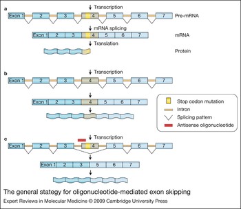
In the various forms of muscular dystrophy, as well as other genetic diseases, mutations give rise to premature stop codons, and splice-site mutations or deletions/duplications can shift the reading frame of a transcript or cause aberrant splicing, resulting in little or no functional protein. Exon skipping addresses these disorders at the post-transcriptional level, by using the process of transcript maturation to remove problematic exons (Fig. 3). Sites within the newly synthesised pre-mRNA that signal splicing at the start of the exon are blocked, and the whole exon containing the stop codon or frameshift mutation, along with its flanking introns, are thereby removed. In the case of dystrophin, multiexon deletion in the rod domain has been found in mild and asymptomatic BMD (Ref. Reference Monaco7). Thus exons in this domain with a stop codon or frameshift mutation may be removed using exon skipping. Translation then generates an in-frame, albeit truncated, gene product that retains many critical functions. Studies of rare, dystrophin-positive ‘revertant’ fibres in some DMD patients first indicated that this might be a feasible therapeutic approach (Ref. Reference Sherratt122). The revertant fibres contained a restored dystrophin reading frame, although it remains unclear whether this was a result of secondary mutations or spontaneous alternative splicing (Ref. Reference Partridge, Lu, Tsuchida and Takeda123).
The general strategy for oligonucleotide-mediated exon skipping. (a) A hypothetical stop codon mutation in exon 4 is translated into a nonfunctional truncated protein. (b) Normal splicing results in translation of full-length protein. (c) The antisense oligonucleotide (AON) blocks the splicing site for exon 4, resulting in its removal during the processing of the pre-mRNA into a mature mRNA transcript. In the case of the mdx mouse model of Duchenne muscular dystrophy, AONs have been used to target the stop codon mutation in exon 23, leading to removal of exon 23 from the pre-mRNA and joining of exons 22 and 24 (Ref. Reference Mann131). Similarly, in Duchenne and Becker muscular dystrophies, targeting of dystrophin in this manner could lead to removal of exons containing stop codon or frameshift mutations. The translated protein, although internally deleted, will ideally retain critical functional domains. In myotonic dystrophy type 1 (DM1), chloride channels have been targeted to remove an exon normally expressed only in the neonatal isoform, but which is retained in DM1 muscles, to prevent generation of a dysfunctional protein in adult muscle (Ref. Reference Wheeler3). Recent studies have demonstrated that the specific sequence targeted within a pre-mRNA is critical to the success of an AON (Ref. Reference Popplewell143).

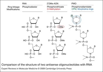
Synthetic antisense oligonucleotides (AONs) were developed for specific exons within dystrophin that can block transcript splicing sites in vitro (Refs Reference Dominski and Kole124, Reference Takeshima125, Reference Pramono126). Most of the early work on AONs for therapeutic benefit was in the form of 2'-O-methyl-phosphorothioate AONs (2'OMeAONs) (Fig. 4), which are more stable than nonmethylated RNA (Ref. Reference Shibahara127). 2'OMeAONs restore the reading frame of dystrophin in primary muscle cells from mdx mice (Ref. Reference Dunckley128). This was also achieved in cells from cxmd dogs (Ref. Reference McClorey129), human cells derived from DMD patients (Ref. Reference van Deutekom130) and in vivo, in mdx mice by intramuscular administration (Ref. Reference Mann131). Other muscular disorders have also successfully used exon skipping to correct abnormal gene expression. In myotonic dystrophy, AONs have been used to correct aberrant splicing in chloride channel 1 gene (CLCN1) pre-mRNA and the associated myotonia (Ref. Reference Wheeler3), demonstrating the broad applications of this tool.
Comparison of the structure of two antisense oligonucleotides with RNA. Antisense oligonucleotides (AONs) are designed to be complementary to specific sequences within mRNA transcripts, and have various chemical modifications that distinguish them from RNA. Specific modifications confer resistance to degradation and ability to penetrate cell membranes. RNA (left panel) contains phosphodiester linkages between each ribose ring, whereas 2′O-methylated (2′OMe) AONs (middle panel) have phosphorothioate linkages (the oxygen is replaced with a sulphur atom). Phosphorodiamidate morpholino oligomers (PMOs, right panel) have morpholine rings instead of ribose rings, often contain conjugated cell-penetrating moieties (CPM, R group in structure) to facilitate cell entry and subunits are linked by phosphorodiamidates (the oxygen is replaced with a nitrogen group). B, RNA nucleobases.

AON chemistry
AON modifications have undergone many iterations, resulting in various chemistries affecting efficiency and delivery to cells in vitro and in vivo (Ref. Reference Summerton and Weller132). Morpholino oligomers show promise for in vivo applications (Ref. Reference Yokota133). Antisense morpholino oligomers, commonly called PMOs (for phosphorodiamidate morpholino oligomers), resemble 2'OMeAONs, but have several key differences, as shown in Fig. 4. PMOs incorporate morpholine rings in place of the ribose sugar rings of RNA and are nonionic, which avoids nonspecific electrostatic interactions within the cell. Replacement of the phosphodiester linkage with phosphorodiamidate imparts resistance to nuclease degradation (Ref. Reference Summerton and Weller132). Because PMOs are non-ionic, and therefore inefficient at penetrating cell membranes (Ref. Reference Sazani134), they are commonly conjugated to cell-penetrating cationic moieties (Fig. 4) (Refs Reference Lebleu135, Reference Wu136). Peptide nucleic acids represent another type of chemistry developed and tested in vivo (Ref. Reference Ivanova137). Aside from sequence specificity and efficient exon skipping, clinical considerations for AON chemistries must include the potential for toxicity or induction of a host immune response, biostability, penetration into striated muscle cells and the cost of large-scale synthesis for systemic delivery.
Administration
System-wide targeting of morpholino oligomers to skeletal muscle was initially achieved in the mdx mouse with no observed immune response or toxicity, but also without targeting of the heart (Refs Reference Alter138, Reference Fletcher139) – outcomes observed previously with 2'OMeAONs (Ref. Reference Lu140). Recently, several groups have successfully targeted skeletal muscle system-wide, as well as the heart in the mdx mouse, upon incorporation of peptide or chemical moieties to facilitate cell uptake (Refs Reference Wu136, Reference Jearawiriyapaisarn141, Reference Wu142).
AON design itself must be optimised for the target sequence within a given mRNA, because of the secondary structure in single-stranded RNA and splice signal sites (Ref. Reference Popplewell143). Indeed, this might explain why multiple AONs, or an oligo ‘cocktail’, seem to be more efficient than a single AON at targeting a single site (Ref. Reference Mitrpant144). Oligo cocktails might be important during the development of a treatment for DMD, to achieve multiexon skipping and thereby cover the majority of DMD mutations with a single treatment.
Another systemic approach for AONs uses rAAV vectors for intravascular delivery. rAAVs can be used to deliver DNA encoding an AON linked to a small nuclear RNA sequence for nuclear targeting of the transcript for modification of dystrophin (Ref. Reference De Angelis145). Treatments show therapeutic improvement in the mdx mouse, with reduced serum creatine kinase and improved skeletal muscle function (Refs Reference Goyenvalle146, Reference Denti147). rAAV vector tropism also allows certain serotypes to reach the heart (Ref. Reference Denti147). Another advantage of rAAV vector delivery of AONs is the persistence of rAAV episomes (Ref. Reference Schultz and Chamberlain74). Longer-term in vivo AON expression from episomes would be preferred over more frequent, lifelong treatment with PMOs. For rAAV AONs, however, potential immune responses as well as scalability of vector production must also be considered.
AON safety
Although it is debatable which AON chemistry will be the most effective (Refs Reference Wu142, Reference Mitrpant144, Reference Heemskerk148), it must be determined whether the most effective are actually safe for human use. The various modifications require careful investigation of potentially harmful degradation products or codelivery agents, immune responses, and nonspecific actions. Owing to inherent resistance to degradation, some morpholinos might be especially harmful if they have low specificity for RNA targets, and at high doses these effects could be amplified (Ref. Reference Yokota133).
Cell therapy for muscular dystrophies
Approaches
Cell transplantation as a therapeutic tool is a promising avenue for treatment of muscular dystrophies (Fig. 2d). Transplanted cells must be able to fuse with existing myofibres or form new ones, and transplanted cell nuclei within those myofibres must express the missing gene product. In addition to regeneration of myofibres, a major goal of stem cell therapies is reconstitution of the satellite cell niche, which might promote future functional regeneration of the muscle.
Cell transplantation might either be allogeneic (donor-derived) or autologous (patient-derived). Although allogeneic transplantation from a wild-type donor does not require genetic manipulation to reintroduce functional dystrophin, the risk for graft rejection remains. Autologous transplantation requires genetic manipulation, potentially by using lentiviral vectors to permanently reintroduce the defective gene. Inherent risks in using lentiviral vectors apply, but the risk of immunogenic graft rejection is much lower. With either approach, expansion and culturing of the cell population will probably be necessary, requiring careful control of conditions to preserve muscle engraftment ability. For a more extensive review of approaches and immune issues associated with cell therapy, see (Ref. Reference Skuk and Tremblay149). Developing systemic routes of administration in cell transplantation will be an important issue, as will identifying an adequate cell source for scalable autologous transplantation in humans. Ultimately, the most promising approach for muscular dystrophy cell therapy will be the use of accessible patient-derived myogenic precursors capable of efficient engraftment into skeletal muscle following ex vivo expansion and genetic correction. A variety of cell types have been investigated for their potential to fulfil these criteria.
Cell types
The most widely studied cell type for muscular dystrophy cell therapy is the satellite cell. Transplanted satellite cells from wild-type donors engraft into skeletal muscle (Ref. Reference Skuk150), but massive cell death, limited migration from injection sites and immune rejection of allogeneic cells is observed (Refs Reference Skuk151, Reference Montarras152, Reference Peault153), even with genetic matching (Ref. Reference Huard31). Isolation and autologous transplantation of satellite cells avoids host-versus-graft immune rejection. However, in later stages of muscle degeneration, fewer myogenic progenitors can be isolated, and expansion of cells in culture significantly reduces their engraftment capacity (Refs Reference Webster and Blau154, Reference Fan155). Many recent approaches aim to improve muscle precursor cell isolation with the rationale of avoiding in vitro expansion to determine therapeutic potential (Ref. Reference Montarras152). However, immune responses and the cost of antibodies commonly used in these isolation techniques prevent direct clinical applications in humans (Ref. Reference Tremblay and Skuk156). Nevertheless, these studies could potentially lead to new discoveries on how to maintain myogenic cell populations in a particular progenitor state in vitro (Refs Reference Deasy157, Reference Collins158), and how to increase transplantation engraftment success. A further complication of these studies is that myogenic progenitors derived from muscle are typically a heterogeneous population, displaying unique properties depending on isolation methods and culture conditions (Ref. Reference Cornelison159). It is also unclear whether satellite cells themselves might derive from a satellite cell precursor within muscle or from a circulating progenitor (Ref. Reference Collins158).
Muscle-derived progenitors that can engraft in muscle after intravascular delivery have also been found; this is a crucial step to making cell-based therapies a feasible treatment for DMD. Such progenitor cells include muscle side-population cells (Refs Reference Bachrach99, Reference Gussoni160) and cells isolated based on expression of specific markers, such as CD34, Sca1 (Ref. Reference Lee161) and CD133 (Refs Reference Peault153, Reference Torrente162). Bone-marrow-derived progenitors, which show migration and engraftment in muscle and modest recruitment to muscle from the circulation (Ref. Reference Ferrari163), can be enriched and expanded quickly with specific sets of factors in vitro for therapeutic applications (Ref. Reference Dezawa164). The mesoangioblast (Ref. Reference Sampaolesi165) has shown impressive levels of engraftment and functional recovery in a mouse model of limb girdle muscular dystrophy (Ref. Reference Sampaolesi165) and in dystrophic dogs after arterial delivery (Ref. Reference Sampaolesi166). Mesoangioblasts have been suggested to derive from the pericyte, which is a microvessel-associated cell type (Ref. Reference Dellavalle100). To understand the clinical relevance and limitations of mesoangioblasts or pericytes in muscular dystrophy, it will be crucial to develop reproducible isolation and expansion methods for them and to better characterise their skeletal muscle regenerative capacity.
Conversion of alternative cell types into myogenic precursors has recently become a plausible approach. Promising cell types that can be induced into the myogenic lineage include embryonic stem (ES) cells (Refs Reference Barberi167, Reference Darabi168), embryonic and postnatal-derived fibroblasts (Ref. Reference Kimura169), and induced pluripotent stem (iPS) cells derived from adult fibroblasts (Refs Reference Takahashi170, Reference Yu171). Although ES cells have shown promise in development of cell therapies, some mouse and human ES cell lines might be predisposed to chromosomal abnormalities that could limit their application in cell replacement therapy (Refs Reference Rebuzzini172, Reference Catalina173). iPS cells are generated by lentiviral-mediated expression of a defined set of transcription factors. Once derived, they display characteristics that are remarkably similar to ES cells, including expression of ES cell markers, teratoma formation and the ability to contribute to generation of chimaeric mice following injection into a blastocyst. Use of iPS cells avoids the ethical concerns and risk of immunorejection with human ES cells, and has shown promising therapeutic potential as a pluripotent stem cell source (Ref. Reference Hanna174). Patient-specific iPS cells could be derived from highly accessible and expandable fibroblast populations, but the process of reprogramming and induction into the myogenic lineage must be streamlined for clinical feasibility, and carefully controlled to avoid the formation of tumours.
Future prospects for experimental muscular dystrophy therapies
Despite current limitations, many recent developments point to the promise of viral-vector-mediated gene therapy. Increased understanding of viral transduction mechanisms have allowed generation of hybrid vectors that target desired tissues with high efficiency and long-term expression (Refs Reference Schultz and Chamberlain74, Reference Dickson175, Reference Goncalves176). We envision that further tailoring with hybrid or chimaeric vectors could lead to more exclusive tissue-specific tropism, thereby improving safety while retaining adequate carrying capacity for most defective genes. In addition, modulation of transgene promoters and enhancers could improve muscle-specific expression for virtually all gene-transfer methods (Ref. Reference Salva177). Immune responses remain a critical issue, and evasion will be an important aspect of viral-mediated therapies (Ref. Reference Zaldumbide and Hoeben178). A promising approach for DMD treatment entails multisite intravascular delivery of rAAV vectors carrying microutrophin cassettes. Transient immune suppression could be used to block cellular immunity against the rAAV vector, whereas use of utrophin cDNAs could avoid such responses against dystrophin. Development of optimal methods for delivery and immune suppression, as well as design of microutrophin cDNA and transcriptional regulation of the cassette, are active areas of study that hold promise for clinical intervention. Exon skipping is a rapidly developing potential therapy, and further modifications to the approach might make it possible to target a large number of the mutations and deletions observed in DMD and BMD (Ref. Reference van Deutekom179). However, further testing is necessary to determine optimal chemistries for the safety and feasibility of systemic delivery. Questions also remain as to the functionality of the truncated proteins that would arise from some of these induced splicing events (Ref. Reference Harper26).
Summary and conclusions
Because ongoing clinical trials are currently focused mostly on safety, intramuscular delivery is the chosen delivery route. Local administration might be of therapeutic benefit to individual muscles or small groups of muscles; however, striated muscle must eventually be targeted systemically for an effective therapy. Combinations of local, regional and systemic routes of administration might also be viable therapeutic options to achieve system-wide targeting of striated muscle. We should consider gene transfer, gene repair, and aspects of regenerative medicine to be complementary approaches – not at all mutually exclusive – that could potentially be most effective in a wide array of muscular dystrophies and other disorders when used in conjunction. Although underestimation of challenges to gene therapy initially led to some disappointing clinical trials, there has been a recent resurgence in interest as new and exciting discoveries in the field have broken though barriers to viral, nonviral and cell-mediated therapies, bringing us a few steps closer to safe and effective treatments for the muscular dystrophies.
Acknowledgements and funding
We thank members of the Chamberlain lab for helpful discussions and the peer reviewers for their thoughtful comments. Our research is supported by grants from the National Institutes of Health (R37AR040864 and P01NS46788) and the Muscular Dystrophy Association (USA).