Introduction
Vestibular schwannomas are benign neoplasms arising from Schwann cells of the VIIIth cranial nerve. They are the commonest tumours of the internal acoustic meatus, they account for 80–90 per cent of tumours of the cerebellopontine angle, and they are the third most common non-malignant brain tumour in adults.Reference Goldbrunner, Weller, Regis, Lund-Johansen, Stavrinou and Reuss1,Reference Maldonado S, Naples, Fathy, Eliades, Lee and Brant2 Vestibular schwannoma incidence shows global variation. For instance, the vestibular schwannoma incidence rate in the USA is 1.09 per 100 000, increasing to 2.93 per 100 000 for 65–74-year olds;Reference Kshettry, Hsieh, Ostrom, Kruchko and Barnholtz-Sloan3 however, a Danish epidemiological study showed a steady increase in incidence from 1976 to 2015, rising from 3 per million to approximately 34 per million.Reference Reznitsky, Petersen, West, Stangerup and Cayé-Thomasen4 The latter study showed that the mean tumour size at diagnosis decreased in this time from 26 mm to 7 mm. The increased incidence and smaller size at diagnosis are largely a result of improved diagnostic magnetic resonance imaging (MRI), earlier screening, and the finding of more tumours in older people in a setting of increasing population age.Reference Reznitsky, Petersen, West, Stangerup and Cayé-Thomasen4–Reference Kleijwegt, Ho, Visser, Godefroy and van der Mey7
Vestibular schwannomas typically arise sporadically and unilaterally; bilateral vestibular schwannomas are the hallmark of neurofibromatosis type 2.Reference Goldbrunner, Weller, Regis, Lund-Johansen, Stavrinou and Reuss1,Reference Hannan, Lewis, O'Leary, Donofrio, Evans and Roncaroli8 Mutations in the NF2 tumour suppressor gene on chromosome 22 are thought to be the primary cause of Schwann cell neoplasia.Reference Hannan, Lewis, O'Leary, Donofrio, Evans and Roncaroli8 Most patients present with unilateral subjective hearing loss (94 per cent), tinnitus (83 per cent), and a varying frequency of vertigo and imbalance.Reference Goldbrunner, Weller, Regis, Lund-Johansen, Stavrinou and Reuss1 Large tumours can have mass effect on the trigeminal nerve, the facial nerve and the brainstem, and can cause hydrocephalus.Reference Goldbrunner, Weller, Regis, Lund-Johansen, Stavrinou and Reuss1 More lateral tumours are associated with decreased facial nerve function.Reference Dunn, Bi, Mukundan, Delman, Parish and Atkins9 As such, whilst they are benign, they can be associated with significant morbidity. Diagnosis confirmation and follow-up surveillance of vestibular schwannoma are carried out with high-resolution T2-weighted and contrast-enhanced T1-weighted MRI scanning.Reference Dunn, Bi, Mukundan, Delman, Parish and Atkins9
The management of vestibular schwannomas is complex and requires careful consideration of their natural history. Around one-third of patients who present with a sporadic vestibular schwannoma will demonstrate measurable growth, and the remaining two-thirds may not.Reference Dunn, Bi, Mukundan, Delman, Parish and Atkins9,Reference Lahlou, Rodallec, Nguyen, Sterkers and Kalamarides10 Decisions regarding management are also based on the following individual patient factors: age, functional status, presenting symptoms, patient preference, tumour size, location and growth rate.Reference Foley, Maweni, Jaafar, McConn, Javadpour and Rawluk11 The main options for management include conservative serial MRI scanning with clinical surveillance, stereotactic radiotherapy, and microsurgery.Reference Goldbrunner, Weller, Regis, Lund-Johansen, Stavrinou and Reuss1,Reference Maldonado S, Naples, Fathy, Eliades, Lee and Brant2,Reference Sethi, Borsetto, Cho, Gair, Gamazo and Jefferies12
Previous studies have attempted to define the factors that contribute to whether or not a presenting vestibular schwannoma is likely to grow; most of the available studies include small numbers or relatively short follow-up periods.Reference Sethi, Borsetto, Cho, Gair, Gamazo and Jefferies12,Reference Paldor, Chen and Kaye13 There is great heterogeneity in the behaviour of the newly diagnosed vestibular schwannoma, with complex and varying considerations required for optimal management, and global variations in practices. Therefore, the present study aimed to determine whether there are any relevant factors within the natural history of growing versus non-growing vestibular schwannoma, based on a 20-year cohort.
Materials and methods
Basic settings and patient selection
We carried out a retrospective case series in a tertiary university centre. This project was approved by the research ethical committee as a retrospective audit; a Caldicott guardian was also appointed.
We utilised the vestibular schwannoma database of our centralised skull base service, which covers a catchment of 2.2 million patients. This dataset extends over a 20-year period. We identified the patients with a known follow-up period and with known natural history of the vestibular schwannoma based on serial MRI. There were 735 patients included in the database, of whom 288 had a growing vestibular schwannoma and 320 had a non-growing vestibular schwannoma. The data were collected retrospectively from the medical notes and imaging reviews. We defined growing vestibular schwannomas as having a difference in size of at least 1.5 mm in their serial imaging. Finally, we excluded patients with neurofibromatosis type 2 because of the known genetic background.
Data collection
The following information was compared for the growing and the non-growing groups: patient age at time of diagnosis, gender, size of vestibular schwannoma at diagnosis, and length of follow up. Patients were excluded from the study if there was missing information regarding their follow up or tumour size. We also examined the presence or absence of balance symptoms at the time of diagnosis based on the medical notes. All the included patients had adequate documentation of any balance symptoms or dizziness based on the patients’ feedback, clinical examination and/or vestibular testing.
Vestibular schwannoma measurement
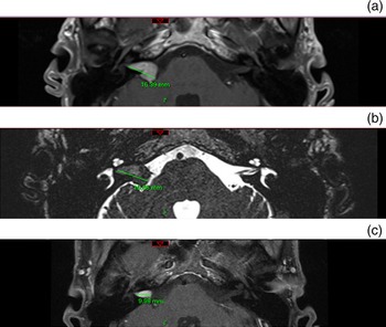
Linear measurements of vestibular schwannoma size were carried out on our online imaging archive system, by experienced radiologists, as seen in Figure 1. The size taken for analysis was the maximum measured length of the tumour; whilst we typically measure only the maximum intracranial diameter, we included the maximum length of the vestibular schwannoma in order to incorporate purely intracanalicular tumours. This value was used before any interventions took place, as these disrupt the natural history. As mentioned above, patients were assigned to the growing vestibular schwannoma cohort if there was at least a 1.5 mm difference in linear measurements on their serial MRI scans.
Linear measurements of vestibular schwannomas. (a) Right vestibular schwannoma on axial plane in post-gadolinium T1-weighted 1.5 Tesla magnetic resonance imaging (MRI). (b) Right vestibular schwannoma on axial plane in 1.5T fast imaging employing steady-state acquisition (‘FIESTA’) MRI. (c) Measurement of a purely intracanalicular vestibular schwannoma on axial plane in post-gadolinium T1-weighted 1.5T MRI. P = posterior

Statistical analysis
Descriptive statistics were obtained to gain an overview of the variations between growing and non-growing vestibular schwannomas, namely mean, median, maximum and minimum values, and standard deviation. Mann–Whitney testing was carried out on Minitab® statistical software, version 17, to compare the two non-parametric data groups. This assessed whether there were statistically significant differences in the median values for size at diagnosis, patient age and follow-up duration, between the two cohorts. The statistical significance of the presence or absence of balance symptoms in the two cohorts was assessed with a chi-square test. The level of statistical significance was set at 0.05.
Results
Patient age and follow up
Following application of our inclusion criteria, there were 215 patients in the growing vestibular schwannoma cohort (93 male, 122 female) and 228 in the non-growing cohort (114 male, 114 female). The ages of the cohorts were similar. The mean age of patients with growing vestibular schwannoma was 64.96 years and the median was 66 years. The mean and median ages of patients with a non-growing vestibular schwannoma were 64.86 years and 65 years respectively.
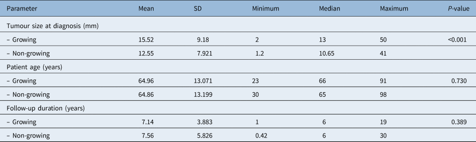
Regarding follow-up duration, the mean time for the growing vestibular schwannoma cohort was 7.14 years and the median was 6 years. For non-growing vestibular schwannoma cohort, the mean follow-up time was 7.56 years and the median was also 6 years. Mann–Whitney testing confirmed that there were no statistically significant differences in the medians for age (p = 0.730) and follow-up duration (p = 0.389). The data for both parameters are shown in Table 1.
Tumour size at diagnosis, patient age and follow-up duration in growing and non-growing vestibular schwannoma cohorts*

* Comparison between growing (n = 215) and non-growing (n = 228) vestibular schwannoma cohorts shown in terms of descriptive statistics and p-values from Mann–Whitney testing. SD = standard deviation
Vestibular schwannoma size
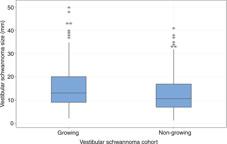
We found that growing vestibular schwannomas were larger at the time of diagnosis than non-growing tumours. For the growing cohort, the mean size at diagnosis was 15.52 mm and the median was 13.00 mm (n = 215). The non-growing cohort had a mean size of 12.55 mm and a median of 10.65 mm (n = 228), as seen in Figure 2 and Table 1. The increased size of vestibular schwannomas in the growing cohort was statistically significant (Mann–Whitney, p < 0.001 at 95 per cent confidence interval).
Boxplot showing vestibular schwannoma sizes at diagnosis for the growing and non-growing vestibular schwannoma cohorts. Growing vestibular schwannomas are larger at diagnosis than non-growing vestibular schwannomas.

Presence of balance symptoms
Balance symptoms were significantly more common at diagnosis in the growing vestibular schwannoma cohort than in the non-growing cohort. In the growing cohort, 175 out of 288 patients presented with balance disorders (60.76 per cent). There were 73 patients without balance issues (25.35 per cent) and 40 patients with no records on this factor (13.89 per cent). From the non-growing vestibular schwannoma cohort, 124 out of 320 patients had balance issues (38.75 per cent) and 96 patients had none (30 per cent). The remaining 100 patients had no available records (31.25 per cent). The difference in the presence of balance symptoms between the growing and non-growing vestibular schwannoma cohorts was statistically significant (chi-square test, p < 0.001).
Discussion
Main findings
The factors present at diagnosis that affect vestibular schwannoma outcomes have been explored in the literature through retrospective cohort studies and reviews. The present study adds to this with a large database and robust follow-up information. We found that growing vestibular schwannomas are larger at diagnosis than non-growing vestibular schwannomas; this result was statistically significant. Another key finding was that balance symptoms were more likely to be present at diagnosis in the growing vestibular schwannoma cohort (60.67 per cent) than in the non-growing cohort (38.75 per cent). This result was statistically significant (chi-square test, p < 0.001).
Whilst balance problems can be present in both growing and non-growing vestibular schwannoma patients, this symptom appears to be significantly more common in those with growing tumours; thus, deterioration in balance should be considered in patients with vestibular schwannoma.
Tumour size at diagnosis
Our results indicate that tumour size at diagnosis may be useful as a predictor for vestibular schwannoma growth. Whilst vestibular schwannomas can grow regardless of their initial size, the tendency of larger tumours to grow has been previously highlighted in the literature. In a retrospective cohort study published around the turn of the century, larger tumours at presentation were found to be statistically significantly more likely to grow.Reference Fucci, Buchman, Brackmann and Berliner14 Moreover, a study of 433 vestibular schwannoma patients agreed with this finding and added that larger tumours also have a faster growth rate.Reference Nutik and Babb15 More recently, using volumetric rather than linear growth rates, a retrospective study of 212 untreated vestibular schwannoma patients found that larger tumours tended to have more absolute growth than smaller ones.Reference Schnurman, Nakamura, McQuinn, Golfinos, Roland and Kondziolka16 A French tertiary centre of 336 vestibular schwannoma patients reported – in line with the present study – that tumour growth is associated with a larger vestibular schwannoma; however, their parameter for a large vestibular schwannoma was smaller than 7 mm.Reference Fieux, Pouzet, Bonjour, Zaouche, Jouanneau and Tringali17
In contrast, a number of studies have concluded that tumour size has no correlation with growth, and inferred that it is not a predicting factor. A recent small, retrospective study of 62 patients, with similar aims to the present study, specifically examined the possible predictive factors for natural vestibular schwannoma growth; it found no factors that significantly correlated with tumour growth, including balance disturbances and size at diagnosis.Reference D'Haese, Parmentier, Keppler, Van, Van and Bauters18 A larger single-centre, prospective study with 355 patients agreed with this finding.Reference Varughese, Breivik, Wentzel-Larsen and Lund-Johansen19 Regarding predictors of growth, it was determined that initial tumour size did not affect its growth. In fact, the authors of that study could not find any parameters correlating with growth, including age, sex, hearing loss, tinnitus and vertigo.Reference Varughese, Breivik, Wentzel-Larsen and Lund-Johansen19
Review articles have attempted to shed more light on the debate over initial tumour size. A 2010 review of 34 articles analysed the natural history of vestibular schwannomas with regard to hearing outcomes.Reference Sughrue, Yang, Aranda, Lobo, Pitts and Cheung20 It included 982 patients, and one of its interesting conclusions was that tumour size at initial presentation had no effect on the rate of interventions. They did not, however, explore specifically whether the tumours from previous studies were growing or non-growing. A 2016 paper reviewed 20 articles that analysed initial tumour size as a predictive factor for growth.Reference Paldor, Chen and Kaye13 They found 13 articles with 1557 patients that noted no correlation between initial tumour size and subsequent growth. However, they showed 6 articles including 772 patients that did show a positive correlation, and even one where larger tumours were negatively correlated with growth. They concluded that initial tumour size is not a strong predictor of growth.
Nevertheless, the data from the present study add to the debate, supporting initial tumour size as a predictor of growth. The most recent review on factors associated with the growth of a vestibular schwannoma, published in May 2021, highlighted the lack of consensus within the literature about tumour size correlating with growth.Reference Whitley, Benedict, Tringali, Gurusinghe, Roberts and Fieux21 However, its authors agree with the present study that initial tumour size has significance as a factor that can indicate whether a vestibular schwannoma will grow or not.
Age and follow up
Patient age and follow-up time were not statistically significantly different between the growing and non-growing vestibular schwannoma cohorts (p = 0.730 and p = 0.389, respectively). Regardless, knowledge of the patients’ current age and follow-up status helped us to rule out confounding variables that could be contributing to differences between the two cohorts. The fact that both factors, age and follow-up duration, are not statistically significantly different adds power to the differences seen in terms of size at diagnosis and balance symptoms.
Age at diagnosis is a debated predictive factor for outcomes of a new vestibular schwannoma diagnosis. A US study with a similar cohort to the present study, comprising 816 patients aged over 18 years, found that older age was associated with smaller tumour size.Reference Ostler, Killeen, Reisch, Barnett, Kutz and Isaacson22 Furthermore, a recent retrospective study of 836 vestibular schwannoma patients who were initially treated with conservative management revealed no significant relationship between gender, tumour size, tumour side and frequency of symptoms, but it did note larger tumour sizes for those aged over 65 years at diagnosis. The authors added that tumours with a cystic component were also related to age over 65 years, so this might account for the discrepancy.Reference Kleijwegt, Bettink, Malessy, Putter and van der23 Contrarily, and emphatically, recent reviews of factors associated with vestibular schwannoma growth did not find age at diagnosis to be a predictor for tumour growth.Reference Paldor, Chen and Kaye13,Reference Whitley, Benedict, Tringali, Gurusinghe, Roberts and Fieux21 Therefore, the age at diagnosis of this cohort would likely have also not been of statistical significance. This is an important finding, as it indicates that younger adult patients with vestibular schwannoma do not have a significantly higher chance of a growing tumour.
Balance symptoms
In the present study, balance symptoms were more likely in the growing vestibular schwannoma cohort (60.67 per cent) than in the non-growing cohort (38.75 per cent, p < 0.001). In a 2016 review, Paldor et al. analysed three studies that evaluated balance and disequilibrium as predictors for vestibular schwannoma growth.Reference Paldor, Chen and Kaye13 Of these studies, two found balance disturbances to be a statistically significant predictor of tumour growth in cohorts of 94 and 240 patients.Reference Jethanamest, Rivera, Ji, Chokkalingam, Telischi and Angeli24,Reference Timmer, Artz, Beynon, Donders, Mulder and Cremers25 Another retrospective study of 102 patients found that balance was not a predictor of growth.Reference Hoistad, Melnik, Mamikoglu, Battista, O'Connor and Wiet26 At this stage, there was an insufficient evidence base to draw any meaningful conclusions, and further work was required. Since then, two single-centre retrospective studies have added more weight to the theory that disequilibrium predicts tumour growth. The first analysed 564 conservatively managed vestibular schwannoma patients, of whom 46.5 per cent in the growing cohort and only 33.5 per cent in the non-growing cohort presented with disequilibrium (p = 0.002).Reference Hunter, Francis, O'Connell, Kabagambe, Bennett and Wanna27 A second study analysed the differences in growth observed on linear and volumetric measurements.Reference Lees, Tombers, Link, Driscoll, Neff and Van Gompel6 In both of these tested methodologies, disequilibrium was associated with growing vestibular schwannoma (p = 0.003, p = 0.012).Reference Lees, Tombers, Link, Driscoll, Neff and Van Gompel6
The results of the present study add to the literature on the theorised association between disequilibrium and vestibular schwannoma growth. When comparing all the data, it is evident that balance symptoms are not specific to growing vestibular schwannoma only, and that disturbances are seen in non-growing cohorts too. However, if a patient presents with balance symptoms, it is statistically significantly more likely that they have a growing vestibular schwannoma. This information could be useful in decision making and predicting patients’ outcomes.
Limitations and strengths
The retrospective nature of the present study and the associated bias are the main limitations. Additionally, as we covered a long time period, we were unable to accurately record and analyse further parameters.
• Literature shows heterogeneity in the behaviour of newly diagnosed vestibular schwannomas
• This retrospective series covered a 20-year period and nearly 800 sporadic vestibular schwannomas
• It showed that growing vestibular schwannomas tend to be larger than non-growing tumours at diagnosis
• Balance symptoms were more common in patients with growing vestibular schwannomas than in patients with stable tumours
• Whilst non-growing vestibular schwannomas can still cause vestibular issues, balance problems are more frequent in those with growing vestibular schwannomas
On the other hand, we only included factors that we could accurately record; this adds to the strengths of the study. Moreover, we investigated a large cohort, with our study being powered at over 85 per cent, which increases its scientific merit. Given the discrepancy in the literature, despite any limitations, our study adds to the understanding of the natural history of vestibular schwannoma.
Conclusion
Management of vestibular schwannomas has changed in recent years and we now have a better understanding of their natural history. The present study highlights that tumour size and the presence of balance symptoms at diagnosis are both potential predictors of whether a vestibular schwannoma will grow or not. It is important to combine the plethora of good-quality single-tertiary-centre studies into a formal review to assess whether these variables could be of value when estimating patient prognosis after diagnosis.
Competing interests
None declared