Implications
Swine is relevant for agriculture and livestock economy. In vitro embryo production can be an effective system for boar fertility evaluation and germoplasm cryopreservation, but also for biomedical research. However, nuclear and cytoplasmic maturation asynchrony and the high intracellular lipid content in porcine oocyte are still limiting factors for the application of such biotechnologies in this species. The present work increases understanding on the effect of two substances, trans-10, cis-12 conjugated linoleic acid and forskolin, capable of lipid modulating to improve oocyte developmental competence and embryo yield.
Introduction
Pigs are important not only as livestock, but also as an experimental model for humans in biomedical research. Obesity and its physiological consequences are increasingly prevalent among women of reproductive age and are associated with infertility; porcine oocytes and embryos are excellent models to study this paradigm (Gesink et al., Reference Gesink, Maclehose and Longnecker2007; Wang et al., Reference Wang, Rodriguez-Osorio, Jean Feugang, Jung, Garrison, Wolgemuth, Greer, Crenshaw and Memili2009). The negative effect of an excessive intracellular lipid content of porcine oocytes and embryos on assisted reproductive technologies has been extensively reported (Dobrinsky et al., Reference Dobrinsky, Nagashima, Pursel, Long and Johnson1999; Men et al., Reference Men, Agca, Riley and Critser2006; Pereira and Marques, Reference Pereira and Marques2008). Lipid droplets (LD) are organelles constituted primarily by triacylglycerols and cholesteryl esters surrounded by a monolayer of phospholipids with embedded integral and peripheral proteins, occupying a considerably mass in the eukaryotic cells, particularly in the mammalian oocytes and embryos (Fujihira et al., Reference Fujihira, Kinoshita, Sasaki, Ohnishi, Ishikawa, Ohsumi and Fukui2004; Walther and Farese, Reference Walther and Farese2009; Zehmer et al., Reference Zehmer, Huang, Peng, Pu, Anderson and Liu2009). LD in porcine oocytes form a dark mass reaching almost all cytoplasm, but their function and importance are scarcely known. This dark appearance of porcine oocytes and embryos, compared with others species, has been attributed to an increased lipid content (McEvoy et al., Reference McEvoy, Coull, Broadbent, Hutchinnson and Speake2000; Genicot et al., Reference Genicot, Leroy, Van Soom and Donnay2005). On the other hand, the intermediate filaments of the cytoskeleton interact directly with these special organelles (Zehmer et al., Reference Zehmer, Huang, Peng, Pu, Anderson and Liu2009). Therefore, it is not surprising that during gamete and embryo cryopreservation, morphological variations occurring in LD as consequence of physical changes of lipids lead to irreversible damages in the cytoskeleton (Fujihira et al., Reference Fujihira, Kinoshita, Sasaki, Ohnishi, Ishikawa, Ohsumi and Fukui2004; Pereira and Marques, Reference Pereira and Marques2008).
Recently, two chemicals, trans-10, cis-12 conjugated linoleic acid (t10,c12 CLA) and forskolin, intracellular lipid modulators, were shown to improve porcine and bovine embryo survival after cryopreservation (Men et al., Reference Men, Agca, Riley and Critser2006; Pereira et al., Reference Pereira, Baptista, Vasques, Horta, Portugal, Bessa, Silva, Pereira and Marques2007 and Reference Pereira, Carvalhais, Pimenta, Baptista, Vasques, Horta, Santos, Marques, Reis, Pereira and Marques2008). Indeed, in vitro-produced bovine embryos cultured with 100 μM t10,c12 CLA were characterized by a reduced lipid accumulation and an improved blastocyst cryosurvival (Pereira et al., Reference Pereira, Baptista, Vasques, Horta, Portugal, Bessa, Silva, Pereira and Marques2007 and Reference Pereira, Carvalhais, Pimenta, Baptista, Vasques, Horta, Santos, Marques, Reis, Pereira and Marques2008). Moreover, alterations in the lipid profile of bovine oocytes matured with this CLA isomer were associated with an enhanced blastocyst quality (Lapa et al., Reference Lapa, Marques, Alves, Vasques, Baptista, Carvalhais, Silva Pereira, Horta, Bessa and Pereira2011). In pig, t10,c12 CLA exerts specific effects on adipocytes by diminishing fat deposition through lipogenesis reduction, lipolysis and fatty acid oxidation increase (Corino et al., Reference Corino, Pastorelli, Douard, Rossi, Musella and Mourot2006). Although this CLA isomer has never been tested in porcine oocytes, it is expected to induce alterations in cytoplasmic lipid content during in vitro maturation (IVM).
Forskolin, an adenylyl cyclase stimulator, at 10 μM dose is an effective lipolytic substance capable of improving oocyte and embryo cryosurvival (Men et al., Reference Men, Agca, Riley and Critser2006; Fu et al., Reference Fu, Wu, Li, Hou, Zhou, Wang and Zhu2011). However, despite the contribution of forskolin to lipid reduction, it can simultaneously arrest meiotic resumption during porcine oocytes IVM in a dose-dependent response. At 10 μM dose, the meiotic arrest was fully reversible (Laforest et al., Reference Laforest, Pouliot, Guéguen and Richard2005). Moreover, by delaying spontaneous meiotic resumption, forskolin can increase oocyte development potential owing to a better synchronization of nuclear and cytoplasmic maturation (Thomas et al., Reference Thomas, Thompson, Armstrong and Gilchrist2004). Thus, the possibility of embracing both forskolin effects on meiosis synchronization and lipid content reduction in porcine oocytes should be investigated using different supplementation time periods.
The present study investigates the effect of supplementing maturation media of porcine oocytes with 100 μM t10,c12 CLA or 10 μM forskolin, during different time periods, on oocyte intracellular lipid content and maturation process. The developmental potential of those oocytes for cleavage was also assessed.
Material and methods
Oocyte recovery and IVM
Otherwise stated, all products were purchased from Sigma, Sintra, Portugal. Prepubertal gilt ovaries were obtained from a local slaughterhouse. At the laboratory, medium-sized follicles (3 to 6 mm) were aspirated, cumulus–oocyte complexes (COC) collected and washed in Dulbecco's phosphate-buffered saline medium (mDPBS; Gil et al., Reference Gil, Abeydeera, Day, Vazquez, Roca and Martinez2003). Oocytes with a dark, evenly granulated cytoplasm and surrounded by a compact cumulus cell mass were selected and washed in maturation medium, the BSA-free North Carolina State University medium (NCSU-23; Petters and Wells, Reference Petters and Wells1993) supplemented with 10% (v/v) porcine follicular fluid, 0.1 mg/ml cysteine, 10 ng/ml epidermal growth factor and 100 μM glutathione (GSH). Oocytes were cultured in pre-equilibrated maturation medium (3 ml) containing 10 IU/ml equine chorionic gonadotrophin (eCG; Intervet, Agualva-Cacem, Portugal) and 10 IU/ml human chorionic gonadotrophin (hCG; Pregnyl, Organon, Portugal) for 22 to 24 h and then for another 22 to 24 h without hormones, at 39°C in an atmosphere of 5% CO2 in air.
Fertilization and embryo culture
In vitro fertilization and embryo culture were performed according to Gil et al. (Reference Gil, Abeydeera, Day, Vazquez, Roca and Martinez2003). Briefly, denuded oocytes were washed in modified Tris-buffered fertilization medium. Oocytes were then placed in 50 μl droplets of this medium under mineral oil (30 oocytes/drop). Frozen-thawed boar sperm (100 μl) were washed in mDPBS by centrifugation (1900 g/3 min). The sperm pellet was resuspended in fertilization medium to obtain a final concentration of 1 × 10⁶ spz/ml and added to oocytes. After 6 h of co-incubation, presumptive zygotes were washed in embryo culture medium (NCSU-23 with 0.4% BSA) and placed in a 4-well multidish for embryo culture during 48 h at 39°C and 5% CO2 in humidified air.
Assessment of oocyte maturation and cleavage rates
Meiotic progression assessment was performed at 0 h, between 22 to 24 h and 44 to 48 h of IVM. COC were gently pipetted in a 1% sodium citrate solution to remove cumulus cells. Denuded oocytes were mounted on slides, fixed in an acetic acid : ethanol solution (1 : 3) for 48 to 72 h, then stained with 1% lacmoid in 45% (v/v) acetic acid before examination under a phase-contrast microscope (×400). Oocytes were classified according to their chromosome configuration as: germinal vesicle (GV); germinal vesicle breakdown (GVBD) till metaphase-I (MI); and anaphase (Anaph.) to metaphase-II (MII). Oocytes in the latest phase were considered matured. Fixed oocytes presenting a broken oolemma or abnormal cytoplasmic appearance were discharged.
Cleavage rates (cleaved embryos/inseminated oocytes × 100) were assessed at 48 h after oocyte insemination.
Lipid content evaluation
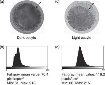
Fresh oocyte samples, before IVM (0 h), at 22 to 24 h and 44 to 48 h of IVM, were examined under Nomarski differential interference contrast microscope for lipid content evaluation (Pereira et al., Reference Pereira, Carvalhais, Pimenta, Baptista, Vasques, Horta, Santos, Marques, Reis, Pereira and Marques2008). Oocytes were photographed using an Olympus camera (Olympus Portugal, Lisboa, Portugal) attached to the microscope and analysed using ImageJ software (version 1.44q, National Institutes of Health, USA). A total of 100 LD randomly distributed at different focal planes were evaluated per oocyte. Total area occupied by LD, denominated fat area, and the gray mean value within fat area of each oocyte were also measured with the same equatorial focal plane, brightness and contrast. This mean value was calculated by converting each pixel to a gray scale by ImageJ software (Figure 1). On the basis of the gray mean value within the fat area at 44 to 48 h of IVM, oocytes were classified into two categories: dark (<112 pixels/μm2) or light (>112 pixels/μm2) gray. In addition, cytoplasmic and total (including zona pellucida) areas of each oocyte were recorded. Fat oocyte index was calculated (fat area/total oocyte area × 100).
Fat areas (arrows) of porcine oocytes evaluated by their gray mean values after Nomarski microscopy observation at the equatorial plan: a dark oocyte from control group after 44 to 48 h of in vitro maturation (IVM) (a) and the correspondent gray value histogram (b); a light oocyte from the trans-10, cis-12 conjugated linoleic acid treatment after 44 to 48 h of IVM (c) and the correspondent gray value histogram (d). Gray scale: 0 pixels/μm2 – black to 255 pixels/μm2 – white.

Experimental design
Three experiments (three replicates each) were designed. Trans-10, cis-12 octadecadienoic acid (100 μM t10,c12 CLA, Matreya, Pleasant Gap, Pennsylvania, USA; Pereira et al., Reference Pereira, Baptista, Vasques, Horta, Portugal, Bessa, Silva, Pereira and Marques2007) and forskolin (10 μM, Men et al., Reference Men, Agca, Riley and Critser2006) were first diluted in ethanol because of its hydrophobicity and added to oocytes in the maturation media using different supplementation times according to the following experiments. The final concentration of ethanol was adjusted to 0.1% in all treatment groups.
Experiment 1
The objective of this experiment was to study the effect of t10,c12 CLA and forskolin, administered during total IVM period on porcine oocyte lipid content, maturation and developmental potential. Four experimental groups were designed: (1) Control – COC (n = 373) were matured during 44 to 48 h without supplementation; (2) Ethanol – COC (n = 337) were matured during 44 to 48 h supplemented with 0.1% ethanol; (3) t10,c12 CLA – COC (n = 322) were matured during 44 to 48 h with 100 μM t10,c12 CLA; (4) Forskolin – COC (n = 353) were matured during 44 to 48 h with 10 μM forskolin. After maturation, COC were inseminated (n = 816) with frozen-thawed boar semen and cleavage rates recorded. At 22 to 24 h and 44 to 48 h of IVM, samples of oocytes were assessed for meiosis progression (n = 493) and lipid content evaluation (n = 76). Immature oocytes (n = 97, 87 for meiosis progression and 10 for lipid content evaluation, respectively) were also evaluated.
Experiment 2
In Experiment 2, supplementation period of 100 μM t10,c12 CLA and 10 μM forskolin was reduced to the first 22 to 24 h followed by 22 to 24 h of standard protocol of IVM and their effect on porcine oocyte lipid content, maturation and developmental potential also studied. Four experimental groups were also established: (1) Control – COC (n = 373) were matured during 44 to 48 h without supplementation; (2) Ethanol – COC (n = 432) were matured during the initial 22 to 24 h with 0.1% ethanol; (3) t10,c12 CLA – COC (n = 330) were matured with 100 μM 10t,12c CLA during the initial 22 to 24 h; (4) Forskolin – COC (n = 354) were matured during the initial 22 to 24 h with 10 μM forskolin. After 44 to 48 h of maturation, COC were inseminated (n = 908) and cleavage rates recorded. At 22 to 24 h and 44 to 48 h, IVM samples of oocytes were assessed for meiosis progression (n = 506) and lipid content evaluation (n = 75). Immature oocytes (n = 97) were also evaluated.
Experiment 3
In the previous experiments, supplementation periods of forskolin showed deleterious effects on oocyte maturation and cleavage rates. A third experiment was performed to investigate whether a time reduction in forskolin supplementation up to the initial 2 h of IVM could avoid these effects, simultaneously maintaining its lipolytic effects. t10,c12 CLA and ethanol were not tested in this trial, as in previous experiments the former did not interfere with oocyte meiotic progression and the latter did interfere neither with oocyte meiotic progression nor with lipid content. Therefore, two experimental groups were performed: (1) Control – COC (n = 291) were matured without supplementation; (2) Forskolin – COC (n = 299) were matured with 10 μM forskolin during the initial 2 h culture. After 44 to 48 h of maturation, COC (n = 238) were inseminated and cleavage rates recorded. At 22 to 24 h and 44 to 48 h, IVM samples of oocytes were assessed for meiosis progression (n = 307) and lipid evaluation (n = 45). Immature oocytes (n = 103) were also evaluated.
Statistical analysis
All results are expressed as least square means (± standard error of means in text). In each experiment, data representing three replicates of cleavage and oocyte nuclear configuration, as well as of oocyte lipid content evaluation, were analysed using the MIXED procedure of Statistical Analysis System (SAS Institute, Cary, NC, USA). The mixed linear model included treatment or treatment and IVM time periods (0 h, 22 to 24 h and 44 to 48 h) as fixed effects for cleavage and oocyte nuclear configuration evaluation, respectively, and replicates as random effect. For lipid content evaluation, treatment and IVM time periods were also considered fixed effects, but the oocyte nested in treatment was the random effect. LD were treated as repeated measures within oocyte. When significant effects were identified, differences between individual treatments were determined by the PDIFF multiple comparison procedure.
Data from Nomarski records were analysed using a multiple regression analysis (Proc Path of SAS), testing whether the fat gray tones were correlated to other individual or associated variables. An appropriate multicolinearity test (Proc Reg) was performed. The χ 2 test (Proc Freq) was used to compare the distribution of oocytes into categories (dark and light gray) of fat gray value. Differences were considered significant when P ⩽ 0.05.
Results
Fat gray value was independent from oocyte total (r = 0.07, P = 0.48), fat (r = −0.02, P = 0.84) and LD (r = −0.04, P = 0.58) areas and fat oocyte index (r = −0.14, P = 0.10). Instead, fat area and fat oocyte index were highly correlated (r = 0.81, P < 0.0001). This association explained 4% of fat gray value (tolerance value = 0.35, variance inflation = 2.8). The path coefficient from fat area to fat oocyte index was +1.50.
Experiment 1
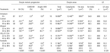
Data from meiosis progression of porcine oocytes supplemented with t10,c12 CLA and forskolin during 44 to 48 h of IVM are represented in Table 1. As expected, the GV configuration was predominant in immature oocytes. At 22 to 24 h of IVM, 93.4 ± 7.9% (P < 0.0001) of oocytes treated with forskolin remained at GV, whereas oocytes from control, ethanol and t10,c12 CLA treatments mainly reached GVBD-MI (P < 0.0001). Moreover, at the end of IVM, only 5.1 ± 5.1% of forskolin-treated oocytes reached Anaph.-MII compared with 72.6 ± 5.0%, 65.1 ± 5.1% and 61.0 ± 5.0% from control, ethanol and t10,c12 CLA treatments (P < 0.0001), respectively. At each time period, no differences (P > 0.05) were identified in nuclear configuration among oocytes from control, ethanol and t10,c12 CLA treatments.
Effect of t10,c12 CLA and forskolin supplementation during 44 to 48 h of in vitro maturation on porcine oocyte meiosis progression, oocyte and LD areas, in three independent replicates

t10,c12 CLA = trans-10, cis-12 conjugated linoleic acid; LD = lipid droplets; GV = germinal vesicle; GVBD-MI = germinal vesicle breakdown up to metaphase I; Anaph.I-MII = anaphase I up to metaphase II; r.s.d. = residual standard deviation.
Oocyte areas: total (cytoplasmic plus zona pellucida); cytoplasmic (cytoplasmic, without zona pellucida); fat area (total area occupied by LD); fat index (fat area/oocyte total area × 100).
LD area: lipid droplets area within oocytes.
Significant differences were identified on oocyte meiosis progression and oocyte areas.
Data were analysed using the MIXED procedure of Statistical Analysis System considering treatment and in vitro maturation time periods as fixed effects.
a,b,c,d,eValues (least square means) with different superscripts letters in the same column are significantly different (P ⩽ 0.05).
The cleavage rate of forskolin-matured oocytes was 0.0%, different (P ⩽ 0.0001) from those of control (55.4 ± 4.1%), ethanol (58.9 ± 4.2%) and t10,c12 CLA (45.6 ± 4.2%) treatments.
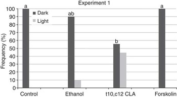
At the end of IVM, total areas of oocytes from control (P = 0.04), ethanol (P = 0.008) and t10,c12 CLA (P = 0.006) treatments were larger than those of immature oocytes (Table 1), whereas areas of forskolin-treated oocytes were similar to those of immature oocytes. Cytoplasmic areas of forskolin-treated oocytes were the smallest at 22 to 24 h (P ⩽ 0.03) and also at 44 to 48 h (P ⩽ 0.05) of IVM. At the end of maturation, cytoplasmic areas of t10,c12 CLA and ethanol-treated oocytes were larger (P ⩽ 0.04) than those of immature but similar to control. Fat areas were smaller in immature and forskolin-treated oocytes (22 to 24 h and 44 to 48 h) than in control at 22 to 24 h (P ⩽ 0.03) and ethanol at 44 to 48 h (P ⩽ 0.0002) of IVM. No differences were identified among groups in fat oocyte index and LD areas. However, after 44 to 48 h of culture, t10,c12 CLA oocytes were lighter than those of control (P = 0.03) and forskolin (P = 0.04) treatments (Figure 2).
Effect of trans-10, cis-12 conjugated linoleic acid (t10,c12 CLA) and forskolin, during 44 to 48 h of in vitro maturation supplementation, on porcine oocytes lipid content. Oocytes were classified into two categories: dark (<112 pixels/μm2) or light (>112 pixels/μm2) gray according to the gray mean value within fat area. a ≠ b, significant differences (P ⩽ 0.04) were observed in gray mean values within fat area of t10,c12 CLA oocytes compared to control or forskolin ones.

Experiment 2
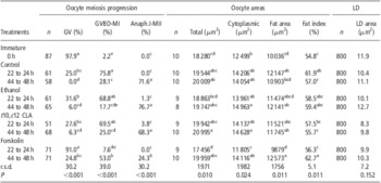
Results from meiosis progression of porcine oocytes supplemented with t10,c12 CLA and forskolin during the initial 22 to 24 h of IVM are shown in Table 2. In immature oocytes, GV configuration was predominant. Forskolin-treated oocytes remained mainly at GV stage at 22 to 24 h of IVM (91.0 ± 7.3%, P < 0.0001). On the contrary, the majority of oocytes from control, ethanol and t10,c12 CLA treatments were at GVBD-MI (P < 0.0001). At 44 to 48 h of IVM, 24.3 ± 5.4% of oocytes treated with forskolin during 22 to 24 h reached Anaph.-MII. This rate continues lower (P < 0.0001) than those from control, ethanol and t10,c12 CLA treatments. At each time period, no differences (P > 0.05) were identified in nuclear configuration among oocytes from control, ethanol and t10,c12 CLA treatments.
Effect of t10,c12 CLA and forskolin supplementation during 22 to 24 h of in vitro maturation on porcine oocyte meiosis progression, oocyte and LD areas, in three independent replicates

t10,c12 CLA = trans-10, cis-12 conjugated linoleic acid; LD = lipid droplets; GV = germinal vesicle; GVBD-MI = germinal vesicle breakdown up to metaphase I; Anaph.I-MII = anaphase I up to metaphase II; r.s.d. = residual standard deviation.
Oocyte areas: total (cytoplasmic plus zona pellucida); cytoplasmic (cytoplasmic, without zona pellucida); fat area (total area occupied by LD); fat index (fat area/oocyte total area × 100).
LD area: lipid droplets area within oocytes.
Significant differences were identified on oocyte meiosis progression and oocyte areas.
Data were analysed using the MIXED procedure of Statistical Analysis System considering treatment and in vitro maturation time periods as fixed effects.
a,b,c,d,eValues (least square means) with different superscripts letters in the same column are significantly different (P ⩽ 0.05).
The cleavage rate of oocytes matured with forskolin (8.3 ± 3.3%) was lower (P ⩽ 0.0001) than those of control (54.5 ± 3.0%), ethanol (44.9 ± 3.0%) and t10,c12 CLA (50.5 ± 4.2%) treatments.
After 44 to 48 h of maturation, total areas of oocytes from control and t10,c12 CLA treatments were larger (P ⩽ 0.05) than those of immature and forskolin at 22 to 24 h of maturation (Table 2). These latter oocytes presented also the smallest cytoplasmic areas (P ⩽ 0.02). After 44 to 48 h of maturation with forskolin supplementation during 22 to 24 h, oocyte areas (total and cytoplasmic) were not different from those of other groups at the same time period. At 22 to 24 h of IVM, oocytes cultured with forskolin presented smaller fat areas than control (P = 0.006) and t10,c12 CLA (P = 0.05). However, the forskolin group showed a larger fat area than control (P = 0.04) at 44 to 48 h of IVM. Oocyte fat areas of immature (0 h) and forskolin groups at 22 to 24 h were also smaller (P ⩽ 0.03) than those of forskolin and ethanol oocytes at 44 to 48 h of IVM (Table 2). Fat oocyte index of forskolin treatment was lower (P = 0.02) than control at 22 to 24 h. At 44 to 48 h of IVM, the former treatment results became superior to control (P = 0.02) and also to forskolin (P = 0.009) oocytes at 22 to 24 h. No differences were identified between oocyte categories (dark and light gray) or LD areas among groups.
Experiment 3
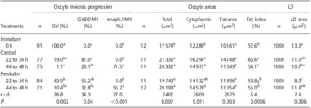
By reducing the time of forskolin exposure to 2 h, oocyte meiotic progression at 22 to 24 h and 44 to 48 h was similar (P > 0.05) to those of control (Table 3). No differences were identified in cleavage rates between treatments.
Effect of forskolin supplementation during the initial 2 h of in vitro maturation on porcine oocyte meiosis progression, oocyte and LD areas, in three independent replicates.

LD = lipid droplets; GV = germinal vesicle; GVBD-MI = germinal vesicle breakdown up to metaphase I; Anaph.I-MII = anaphase I up to metaphase II; r.s.d. = residual standard deviation.
Oocyte areas: total (cytoplasmic plus zona pellucida); cytoplasmic (cytoplasmic, without zona pellucida); fat area (total area occupied by LD); fat index (fat area/oocyte total area × 100).
LD area : lipid droplets area within oocytes.
Significant differences were identified on oocyte meiosis progression and oocyte and LD areas.
Data were analysed using the MIXED procedure of Statistical Analysis System considering treatment and in vitro maturation time periods as fixed effects.
a,b,cValues (least square means) with different superscripts letters in the same column are significantly different (P ⩽ 0.05).
At the end of IVM, cytoplasmic and total areas of oocytes from both groups were similar among themselves and larger (P ⩽ 0.01) than those of immature oocytes (Table 3). Exception made for cytoplasmic areas of forskolin oocytes, these measurements at 22 to 24 h of IVM were also larger (P ⩽ 0.04) than those of immature oocytes. Control oocytes matured during 22 to 24 h presented the largest (P ⩽ 0.03) fat area (Table 3). Exception made for forskolin oocytes at 22 to 24 h, similar results (P ⩽ 0.03) were obtained for fat oocyte index. This index presented no differences between treatments at the end of IVM. In addition, no differences were identified between oocyte categories (dark and light gray). Although reducing the time exposure to forskolin (2 h), at 22 to 24 h of IVM, these oocytes had smaller LD areas than those of control (P = 0.03). These LD were also smaller than those of immature (P = 0.002) and forskolin oocytes at the end of IVM (P = 0.03). LD areas of immature oocytes were larger (P = 0.02) than control ones at 44 to 48 h (Table 3).
Discussion
The present study demonstrated that the supplementation of 100 μM t10,c12 CLA or 10 μM forskolin during different time periods distinctively interfere on maturation progress and lipid content of porcine oocytes. Data presented here suggest for the first time that t10,c12 CLA did not affect meiosis progression or competence for cleavage of porcine oocytes, although matured oocytes were lighter when this CLA isomer was supplemented during the entire maturation period. In opposition, forskolin presence during 44 to 48 h or 22 to 24 h impaired oocyte maturation and their competence for cleavage. However, by decreasing forskolin supplementation interval to the initial 2 h of IVM, these deleterious effects disappeared though morphological changes in LD and oocyte fat areas were identified during maturation progression.
Previous reports of continuing exposure of porcine oocytes to forskolin along maturation have demonstrated an increase of intracellular level of cyclic adenosine monophosphate (cAMP) thus inhibiting GVBD (Racowsky, Reference Racowsky1985; Xia et al., Reference Xia, Kikuchi, Noguchi and Izaike2000). In accordance, since at 44 to 48 h of culture, GV configuration continued to be predominant in forskolin-treated oocytes, remaining their areas similar to immature ones, these oocytes were incapable of normal fertilization and cleavage (0%). In contrast, Fu et al. (Reference Fu, Wu, Li, Hou, Zhou, Wang and Zhu2011) achieved a cleavage rate of 64.5 ± 3.6% in parthenogenetically activated porcine oocytes cultured with 10 μM forskolin during the entire IVM period. An increase in cytoplasmic calcium during parthenogenic activation was referred to be sufficient to induce low to moderate extents of oocyte activation events such as cell cycle resumption (Gardner and Evans, Reference Gardner and Evans2006). Differences between these two mechanisms, parthenogenesis and cleavage, might explain the above discrepancies.
The inhibitory effect of forskolin during maturation seems to be transiently depending on the supplementation time and dose. However, at higher doses above 10 μM, forskolin presents a toxic effect on COC metabolism (Racowsky, Reference Racowsky1985; Xia et al., Reference Xia, Kikuchi, Noguchi and Izaike2000). Our findings showed that by reducing forskolin exposure to the initial 22 to 24 h of maturation, GVBD-MI nuclear status became predominant at the end of IVM. Despite of a maturation rate of 24.3 ± 5.4%, only 8.3 ± 3.3% of inseminated oocytes cleaved. Moreover, forskolin affected not only nuclear progression, but also oocyte cytoplasmic and total areas. Therefore, the transient delay in nuclear meiosis progress of porcine COC matured in the presence of 10 μM forskolin identified initially by Racowsky (Reference Racowsky1985) might be extended to cytoplasmic maturation. However, when this supplementation was reduced to the first 2 h, meiosis progression, oocyte areas and cleavage rates were similar to those of control group. Nonetheless, several authors suggest that forskolin might be applied during oocytes IVM to improve synchronization of nuclear and cytoplasmic maturation, simultaneously increasing the homogeneity and developmental potential of matured oocytes (Racowsky, Reference Racowsky1985; Thomas et al., Reference Thomas, Thompson, Armstrong and Gilchrist2004). Further studies are needed to clarify forskolin positive action on oocyte developmental competence for embryo production and quality.
Porcine oocytes present variations in both morphology and amount of cytoplasmic LD during in vivo or in vitro maturation (Kikuchi et al., Reference Kikuchi, Ekwall, Tienthai, Kawai, Noguchi, Kaneko and Rodriguez-Martinez2002). In the present study, we investigated the extent of these variations by measuring LD areas and total area occupied by LD, the fat areas, as well as the relationship of fat area with total oocyte area through the fat oocyte index, before and during porcine oocyte maturation. Moreover, oocytes exhibit distinct colour tones of cytoplasm due to LD content: dark colour and opaqueness in lipid rich ones contrasting with the brightness and transparency of leaner oocytes (Fujihira et al., Reference Fujihira, Kinoshita, Sasaki, Ohnishi, Ishikawa, Ohsumi and Fukui2004). Furthermore, Isachenko et al. (Reference Isachenko, Isachenko, Rahimi, Mallmann, Krivokhrchenko and Nawroth2003) identified two kinds of LD in porcine oocytes: homogenous, dark looking vesicles and ‘gray’ looking droplets with electron-lucent streaks. Silva et al. (Reference Silva, Báo, Jivago and Lucci2011) suggested that dark LD may change to gray after lipid utilization. By measuring the fat gray value of porcine oocytes using a Nomarski microscope and ImageJ software, it was possible to distinguish different colour tones of fat areas reflecting alterations in lipid content. Differences in the gray tones might suggest not only alterations of the composition, amount and morphology of LD but also its aggregation or dispersion in the ooplasm. Several authors confirmed that as maturation progresses, the ooplasm can have different shades of gray caused by uneven distribution of organelles, specifically mitochondria and LD, which reside in close proximity (Kikuchi et al., Reference Kikuchi, Ekwall, Tienthai, Kawai, Noguchi, Kaneko and Rodriguez-Martinez2002; Ambruosi et al., Reference Ambruosi, Lacalandra, Iorga, De Santis, Mugnier, Matarrese, Goudet and Dell'Aquila2009). Here it was shown that after 44 to 48 h of culture, t10,c12 CLA oocytes were lighter (dark gray = 55.6% and light gray = 44.4%) than those of control and forskolin treatments (dark gray = 100%). Reports confirm the reduction of lipid accumulation in in vitro embryos (Pereira et al., Reference Pereira, Baptista, Vasques, Horta, Portugal, Bessa, Silva, Pereira and Marques2007 and Reference Pereira, Carvalhais, Pimenta, Baptista, Vasques, Horta, Santos, Marques, Reis, Pereira and Marques2008) or the decrease in lipogenesis of porcine adipose explants (José et al., Reference José, Gama and Lanna2008) after culture with this CLA isomer. Moreover, the presence of t10,c12 CLA during maturation improved bovine oocyte developmental competence (Lapa et al., Reference Lapa, Marques, Alves, Vasques, Baptista, Carvalhais, Silva Pereira, Horta, Bessa and Pereira2011). Although these latter results at present have not been confirmed for porcine oocytes, t10,c12 CLA during maturation clearly interfered with LD organization and probably with their composition reflected by a lighter colour tone of their cytoplasm. The mechanism of action of t10,c12 CLA is not clearly known, demanding additional investigation.
Even though forskolin stimulates lipolysis (Men et al., Reference Men, Agca, Riley and Critser2006), the fat gray value of forskolin-treated oocytes were not different from control independently of time exposure or meiosis progression. On the contrary, Fu et al. (Reference Fu, Wu, Li, Hou, Zhou, Wang and Zhu2011) showed that 10 μM forskolin effectively reduced the fluorescence intensity of porcine oocytes stained with Nile Red as well as the number of LD during IVM. Data presented here demonstrated no differences in LD areas when this lipolytic agent was present during the entire maturation period or till 22 to 24 h, but fat areas were reduced (exp. 1: control =12003 μm2v. forskolin = 9769 μm2, P ⩽ 0.03; exp. 2: control = 12147 μm2v. forskolin = 9879 μm2, P = 0.006). It seems that by blocking meiosis progression through the cAMP intracellular level increase (Racowsky, Reference Racowsky1985; Xia et al., Reference Xia, Kikuchi, Noguchi and Izaike2000), forskolin interferes also in the characteristic distribution of LD in ooplasm during the maturation process, resembling the immature oocyte organization. Similar to what happened to the total and cytoplasmic oocyte areas, the fat areas did not grow. Therefore, the oocyte fat indexes were similar to control. The mechanism by which forskolin interfered with LD organization is not well understood but its lypolitic activity in porcine oocytes as well as in embryos has already been identified (Men et al., Reference Men, Agca, Riley and Critser2006; Fu et al., Reference Fu, Wu, Li, Hou, Zhou, Wang and Zhu2011). Herein, even when forskolin exposure was only at the initial 2 h of culture, these oocytes at 22 to 24 h had smaller LD and fat areas compared with the control group, although at 44 to 48 h of meiosis these measurements were similar to those of control.
In conclusion, the present study demonstrates that the distribution and morphology of cytoplasmic LD during porcine oocyte maturation can be modified by t10,c12 CLA or forskolin reducing its lipid content in a time-dependent way. However, at the doses tested, whereas t10,c12 CLA did not seem to interfere with oocyte meiosis progression or developmental competence, forskolin should be supplemented for only 2 h to prevent the negative effects on these processes. On the other hand, fat gray value seems a potential tool to evaluate the lipid content of a single porcine oocyte.
Acknowledgements
The ‘Fundação para a Ciência e Tecnologia’ is greatly acknowledged for funding a grant for E.G. Prates (SFRH/BD/42359/2007).