Introduction
Parthenogenetic activation of mammalian oocytes has been induced by a wide variety of physical or chemical stimuli in vitro (Kaufman, Reference Kaufman1983) and the optimal activation protocol is dependent upon different species. Low temperatures are able to trigger parthenogenesis in rabbits (Chang, Reference Chang1954), sheep (Moor & Crosby, Reference Moor and Crosby1985) and cattle (Powell & Barnes, Reference Powell and Barnes1992). Electrical pulses are widely used for activation of oocytes in mammals, such as rabbit (Ozil, Reference Ozil1990), mouse (Vitullo & Ozil, Reference Vitullo and Ozil1992), cattle (Collas et al., Reference Collas, Fissore, Robl, Sullivan and Barnes1993a), sheep (Tacitus, Reference Tacitus, Moore and Jackson1937) and pig (Grupen et al., Reference Grupen, Verma, Du, McIlfatrick, Ashman and Nottle1999). Ethanol was the first chemical to be used to activate mammalian eggs. Mouse (Cuthbertson et al., Reference Cuthbertson, Whittingham and Cobbold1981; Cuthbertson, Reference Cuthbertson1983; Winston & Maro, Reference Winston and Maro1995), bovine (Nagai, Reference Nagai1987; Presicce & Yang, Reference Presicce and Yang1994; Yang et al., Reference Yang, Presicce, Moraghan, Jiang and Foote1994), ovine (Loi et al., Reference Loi, Ledda, Fulka, Cappai and Moor1998) and porcine oocytes (Didion et al., Reference Didion, Martin and Markert1990) have been efficiently activated by ethanol, but parthenogenetic development of the oocytes was found to be very poor in rabbits (Onodera & Tsunoda, Reference Onodera and Tsunoda1989) and humans (Winston et al., Reference Winston, Johnson, Pickering and Braude1991). Parthenogenetic activation is initiated by the stimuli that evoke calcium transients in the oocyte. Electrical pulses (Ducibella et al., Reference Ducibella, Huneau, Angelichio, Xu, Schultz, Kopf, Fissore, Madoux and Ozil2002), ethanol (Cuthbertson et al., Reference Cuthbertson, Whittingham and Cobbold1981; Cuthbertson, Reference Cuthbertson1983; Nagai, Reference Nagai1987; Loi et al., Reference Loi, Ledda, Fulka, Cappai and Moor1998), strontium (O'Neill et al., Reference O'Neill, Rolfe and Kaufman1991; Meo et al., Reference Meo, Yamazaki, Leal, de Oliveira and Garcia2005; Tomashov-Matar et al., Reference Tomashov-Matar, Tchetchik, Eldar, Kaplan-Kraicer, Oron and Shalgi2005; Yamazaki et al., Reference Yamazaki, Ferreira, Meo, Leal, Meirelles and Garcia2005) and calcium ionophores (Jones et al., Reference Jones, Carroll and Whittingham1995; Tesarik & Sousa, Reference Tesarik and Sousa1995; Loi et al., Reference Loi, Ledda, Fulka, Cappai and Moor1998) have been proved to induce calcium transients successfully in the oocyte and initiate activation. However, the oocytes activated by these stimuli are unable to develop to blastocysts, because most of the activated oocytes form one pronucleus and release the second polar body after activation (Wang et al., Reference Wang, Machaty, Abeydeera, Prather and Day1998). To prevent the release of a second polar body, cycloheximide (CHX, the protein synthesis inhibitor), 6-dimethylaminopurine (6-DMAP, the histone kinase inhibitor) or cytochalasin B (CB, an inhibitor of microfilaments) was combined with one of the above stimuli to improve parthenogenetic development of mammalian oocytes (Fukui et al., Reference Fukui, Sawai, Furudate, Sato, Iwazumi and Ohsaki1992). The combination is species dependent. For example, ethanol combined with cytochalasins (Kubiak et al., Reference Kubiak, Paldi, Weber and Maro1991; Fukui et al., Reference Fukui, Sawai, Furudate, Sato, Iwazumi and Ohsaki1992), CHX (Presicce & Yang, Reference Presicce and Yang1994) or 6-DMAP (Ledda et al., Reference Ledda, Loi, Bogliolo, Moor and Fulka1996; Loi et al., Reference Loi, Ledda, Fulka, Cappai and Moor1998) and ionophores combined with 6-DMAP (Susko-Parrish et al., Reference Susko-Parrish, Leibfried-Rutledge, Northey, Schutzkus and First1994) or CHX (Tacitus, Reference Tacitus, Moore and Jackson1937) is efficient for parthenogenetic activation and of benefit to subsequent blastocyst development. Strontium, an alternative divalent cation, can effectively induce the artificial activation of mammalian oocytes (Fraser, Reference Fraser1987; Tomashov-Matar et al., Reference Tomashov-Matar, Tchetchik, Eldar, Kaplan-Kraicer, Oron and Shalgi2005) and trigger multiple intracellular calcium spikes in a similar way to that of fertilization (Kline & Kline, Reference Kline and Kline1992). Parthenogenetic development has also been improved by the combination of SrCl2 with CHX (Bos-Mikich et al., Reference Bos-Mikich, Swann and Whittingham1995) or CB (Ma et al., Reference Ma, Liu, Miao, Han, Zhang, Miao, Yanagimachi and Tan2005). However, strontium is much more effective for mouse oocyte activation rather than for bovine oocytes (Meo et al., Reference Meo, Leal and Garcia2004). These findings indicate that parthenogenetic activation pathways vary from one species to another. Because oocyte activation is a crucial step for the success of mammalian somatic cell nuclear transfer (SCNT), it is essential to investigate the optimal protocol for oocyte activation for each species of animals.
The first somatic cell cloned goats were been produced by using electrical activation combined with CB (Baguisi et al., Reference Baguisi, Behboodi, Melican, Pollock, Destrempes, Cammuso, Williams, Nims, Porter, Midura, Palacios, Ayres, Denniston, Hayes, Ziomek, Meade, Godke, Gavin, Overstrom and Echelard1999), however, the activation efficiency for goat oocytes was much lower compared with bovine oocytes (Zakhartchenko et al., Reference Zakhartchenko, Alberio, Stojkovic, Prelle, Schernthaner, Stojkovic, Wenigerkind, Wanke, Duchler, Steinborn, Mueller, Brem and Wolf1999). Only a few reports have been published on goat oocyte activation by different protocols (Ongeri et al., Reference Ongeri, Bormann, Butler, Melican, Gavin, Echelard, Krisher and Behboodi2001; Lan et al., Reference Lan, Han, Wu, Han, Ma, Liu, Chang and Tan2005), although ionomycin combined with 6-DMAP has been successfully employed for activation of SCNT embryos to clone goats (Keefer et al., Reference Keefer, Baldassarre, Keyston, Wang, Bhatia, Bilodeau, Zhou, Leduc, Downey, Lazaris and Karatzas2001; Ongeri et al., Reference Ongeri, Bormann, Butler, Melican, Gavin, Echelard, Krisher and Behboodi2001; Guo et al., Reference Guo, An, Yu, Li, Li, Guo and Zhang2002; Lan et al., Reference Lan, Chang, Luo, Jiang, Han, Wu, Han, Ma and Tan2006). It is still important to develop and improve the current protocols for goat oocyte activation.
The present study set out to investigate the different activation protocols for goat oocytes. Temperature shock, electrical pulses, ethanol, ionomycin and SrCl2 were employed for activation of in vitro matured goat oocytes. Parthenogenetic development was evaluated following activation. The activation of SCNT embryos was also studied using selected activation protocols.
Materials and Methods
Animals and chemicals
All experiments using animals were performed in accordance with the regulations for the administration of affairs concerning experimental animals from the state council of China.
All chemicals were purchased from Sigma Chemical Co. unless stated otherwise.
Culture media
Culture media used for maturation and manipulation of oocytes are based on tissue culture medium 199 (M199) (Gibco BRL Life Technologies). Oocyte maturation medium (MM199) is M199 supplemented with 5% fetal bovine serum (FBS; HyClone), 50 μg/ml gentamycin, 0.075 IU/ml human menopausal gonadotropin (hMG) (Serono Laboratories Inc.), 1 μg/ml 17β-estradiol and 0.38 mM pyruvate. Oocyte culture medium (CM199) is M199 supplemented with 5% FBS, 50 μg/ml gentamycin and 0.38 mM pyruvate. The handling medium (HM199) is M199 supplemented with 3 mg/ml bovine serum album (BSA) and 15 mM HEPES. Embryo culture medium (mCR1aa) is modified from CR1aa (Rosenkrans et al., Reference Rosenkrans, Zeng, GT, Schoff and First1993; Rosenkrans & First, Reference Rosenkrans and First1994) by reducing the NaCl concentration to 109.5 mM and supplementing 1.19 mM NaH2PO4, 1.5 mM d-glucose, 0.80 mM MgCl2, 6 mg/ml BSA, 5% FBS, 1% non-essential amino acids (NEAA, Gibco BRL Life Technologies) and 2% essential amino acids solution (EAA, Gibco BRL Life Technologies) (Guo et al., Reference Guo, An, Yu, Li, Li, Guo and Zhang2002). DMEM (Gibco BRL Life Technologies) plus 10% FBS was used for all cell culture.
Oocyte isolation and maturation in vitro
Dairy goat ovaries were collected in local abattoirs during the breeding seasons and were delivered to the laboratory within 6 h in saline with 0.32 mg/ml gentamycin at 20–28 °C. Cumulus–oocyte complexes (COCs) were isolated by cutting the surfaces of 1–5 mm follicles in phosphate-buffered saline (PBS) (Gibco BRL Life Technologies). Cumulus-enclosed oocytes were washed three times in HM199. Groups of 100–200 COCs were transferred into 2 ml MM199 and cultured at 38.5 °C in a humidified atmosphere of 5% CO2 in air for in vitro maturation (Guo et al., Reference Guo, An, Yu, Li, Li, Guo and Zhang2002).
Parthenogenetic activation and embryo culture in vitro
The oocytes were denuded from cumulus cells by pipetting with 0.2% hyaluronidase in PBS at 20–24 h post maturation. Oocytes with the second polar body were selected and assigned to five groups for parthenogenetic activation. After activation, the oocytes were cultured in 1 ml of mCR1aa at 38.5 °C in a humidified atmosphere of 5% CO2 in air. Cleavage and blastocyst development was observed at 48 h and day 8 of culture in vitro, respectively.
The Group 1 procedure was carried out using temperature shock. In vitro matured oocytes were incubated at 22–24 °C in HM199 in the air for 3 h or 12 h and then cultured in mCR1aa at 38.5 °C in a humidified atmosphere of 5% CO2 in air to allow the embryos to develop in vitro.
The Group 2 procedure was carried out using electrical pulses. In vitro matured oocytes were placed between the electrodes of a 0.5 mm gap fusion chamber overlaid with fusion buffer, containing 0.3 mM mannitol, 100 mM CaCl2, 100 mM MgCl2, 0.5 mM HEPES and 0.05% BSA. Oocyte activation was carried out by two direct current (DC) pulses of 1.2 kV/cm applied for 20 μs at 1 s interval (CRY-3 cell fusion apparatus, Ningbo Scientz Biotechnology Co., Ltd). After being washed with mCR1aa, the oocytes were cultured in mCR1aa containing 2 mM 6-DMAP for 3 h and then cultured in mCR1aa at 38.5 °C in a humidified atmosphere of 5% CO2 in air for further development or immediately were cultured in mCR1aa to allow the embryos to further develop.
The Group 3 procedure was carried out using ethanol. In vitro matured oocytes were exposed to 7% ethanol in HM199 for 7 min, 10% for 5 or 10 min and 20% for 5 min at room temperature, respectively. After the treatment, the oocytes were cultured in mCR1aa with 2 mM 6-DMAP for 3 h and then in mCR1aa, or directly cultured in mCR1aa at 38.5 °C in a humidified atmosphere of 5% CO2 in air for further development.
The Group 4 procedure was carried out with ionomycin plus 6-DMAP. In vitro matured oocytes were exposed to 5 μM ionomycin in HM199 for 4 min at room temperature followed by culture in mCR1aa containing 2 mM 6-DMAP (I-DMAP) for 4 h, 3 h, 2 h or 0.5 h; or in mCR1aa with 2 mM 6-DMAP plus 5 μg/ml CB (I-DMAP-CB) for 4 h, 3 h or 2 h. After activation, the oocytes were washed completely and cultured in mCR1aa at 38.5 °C in a humidified atmosphere of 5% CO2 in air for further development.
The Group 5 procedure was carried out with SrCl2 plus CB. In vitro matured oocytes were cultured for 6, 12, 16 or 24 h in Earle's balanced salt solutions (EBSS) without calcium but with 10 mM SrCl2, 5 μg/ml CB and 6 mg/ml BSA (Sr2+-CB). After activation, the oocytes were washed and then cultured in mCR1aa at 38.5 °C in a humidified atmosphere of 5% CO2 in air for further development.
Goat SCNT and SCNT embryo activation
The oocytes used for nuclear transfer were denuded from cumulus cells by pipetting with 0.2% hyaluronidase in PBS at 18–20 h post maturation. The denuded oocytes were incubated in HM199 at room temperature for 10 min followed by culture in CM199 at 38.5 °C for 30 min to induce the oocytes to protrude membrane partially, at a point where the metaphase II (MII) chromosomes are located (Fig. 1). The oocytes with protruding membranes were selected and enucleated in HM199 containing 5 μg/ml CB. Enucleation was carried out under a microscope by the aid of a micromanipulator (Narishige). The enucleated oocytes were incubated in CM199 at 38.5 °C until somatic cell injection (Guo et al., Reference Guo, An, Yu, Li, Li, Guo and Zhang2002).
In vitro matured goat oocytes treated at room temperature for 30 min. The clearly protruded membranes are shown in the image.

Goat cumulus cells were isolated from in vitro matured oocytes and cultured in DMEM plus 10% FBS. The cells were starved with DMEM plus 0.5% FBS for 2–5 days at passages 5–9 when they were confluent. Before nuclear transfer, the cells were dissociated by 0.25% trypsin/EDTA. The individual cells were disrupted by pipetting with an injection pipette in HM199 containing 10% polyvinylpyrrolidone (PVP) (360 000 kDa) and the karyoplast was injected into the enucleated oocyte (Guo et al., Reference Guo, An, Yu, Li, Li, Guo and Zhang2002). Injected oocytes were cultured in mCR1aa at 38.5 °C for 3–6 h before activation.
Some of the injected oocytes were activated with 5 μM ionomycin in HM199 for 4 min followed by culture in 2 mM 6-DMAP in mCR1aa for 3 h, others were incubated with calcium-free EBSS containing 10 mM SrCl2, 5 μg/ml CB and 6 mg/ml BSA for 12 h. After activation, the embryos were washed and cultured in mCR1aa at 38.5 °C in a humidified atmosphere of 5% CO2 in air to allow further development.
In vivo development of SCNT embryos derived from strontium and ionomycin activation
Three 4-cell embryos developed from SCNT were surgically transferred to each side of the oviduct of recipient dairy goats at day 2 of natural estrus. In vivo development of cloned embryos was examined fortnightly by ultrasonics from day 30 to day 60.
Statistical analyses
Proportions of embryos cleaved to 2-cell and developed to blastocyst stage from various treatments within each experiment were analysed by chi-squared test (http://statpages.org/ctab2×2.html). A p-value less than 0.05 was considered as a significant difference between treatments.
Results
Parthenogenetic activation of goat oocytes by low temperature shock
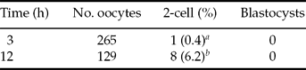
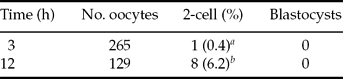
The goat oocytes were insensitive to low temperature under our experimental conditions. Only 6.2% (8/129) and 0.4% (1/265) of oocytes were cleaved when the oocytes were incubated at 22–24 °C (room temperature) for 3 h and 12 h, respectively. None of the embryos developed beyond the 2-cell stage except for some of abnormal fragments when the embryos were further cultured (Table 1). The results indicated that low temperature conditions had a very limited capacity to activate goat oocytes.
Development of goat oocytes treated at room temperature (22–24°C).
a ,b Values with different superscripts within each column differ significantly (p < 0.01).
Parthenogenetic activation of goat oocytes by electrical pulse
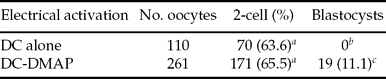
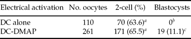
When goat oocytes were delivered with two DC pulses of 1.2 kV/cm for 20 μs alone or combined with 3 h culture in 2 mM 6-DMAP (DC-DMAP), 63.6% (70/110) and 65.5% (171/261) of oocytes were cleaved, respectively. Only 11.1% (19/171) of embryos activated by DC pulses combined with 6-DMAP developed into blastocysts (Table 2). There was no significant difference in the cleavage rates between the two groups (p > 0.05). The results showed that goat oocytes were able to be activated by electrical pulses and the activated oocytes had the potential to develop into blastocysts at a low rate.
Development of goat oocytes activated by electrical pulses.
a ,b,c Values with different superscripts within each column differ significantly (p < 0.01). Percentage of blastocyst development was calculated in relation to the 2-cell embryos.
Parthenogenetic activation of goat oocytes by ethanol
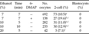
When goat oocytes were activated with 7% ethanol for 7 min, 10% ethanol for 5 min, 10% ethanol for 10 min and 20% ethanol for 5 min, 10.5% (73/692), 11.0% (31/282), 12.9% (30/233) and 7.1% (3/42) of the oocytes were cleaved, respectively. There were no significant differences in cleavage rates between these treatment groups (p > 0.05, Table 3). None of the oocytes developed into blastocysts by ethanol treatments, although 19.6% (27/138) of embryos were cleaved when the oocytes were treated with 7% ethanol for 7 min followed by culture in 2 mM 6-DMAP for 3 h. Our results suggested that goat oocytes at 20–24 h of in vitro maturation were unable to be activated by ethanol.
Development of goat oocytes activated by ethanol.

a ,b,c Values with different superscripts within each column differ significantly (p < 0.05).
+/−, activated oocytes were cultured with/without 2 mM 6-DMAP for a further 3 h.
Parthenogenetic activation of goat oocytes by ionomycin
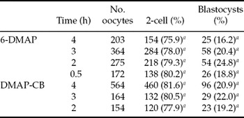
When goat oocytes were exposed to ionomycin for 4 min at room temperature followed by culture in 6-DMAP for 4 h, 3 h, 2 h and 0.5 h, 75.9% (154/203), 78.0% (284/364), 79.3% (218/275) and 80.2% (138/172) of the activated oocytes were cleaved and 16.2% (25/154), 20.4% (58/284), 24.8% (54/218) and 18.8% (26/138) of the cleaved embryos developed into blastocysts after further culture, respectively. While the oocytes were treated with 5 μM ionomycin for 4 min then in DMAP-CB for 4 h, 3 h and 2 h, the cleavage rates were 81.6% (460/564), 80.5% (164/132) and 77.9% (120/154) and blastocyst development rate was 20.9% (96/460), 22.0% (29/132) and 19.2% (23/120), respectively. There was no significant difference between the different treatment groups (p > 0.05, Table 4). This result indicated that goat oocytes were able to be activated by ionomycin combined with 6-DMAP or DMAP-CB and there were no significant effects on cleavage and blastocyst development when using a combination of I-DMAP for 0.5–4 h or I-DMAP-CB for 2–4 h. This finding indicates that even culture for 0.5 h in 6-DMAP is sufficient for in vitro development of normal parthenogenetic blastocysts (Fig. 2a).
Development of goat oocytes activated by ionomycin combined with 6-DMAP or DMAP-CB.

aValues with the same superscript within each column do not differ significantly (p > 0.05). Percentage of blastocyst development was calculated in relation to the 2-cell embryos.
Blastocysts developed in vitro from oocytes parthenogenetically activated by I-DMAP (a) and SrCl2-CB (b). (a) Blastocysts developed from ionomycin activation combined with 0.5 h culture in 6-DMAP. (b) Blastocysts were stained with Hoechst 33342 to show cell nuclei.

Parthenogenetic activation of goat oocytes by SrCl2
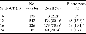
When goat oocytes were activated by incubation in Ca2+-free EBSS supplemented with 10 mM SrCl2 and 5 μg/ml CB for 6 h, 12 h, 16 h and 24 h, the cleavage rate was 2.2% (3/139), 80.4% (436/542), 78.8% (178/226) and 70.6% (60/85) and the blastocyst rate was 0% (0/3), 15.6% (68/436), 10.1% (18/178) and 1.7% (1/60), respectively. Our results showed that goat oocytes were unable to be activated by 6 h culture with SrCl2, but activated by culture with SrCl2 for 12 h and above. There was no significant difference between activation groups (12, 16 and 24 h group), but the blastocyst development rates were significantly higher in the 12 h and 16 h groups compared with the 24 h group (Table 5). The results indicated that in vitro developmental capacity of goat oocytes might be impaired due to damage from extended exposure to SrCl2. The blastocysts derived from strontium activation showed typical cells within the blastocysts (Fig. 2b).
Development of goat oocytes activated by SrCl2-CB.
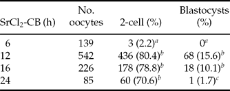
a ,b Values with different superscripts within each column differ significantly (p < 0.01). Percentage of blastocyst development was calculated in relation to the 2-cell embryos.
Developmental potential of goat SCNT embryos activated by SrCl2 and ionomycin
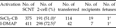
When the cloned embryos were activated by Sr2+-CB and I-DMAP, 51.0% (191/375) and 72.5% (298/411) of embryos developed into the 2-cell stage, respectively. The cleavage rates from Sr2+ and ionomycin activation group differed significantly (p < 0.01) (Table 6). When 114 4-cell embryos activated by Sr2+-CB were transferred into 19 recipients and 42 embryos activated by I-DMAP were transferred into seven recipients, one recipient from each group (1/19 and 1/7) was pregnant by ultrasonographic pregnancy diagnosis. However, none of these fetuses developed to full term, both conceptuses were lost between 45 and 60 days of pregnancy.
In vivo development of goat SCNT embryos activated by SrCl2-CB and I-DMAP.
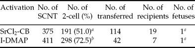
a ,b Values with different superscripts within each column differ significantly (p < 0.01).
Discussion
Oocytes are sensitive to low temperature shock in some mammalian species. For example, unfertilized rabbit eggs (18%) are able to develop into blastocysts in vivo after being stored at 10 °C for 1 day (Chang, Reference Chang1954). Sheep oocytes from germinal vesicle breakdown (GVBD) to metaphase II are also very sensitive to low temperatures (20 °C) (Moor & Crosby, Reference Moor and Crosby1985), even room temperature (23–27 °C) could efficiently trigger bovine oocyte activation comparable to sperm (Stice et al., Reference Stice, Keefer and Matthews1994). However in our study, goat oocytes were found to be difficult to activate at room temperature. In our experiments, only a few oocytes were activated and most of these showed abnormalities and none of them developed beyond the 8-cell stage after further culture. These results suggested that the activation pathway of goat oocyte somehow differed from that of other mammals. Room temperature could not activate goat oocytes efficiently, only induce goat oocytes (more than 70%, data not shown) to protrude the membrane to some extent and this situation would be a benefit to locate MII chromosomes for enucleation in SCNT (Guo et al., Reference Guo, An, Yu, Li, Li, Guo and Zhang2002). Such low temperature treatment did not affect the potential of goat oocytes to reprogramme somatic cells for the purpose of goat cloning. This finding suggests that goat oocytes can be manipulated at room temperature without loss of reprogramming potential for SCNT.
Electrical pulses can fully induce parthenogenetic activation of oocytes in mice, rabbits and cattle. About 78% of mouse, 77% of rabbit (Onodera & Tsunoda, Reference Onodera and Tsunoda1989) and 89% of bovine oocytes (Collas et al., Reference Collas, Sullivan and Barnes1993b) were activated by electrical stimulation and 32% of mouse, 25% of rabbit and 24% of bovine eggs developed to blastocysts after culture in vitro, respectively. Cloned goats have been generated by SCNT derived from cell fusion and activation with 2.2–3.2 kV/cm electrical pulses simultaneously or combination with additional electrical pulses (Baguisi et al., Reference Baguisi, Behboodi, Melican, Pollock, Destrempes, Cammuso, Williams, Nims, Porter, Midura, Palacios, Ayres, Denniston, Hayes, Ziomek, Meade, Godke, Gavin, Overstrom and Echelard1999; Melican et al., Reference Melican, Butler, Hawkins, Chen, Hayden, Destrempes, Williams, Lewis, Behboodi, Ziomek, Meade, Echelard and Gavin2005). However, the oocytes began to undergo cytolysis when 1.2 kV/cm electrical pulses were employed in our experiments. Even so, cleavage of goat oocytes obtained by our gentle electrical activation is similar to the results found from mouse and rabbit experiments, but blastocyst development is uncomparable. We suggest that the nuclear transfer embryos are more tolerant compared with fresh oocytes and that the aged oocytes were much more sensitive to electrical stimulation. Probably, the oocytes became insensitive after manipulation and the tolerances of oocytes derived from in vitro maturation differed from that of oocytes matured in vivo (Das & Majumdar, Reference Das and Majumdar2003). One report showed that activation of goat SCNT embryos was clearly improved by increasing the electrical field strength from 1.67 to 2.33 kV/cm (Shen et al., Reference Shen, Lee, Wu, Huang, Chu, Chang, Kung, Lin, Chen, Shiau, Yen and Cheng2006). Compared with ionomycin in our experiments, electrical stimulation was not a good option for parthenogenetic activation of goat oocytes.
Ethanol is an efficient parthenogenetic stimulus for mouse (Cuthbertson et al., Reference Cuthbertson, Whittingham and Cobbold1981; Cuthbertson, Reference Cuthbertson1983; Winston & Maro, Reference Winston and Maro1995), bovine (Nagai, Reference Nagai1987; Presicce & Yang, Reference Presicce and Yang1994; Yang et al., Reference Yang, Presicce, Moraghan, Jiang and Foote1994), sheep (Loi et al., Reference Loi, Ledda, Fulka, Cappai and Moor1998) and porcine oocytes (Didion et al., Reference Didion, Martin and Markert1990), but it is ineffective for rabbit (Onodera & Tsunoda, Reference Onodera and Tsunoda1989) and human oocytes (Winston et al., Reference Winston, Johnson, Pickering and Braude1991). Contrary to other groups (Ongeri et al., Reference Ongeri, Bormann, Butler, Melican, Gavin, Echelard, Krisher and Behboodi2001; Apimeteetumrong et al., Reference Apimeteetumrong, Thuangsanthia, Leingcharoen, Yiengvisavakul, Harintharanon, Kunavongkrit, Sumretprasong, Vignon and Techakumphu2004), our results here showed that goat oocytes were insensitive to ethanol under our conditions. The reason remains to be investigated further. The ovaries used in our experiments were delivered to the laboratory in 6 h after collection from abattoirs without any superovulation treatments and then washed with 70% ethanol before oocyte isolation. Additionally, all our experiments were carried out at room temperature except for culture in vitro. These procedures might have caused the goat oocytes to lose their sensitivities to ethanol after maturation in vitro and manipulation at room temperature (22–24 °C). Transportation time of ovaries obviously affects parthenogenetic development (Ongeri et al., Reference Ongeri, Bormann, Butler, Melican, Gavin, Echelard, Krisher and Behboodi2001). The result from our previous experiments on bovine parthenogenetic activation was similar to the finding above, the ovaries were delivered to the laboratory less than 3 h before oocyte collection, which resulted in 81.7% oocytes cleaved and 11.1% activated oocytes developed to blastocysts by 7% ethanol activation (Li et al., Reference Li, An, Guo and Zhang2002).
Ionomycin combined with 6-DMAP has been commonly used for parthenogenetic activation in domestic animals (Loi et al., Reference Loi, Ledda, Fulka, Cappai and Moor1998; Ongeri et al., Reference Ongeri, Bormann, Butler, Melican, Gavin, Echelard, Krisher and Behboodi2001; Cheng et al., Reference Cheng, Sun, An, Zhu, Li, Li and Tian2007; Meo et al., Reference Meo, Yamazaki, Ferreira, Perecin, Saraiva, Leal and Garcia2007). Our best result for parthenogenetic activation was also derived from the ionomycin activation group, which is consistent with the result from Ongeri et al. (Reference Ongeri, Bormann, Butler, Melican, Gavin, Echelard, Krisher and Behboodi2001). In our experiments, no difference was observed for cleavage and blastocyst development from the groups that had 4 h, 3 h and 2 h of culture in 6-DMAP or 6-DMAP plus CB. We even had a similar result when using ionomycin combined with 0.5 h of culture in 6-DMAP. Further investigations are required for goat cloning by SCNT with shortened incubation times in 6-DMAP.
To our knowledge, this is the first report on goat oocyte activation by strontium. Previously, strontium was shown to be inefficient for activation of Chinese hamster (Tateno & Kamiguchi, Reference Tateno and Kamiguchi1997), bovine (Meo et al., Reference Meo, Leal and Garcia2004, Reference Meo, Yamazaki, Leal, de Oliveira and Garcia2005, Reference Meo, Yamazaki, Ferreira, Perecin, Saraiva, Leal and Garcia2007) and porcine oocytes (Che et al., Reference Che, Lalonde and Bordignon2007), although it had been shown to be effective for mouse (O'Neill et al., Reference O'Neill, Rolfe and Kaufman1991; Bos-Mikich et al., Reference Bos-Mikich, Swann and Whittingham1995; Kishigami & Wakayama, Reference Kishigami and Wakayama2007) and rat oocytes (Krivokharchenko et al., Reference Krivokharchenko, Popova, Zaitseva, Vil'ianovich, Ganten and Bader2003; Tomashov-Matar et al., Reference Tomashov-Matar, Tchetchik, Eldar, Kaplan-Kraicer, Oron and Shalgi2005). However, oocyte activation could be improved by a combination of strontium with other chemicals, such as ethanol (Meo et al., Reference Meo, Leal and Garcia2004) or ionomycin in cattle (Meo et al., Reference Meo, Yamazaki, Leal, de Oliveira and Garcia2005; Che et al., Reference Che, Lalonde and Bordignon2007) and in pigs (Che et al., Reference Che, Lalonde and Bordignon2007). Our result showed that 12 h of culture in 10 mM strontium was efficient for activation of goat oocytes, but goat (data not shown) and bovine oocytes were unable to be activated by less than 10 h of culture in strontium (Li et al., Reference Li, An, Guo and Zhang2002). Probably, this finding is one of the reasons why bovine and porcine oocytes were unable to be activated. Further studies remain to be carried out for oocyte activation by extended culture time for other species. In our experiments, the morphology of the blastocyst developed from parthenogenetic embryos activated by strontium was better than by I-DMAP. These results suggest that strontium can be an alternative agent for activation of goat parthenogenetic and SCNT embryos, because it can mimic sperm to promote calcium oscillations in mouse egg after fertilization (Kline & Kline, Reference Kline and Kline1992).
The protocols that were efficient for parthenogenetic activation of goat oocytes were evaluated for activation of SCNT embryos. However, no live birth was obtained from this study although fetuses were observed from cultured cumulus cell nuclear transfer embryos activated with ionomycin combined with 6-DMAP or strontium. Because we only successfully produced cloned goats activated by I-DMAP from ear skin fibroblast cells (Guo et al., Reference Guo, An, Yu, Li, Li, Guo and Zhang2002), but not from fresh (data not shown) or cultured cumulus cells, the in vitro and in vivo developmental potential of SCNT embryos from fibroblast cells needs to be tested with strontium activation. These findings suggest that strontium has the potential, similar to that of ionomycin, to activate goat SCNT embryos to develop in vivo to a postimplantation fetus. More experiments are needed to confirm the potential of full-term development of the SCNT embryos derived from strontium activation.
Acknowledgements
The authors thank Dr Xinzhuang Wang and Zelong Liu for their kind help with goat SCNT embryo transfer. This research was funded by the National Natural Science Foundation of China for the Key Program (grant no. 39830280).