

Introduction
TBI is a serious public health issue that presents clinicians across the continuum of care with a challenge to implement evidence-based interventions that would help to improve the overall health of such patients. The risks of poor health begin immediately following TBI, as a typical outcome is cardiovascular deconditioning due to prolonged bed rest and physical, cognitive, and emotional issues (Mossberg, Ayala, Baker, Heard, & Masel, Reference Mossberg, Ayala, Baker, Heard and Masel2007). Furthermore, after a TBI, individuals have been shown to have a low ventilatory threshold, a peak exercise capacity below the metabolic demands for even basic daily tasks (Amonette & Mossberg, Reference Amonette and Mossberg2013), and do not meet the recommended physical activity guidelines (Driver, Reference Driver2008; Driver, Ede, Dodd, Stevens, & Warren, Reference Driver, Ede, Dodd, Stevens and Warren2012). Over time, this inactivity places individuals at significantly greater risk of mortality (five times that of the general population) (Brown et al., Reference Brown, Leibson, Malec, Perkins, Diehl and Larson2004) and morbidity (McDermott et al., Reference McDermott, Moran, Platt, Isaac, Wood and Dasari2007), including diabetes, hypertension, and heart disease (Selassie, Cao, Church, Saunders, & Krause, Reference Selassie, Cao, Church, Saunders and Krause2014). Therefore, clinicians are challenged to identify strategies to increase the cardiorespiratory fitness of people following TBI (Amonette & Mossberg, Reference Amonette and Mossberg2013; Mossberg, Amonette, & Masel, Reference Mossberg, Amonette and Masel2010), which should begin as soon as the patient is medically stable post injury (Jackson et al., Reference Jackson, Turner-Stokes, Culpan, Bateman, Scott, Powell and Greenwood2001).
However, the ability of clinicians to provide activity-based interventions of adequate dosage is challenging due to a short LOS post injury (M stay = 19 days acute care; M stay = 26 days outpatient care). Furthermore, the motor (e.g., issues with strength, coordination, balance, gait) and cognitive disorders (e.g., executive function, attention, memory) experienced following TBI present barriers to patient engagement (Driver et al., Reference Driver, Irwin, Woolsey and Pawlowski2012). This ensures activity-based interventions are highly labour intensive for the therapy team (Wilson, Powell, Gorham, & Childers, Reference Wilson, Powell, Gorham and Childers2006). Unfortunately, limited amount of activity contrasts with recommendations for dosage during rehabilitation, as higher doses of rehabilitation result in quicker functional gains (Shiel et al., Reference Shiel, Burn, Henry, Clark, Wilson, Burnett and McLellan2001) and shorter LOS (Slade, Tennant, & Chamberlain, Reference Slade, Tennant and Chamberlain2002). Conversely, lower activity levels are associated with movement disorders which significantly influence the individual's ability to self-ambulate, thus impacting quality of life and independence (Archer, Reference Archer2012; Archer, Svensson, & Alricsson, Reference Archer, Svensson and Alricsson2012).
Collectively, this information highlights the importance of increased dose (i.e., intensity, duration) of activity immediately following TBI and across the rehabilitation process. Thus, intensity of activity-based treatment is a key component of therapy dosing. This is emphasised in a study by Canning and colleagues whereby inpatients with TBI were assigned to an experimental (i.e., intensive sit-to-stand training) or control group (i.e., usual rehabilitation) (Canning et al., Reference Canning, Shepherd, Carr, Alison, Wade and White2003). The authors determined that the experimental group improved their motor performance (i.e., number of repetitions of sit-to-stand in 3 minutes) by 62% while the control group only improved 18%. While this study highlights the important role of intensity in improving motor performance, to our knowledge, there is no literature examining the intensity of activity during acute inpatient rehabilitation post-TBI. Bhambhani et al. reported no improvement in body composition or peak cardiorespiratory outcomes (i.e., peak exercise time, absolute VO2, oxygen pulse, expired volume, heart rate, respiratory exchange ratio) following participation in a 5-week typical TBI inpatient rehabilitation program (Bhambhani, Rowland, & Farag, Reference Bhambhani, Rowland and Farag2005). However, dose of activity was not assessed and patients in the program were typically sub-acute injury (479 ± 852.9 days since injury) (Gray & Burnham, Reference Gray and Burnham2000). Results from two observational studies involving individuals following stroke also indicate that inpatient therapy does not meet the minimum intensity necessary to lead to improved cardiorespiratory training (Kuys, Brauer, & Ada, Reference Kuys, Brauer and Ada2006; MacKay-Lyons & Makrides, Reference MacKay-Lyons and Makrides2002). Kuys and associates reported that individuals completed an average of 21 minutes/day in physical and occupational therapy activities that could induce a cardiorespiratory training effect, although the intensity (24%) was below the minimum threshold (40%) of heart rate reserve (Kuys et al., Reference Kuys, Brauer and Ada2006). MacKay-Lyons et al. found patients in physical therapy spent 2.8 ± 0.9 minutes a day in the target heart rate zone required to improve aerobic fitness, and only 0.7 ± 0.2 minutes in occupational therapy (MacKay-Lyons & Makrides, Reference MacKay-Lyons and Makrides2002). Neither of these therapy sessions induced a cardiovascular training effect. Finally, Mattlage and associates used accelerometers to measure sedentary time of 32 participants on an acute stroke unit (enrolled 48 hours after admission) (Mattlage et al., Reference Mattlage, Redlin, Rippee, Abraham, Rymer and Billinger2015). Results indicated that individuals spent 93.9 ± 4.1% of the day sedentary and 5.1 ± 2.4% in light activity, and that sedentary behaviour was significantly related to physical performance (r = −0.37; P = 0.05).
In summary, while there are numerous benefits associated with intense therapeutic activity across all stages of the rehabilitation continuum, reported activity levels are generally low. More intensive therapeutic activity across the continuum of care could improve function and therefore reduce the effect of cardiovascular deconditioning following TBI. However, limited data is available describing the activity level of inpatients with a TBI, thus prohibiting the identification and implementation of an appropriate intervention. Therefore, the purpose of this study is to examine the dosage of activity performed by individuals with TBI during acute inpatient rehabilitation by using accelerometers. Hassett and colleagues recommended that accelerometers be used to assess light, moderate, and vigorous activity levels in people post-TBI (Hassett, Moseley, Harmer, & van der Ploeg, Reference Hassett, Moseley, Harmer and van der Ploeg2015).
Method
Participants
Individuals were patients admitted to the brain injury unit in an inpatient rehabilitation hospital in the southwest United States. The inpatient program is multi-disciplinary in nature and includes traditional rehabilitation care such as physiatry, neuropsychology, physical, occupational, recreation, and speech therapy in addition to non-traditional services (e.g., aquatics therapy). Individuals completed 3 hours of daily therapy during their stay.
The current study and data is part of a larger multi-centre project examining sleep efficiency in TBI patients. Inclusion criteria were: moderate to severe TBI, between the age of 18 to 70 years, within 3 months from date of injury, admitted to acute inpatient rehabilitation, and English speaking. Moderate to severe TBI was quantified using the TBI Model System (TBIMS) case definition (Traumatic Brain Injury Model Systems, 2015). This includes damage to brain tissue caused by an external mechanical force, post-traumatic amnesia (PTA) greater than 24 hours, a loss of consciousness greater than 30 minutes, a Glasgow Coma Scale (GCS) score in the Emergency Department of 3–12, and intracranial abnormalities on imaging (Cuthbert et al., Reference Cuthbert, Corrigan, Whiteneck, Harrison-Felix, Graham, Bell and Coronado2012). The exclusion criteria were three or less days of wearing the accelerometer and a concurrent spinal cord injury at the C6 level, as actigraphy cannot reliably detect upper or lower extremity movement with this level of paralysis.
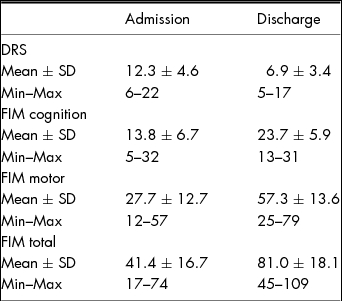
For the purpose of the current study, we were interested in examining the data to better understand physical activity patterns rather than sleep quality. Thus, the project adopts a retrospective, cross-sectional design utilising a convenience sample. A total of 24 participants met the inclusion criteria and were enrolled in the project. Three withdrew due to agitation resulting in a total of 21 participants. The final sample included 9 females and 12 males (M age = 43.8 ± 14.7 years; range 22–68 years) of which 17 were Caucasian, 2 were African American, and 2 were Hispanic. The mean length of time since injury until beginning activity tracking was 40.8 ± 22.1 days (range 11–90 days), LOS during inpatient rehabilitation was 30 ± 14 days (range 8–61 days), and patients had a mean GCS total of 9.3 ± 4.3 (range 3–15). Table 1 shows the Disability Rating Scale (DRS) and Functional Independence Measure (FIM) assessments for patients at admission and discharge. The DRS and FIM admission and discharge scores were not significantly different from typical values reported by the Traumatic Brain and Spinal Cord Injury Model Systems. Based on FIM discharge scores, 10 participants were independent walkers, 10 were supervised walkers, and 1 was unable to walk (M FIM = 5.14 ± 1.14; range 2–6).
Summary of Clinical Assessments at Admission and Discharge

Procedure
Institutional Review Board approval to complete the study was received from the hospital research institute ensuring that all procedures were considered ethical. Screening of patients was completed by a research coordinator to determine eligibility, who then approached suitable patients or their legal representative about participation in a private setting. Patients with an Orientation Log score of 25 or greater provided independent informed consent and proxy consent was obtained from the legally authorised representative if individuals scored less than 25. The Orientation Log measures the patient's orientation to time, place, and situation in a rehabilitation environment, with higher scores indicating increased orientation (Jackson, Novack, & Dowler, Reference Jackson, Novack and Dowler1998). Once consent was received individuals began enrolment in the project.
Measures
Participants were fitted with an Actiwatch-2 (Philips/Respironics, Bend, OR) on their wrist after completing consent. The watch was worn continuously to measure activity until discharge from inpatient rehabilitation. Participants wore the Actiwatch on their least-involved upper extremity and it was only removed for hygiene, bathing, and aquatic therapy. The Actiwatch has been validated to measure activity during sleep (Kushida et al., Reference Kushida, Chang, Gadkary, Guilleminault, Carrillo and Dement2001), and used to measure physical activity behaviours in healthy adults (Berlin, Kop, & Deuster, Reference Berlin, Kop and Deuster2006; Cuddy, Gaskill, Sharkey, Harger, & Ruby, Reference Cuddy, Gaskill, Sharkey, Harger and Ruby2007). The Actiwatch contains a piezo-electric sensor which determines the peak or maximum acceleration of the device from 32 samples per second. The resulting peak activity value from each second is added to an accumulated AC. For the current study, the Actiwatch was configured using a 60-second epoch length, and each 1-second peak activity value was summed to generate a total AC for the 1-minute period. Intensity of activity was then calculated with AC cut points used to analyse National Health and Nutrition Exam Survey (2014) data and included sedentary behaviour (<100 AC/min), inactive behaviour (100–499 counts/minute), light activity (500–2019 count/minute), moderate activity (2020–5998 counts/minute) and vigorous activity (>5999 counts/minute) (Matthews et al., Reference Matthews, Chen, Freedson, Buchowski, Beech, Pate and Troiano2008; Troiano et al., Reference Troiano, Berrigan, Dodd, Masse, Tilert and McDowell2008; Tudor-Locke, Ainsworth, Thompson, & Matthews, Reference Tudor-Locke, Ainsworth, Thompson and Matthews2002; Tudor-Locke, Johnson, & Katzmarzyk, Reference Tudor-Locke, Johnson and Katzmarzyk2009). Data were also used to calculate overall active and sedentary time in minutes. The average length of sedentary and active was calculated, with an epoch of less than 100 ACs considered sedentary and greater than 100 ACs considered active.
Data Analysis
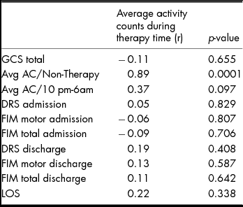
Actigraphy data were initially analysed using Actiware-5 software. All quantitative variables were then summarised in SAS 9.3 (SAS Institute, Cary, NC) using means, standard deviations, and ranges. Categorical variables were summarised with counts and percentages. Pearson correlations were calculated between average ACs during therapy time and GCS total, ACs between 10 pm and 6am, admission and discharge DRS and FIM, and LOS.
AC were separated for each patient into three categories due to the different activities scheduled during those time periods. Specifically, the categories included (1) therapy time (7.30am–3.30 pm), (2) non-therapy time (6am–7.30am and 3.30 pm–10 pm), and (3) sleep (10 pm–6am). During therapy time, individuals had 3 hours of scheduled therapeutic activity including physical, occupational, speech, and recreation therapy. For the period of the non-therapy time, individuals completed self-directed activities such as, but not limited to, using the gym, clubhouse (e.g., TV and game room), visiting with family, and sitting in their room. Between 10 pm–6am individuals were sleeping except for nursing visits during the night.
Results
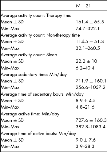
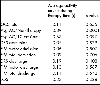
Participants wore the Actiwatch for an average of 8.4 ± 2.0 days (range 4–11 days). The actigraphy data were summarised in Table 2 and correlations between ACs in therapy time and physical functioning and injury variables in Table 3. ACs during therapy and non-therapy time were significantly associated (r = 0.89; p = 0.0001), but none of the remaining associations were significant. Examination of the raw data indicated that the highest maximum AC in one minute across all participants was 2369 (moderate-intensity activity) and the lowest maximum was 479 (inactive behaviour). However, no participant maintained moderate-intensity activity (>2020 ACs) for longer than 2 minutes and the maximum length of time spent consecutively in light intensity activity (500–2019 ACs) was 6 minutes.
Summary of Actigraphy Data

Correlations between Physical Functioning and Injury Variables and Activity Counts during Therapy
Discussion
The purpose of this study was to use accelerometers to better understand the activity level of patients with a TBI undergoing inpatient rehabilitation. Results for this initial sample indicate that activity levels are low, such that patients were typically considered inactive during therapy time and inactive/sedentary during non-therapeutic time. In this retrospective observational study, it is difficult to speculate on causes (i.e., cognition, attention, fatigue, medication) of low activity. However, the overall low activity of our sample during inpatient rehabilitation may not be in the best interest of the patient. In his review paper, Archer suggests exercise promotes adaptive brain processes after TBI in a ‘scaffolding effect’ (Archer, Reference Archer2012). In our sample, the activity during therapy time and non-therapy time was noticeably similar between participants and remarkably low and may be insufficient to activate the neuroreparative processes. Additionally, the low activity is likely insufficient to counteract the physical effects of bed rest and immobility experienced immediately after TBI. For example, in a sample of healthy young men, a daily exercise program of light intensity lasting 25 minutes in the presence of bed rest maintained some components of musculoskeletal function (i.e., muscle size, strength) but not others (i.e., muscle power, bone density) (Mulder et al., Reference Mulder, Clement, Linnarsson, Paloski, Wuyts, Zange and Rittweger2015). Comparatively, the maximum time spent in light intensity activity was 6 minutes for our sample. Therefore, the low activity levels experienced by our patients with TBI during inpatient rehabilitation may not adequately address the neurocognitive or musculoskeletal systems.
Further, if cardiovascular deconditioning is to be addressed across the patient's rehabilitation process, it is critical that activity interventions be implemented that provide continuity between settings (e.g., acute, inpatient, outpatient, community) and include a progression of intensity and duration (Hassett, Moseley, Whiteside, Barry, & Jones, Reference Hassett, Moseley, Whiteside, Barry and Jones2012; Mossberg et al., Reference Mossberg, Ayala, Baker, Heard and Masel2007). However, it is important to note that while patients with TBI have the capacity to engage in a structured exercise program, some individuals’ progression to an intensity to benefit aerobically may be more gradual than others (Jackson et al., Reference Jackson, Turner-Stokes, Culpan, Bateman, Scott, Powell and Greenwood2001). For example, reduced bone mineral density following TBI places individuals at a greater risk of fracture (Banham-Hall, Kothwal, Pipkin, Bentley, & Dickens, Reference Banham-Hall, Kothwal, Pipkin, Bentley and Dickens2013) and should be a consideration when developing the progression of an intensive activity program. Nevertheless, there is a critical need to identify strategies to facilitate physical activity participation early in the rehabilitation process as a preventative measure against the development of chronic disease in patients recovering from a TBI (Mossberg et al., Reference Mossberg, Amonette and Masel2010). In the absence of such strategies, individuals with a TBI will continue to remain at greater risk of disease and disability.
In general, there are a lack of health promotion efforts for individuals following TBI, despite the fact that clear associations between physical activity and dimensions of health (e.g., strength, endurance, quality of life) have been demonstrated (Pawlowski, Dixon-Ibarra, & Driver, Reference Pawlowski, Dixon-Ibarra and Driver2013). A recent systematic audit of the physical activity research specific to people post-TBI found that only 6% of available research evaluated the efficacy of behavioural interventions to promote activity. However, individuals immediately following TBI have reported wanting to know about activity, its role in the rehabilitation process, and how to incorporate activity as part of their recovery (Self, Driver, Stevens, & Warren, Reference Self, Driver, Stevens and Warren2013). Thus, effective programmes that include activity and social-behavioural interventions across the continuum of care are needed to promote activity (Driver, Irwin, Woolsey, & Pawlowski, Reference Driver, Irwin, Woolsey and Pawlowski2012) and reduce the risk of morbidity and mortality.
However, the challenge to increase activity is impacted by the fact that individuals face a growing number of personal and environmental barriers to activity over time (Driver, Reference Driver2009; Driver et al., Reference Driver, Ede, Dodd, Stevens and Warren2012). These challenges begin in the inpatient rehabilitation environment, as patients undergoing inpatient rehabilitation (i.e., TBI, stroke, total knee arthroplasty) have been shown to spend a greater percentage of time focused on gait training when compared to other physical therapy activities (i.e., assessment/evaluation; pre-functional activities; bed mobility; transfers; wheelchair mobility; pre-gait; therapeutic exercise; community mobility) (DeJong et al., Reference DeJong, Hsieh, Putman, Smout, Horn and Tian2011). Further, individuals with TBI were reported to receive less physical therapy treatment compared to other rehabilitation groups (DeJong et al., Reference DeJong, Hsieh, Putman, Smout, Horn and Tian2011). While the primary goal of inpatient rehabilitation is skill development or functional task retraining (i.e., gait), activity at a dosage to promote the scaffolding effect may be beneficial (Archer, Reference Archer2012). The combination of skill development paired with appropriately dosed activity may promote improved motor learning (Mang, Snow, Campbell, Ross, & Boyd, Reference Mang, Snow, Campbell, Ross and Boyd2014). Thus, innovative methods of increasing activity during inpatient rehabilitation are required, particularly with an evolving healthcare system where clinical productivity and patient outcomes are increasingly important factors.
The challenges to provide activity interventions are further limited by both health care and patient-related factors. For example, in the United States, inpatient rehabilitation centres are confronted with limited lengths of stay and diminishing resources as third-party payments decrease. As a result, the therapist is often forced to choose between function-based or activity-based interventions with the goal of maximising the patient's level of independence. In addition, complications after TBI itself may also be a limitation. Individuals with acute TBI often have cognitive impairments including impulsivity, severely limiting their ability to attend or remain on task. Attention can be limited to just minutes at a time, making sustained or intensive activity very challenging. Furthermore, individuals may have extensive physical limitations of neuromuscular (e.g., spasticity, hemiparesis) or orthopedic (e.g., fractures, weight bearing status, heterotopic ossification) origin.
Nevertheless, it is necessary to explore options for increasing activity during inpatient rehabilitation despite these challenges. For example, while therapists may not have the availability to supervise additional interventions, a therapist could prescribe an activity that a therapy technician could oversee (e.g., supervising ergometer training, assisting with basic exercises, or providing repeated practice of functional skills). Nursing staff could continue overseeing the activity program into the evening, when there is typically less opportunity for activity. Also, family members are often seeking ways to be involved in the rehabilitation process and, for some patients, it may be appropriate to train the family to implement the supplemental intervention. Galvin and colleagues demonstrated effectiveness of a family-mediated exercise intervention for people following acute stroke (Galvin, Cusack, O’Grady, Murphy, & Stokes, Reference Galvin, Cusack, O’Grady, Murphy and Stokes2011; Galvin, Stokes, & Cusack, Reference Galvin, Stokes and Cusack2014), which included family members completing additional activity in addition to the exercises completed with therapists. Finally, within therapy sessions, therapists should maximise active time by providing increased repetitions and fewer resting periods.
It is important to note the limitations of the study. First, the sample is small and cross-sectional, thus only representing one sub-group of individuals at a single inpatient rehabilitation hospital. The small sample also limited the type of analyses completed, as regression to predict activity levels could not be completed. Second, the activity data presented during therapy time (7.30am-3.30 pm) were aggregated and included the minimum 3 hours of therapy time and ‘other’ activities (e.g., dressing, lunch, naps, independent activity, showering). Thus, the mean AC for therapy time was influenced by other day-to-day activities typically completed by the individual. Future efforts should be made to assess the length of time spent completing specific activities within therapy and assessing the intensity of activity during those time points. Third, because the study was part of a larger, multi-centre project examining sleep efficiency after TBI, the Actiwatch-2 was used as the accelerometer rather than a more widely used model. Finally, while the cut points used in the current study accurately classify free-living activity in the general population (Matthews et al., Reference Matthews, Chen, Freedson, Buchowski, Beech, Pate and Troiano2008; Troiano et al., Reference Troiano, Berrigan, Dodd, Masse, Tilert and McDowell2008; Tudor-Locke et al., Reference Tudor-Locke, Ainsworth, Thompson and Matthews2002; Tudor-Locke et al., Reference Tudor-Locke, Johnson and Katzmarzyk2009), they have not been validated with people with TBI, which provides scope for future research. The cut points established by Freedson et al. (Freedson, Melanson, & Sirard, Reference Freedson, Melanson and Sirard1998) have been validated with individuals with acquired brain injury (Tweedy & Trost, Reference Tweedy and Trost2005) and used with individuals with a TBI (Hassett et al., Reference Hassett, Moseley, Harmer and van der Ploeg2015). Thus, there is an opportunity for future research efforts to examine the reliability and validity of the free-living activity cut points specific to people with a TBI.
In conclusion, due to the benefits associated with physical activity, it is important to promote participation across the rehabilitation process following TBI. This begins during inpatient rehabilitation, where higher doses of activity are associated with faster functional gains (Shiel et al., Reference Shiel, Burn, Henry, Clark, Wilson, Burnett and McLellan2001) and reduced LOS (Slade et al., Reference Slade, Tennant and Chamberlain2002). However, results from the current study reinforce the notion that the activity level of individuals undergoing inpatient rehabilitation is low (e.g., stroke, spinal cord injury, TBI) (DeJong et al., Reference DeJong, Hsieh, Putman, Smout, Horn and Tian2011; Kuys et al., Reference Kuys, Brauer and Ada2006; MacKay-Lyons & Makrides, Reference MacKay-Lyons and Makrides2002). While the safety of the patient is paramount immediately following injury, it is important that the activity is of the appropriate intensity and progresses as the individual's medical stability increases. If the health of patients following TBI is to be improved, it is important that we identify safe, evidence-based approaches to increasing activity levels across the continuum of care.
Acknowledgements
The authors would like to thank Cindy Dunklin for her assistance with data collection, and to the TBI clinical team for supporting study implementation within the hospital.
Financial Support
The project was supported by the Ginger Murchison Foundation TBI Research Fund. The contents of this manuscript were also developed under a grant from the National Institute on Disability, Independent Living, and Rehabilitation Research (NIDILRR grant number 90DP0045-01-0). NIDILRR is a Center within the Administration for Community Living (ACL), Department of Health and Human Services (HHS). The contents of this manuscript do not necessarily represent the policy of NIDILRR, ACL, HHS, and you should not assume endorsement by the Federal Government.
Conflict of Interest
‘None’.
Ethical Standards
The authors assert that all procedures contributing to this work comply with the ethical standards of the relevant national and institutional committees on human experimentation and with the Helsinki Declaration of 1975, as revised in 2008.