Introduction
Over the last two decades, the greater wax moth, Galleria mellonella (Lepidoptera: Pyralidae), has received significant attention from the scientific community as a model organism (Junqueira et al., Reference Junqueira, Mylonakis and Borghi2021). The larval stage of G. mellonella has become a widely used infection model to study microbial pathogens and test antimicrobial agents, serving as a preclinical step supporting the 3 R principles (replacement, reduction, and refinement) in animal research (Serrano et al., Reference Serrano, Verdial, Tavares and Oliveira2023). Research indicates that alterations in cell-mediated immune responses correspond with variations in the levels of antimicrobial peptides (AMPs) expressed in response to diverse invaders in G. mellonella (Wu et al., Reference Wu, Liu, Ding and Yi2016). Eighteen AMPs, including gallerimycin and galiomycin, have been identified in G. mellonella (Tsai et al., Reference Tsai, Loh and Proft2016). The study on the effects of two Pseudomonas aeruginosa strains on G. mellonella showed that the appearance of AMPs in haemolymph can be triggered not only by pathogen-associated molecular patterns (PAMPs) but also by bacterial extracellular proteases secreted during infection (Andrejko et al., Reference Andrejko, Mak, Siemińska-Kuczer, Iwański, Wojda, Suder, Kuleta, Regucka and Cytryńska2021). AMPs and pattern recognition receptor proteins (PRPs), such as hemolin, which have significant characteristics, are released from the fat body during bacterial infections (Schuhmann et al., Reference Schuhmann, Seitz, Vilcinskas and Podsiadlowski2003). The amount of AMPs and hemolin increases after bacterial infection, and gallerimycin suppresses the growth of filamentous fungi (Ladendorff and Kanost, Reference Ladendorff and Kanost1991; Langen et al., Reference Langen, Imani, Altincicek, Kieseritzky, Kogel and Vilcinskas2006). In a study with G. mellonella, injecting Candida albicans at a lethal dose (2 × 10⁵) increased expression of gallerimycin, galiomycin, inducible metalloproteinase inhibitor (IMPI), and hemolin peptides (Vertyporokh and Wojda, Reference Vertyporokh and Wojda2020). A study found that injecting St. John’s wort (Hypericum perforatum L.) plant extract with a sublethal dose of C. albicans into the G. mellonella larvae caused increased expression of gallerimycin- and hemolin-encoding genes (Genç et al., Reference Genç, Kaya, Günay and Çakaloğlu2024).
Another key insect defence mechanism is melanisation, which is mediated by the phenoloxidase (PO) system. Upon pathogen entry, PO converts tyrosine into reactive quinones, which then polymerise into melanin, forming a barrier around the invader (e.g. viruses, bacteria, fungi, and parasites) (Hillyer, Reference Hillyer2016; Cerenius and Söderhäll, Reference Cerenius and Söderhäll2021; Marieshwari et al., Reference Marieshwari, Bhuvaragavan, Sruthi, Mullainadhan and Janarthanan2023). PO is copper-dependent and synthesised primarily in granulocytes, oenocytoids, or crystal cells, depending on the insect species (Cerenius and Söderhäll, Reference Cerenius and Söderhäll2021; Marieshwari et al., Reference Marieshwari, Bhuvaragavan, Sruthi, Mullainadhan and Janarthanan2023). Plant extracts have been observed to modulate PO activity in G. mellonella, with some enhancing and others inhibiting the response depending on their composition (Kaya and Türkdoğan, Reference Kaya and Türkdoğan2021; Kaya, Reference Kaya2023). Notably, the absence of hemolin reduces PO activation, indicating its regulatory role in initiating the melanisation cascade (Terenius et al., Reference Terenius, Bettencourt, Lee, Li, Söderhäll and Faye2007; He et al., Reference He, Zhou, Cai, Liu, Zhao, Zhang, Wang and Zhang2022).
Insects, although evolutionarily distinct from mammals, share fundamental immune mechanisms (Tojo et al., Reference Tojo, Naganuma, Arakawa and Yokoo2000; Sheehan et al., Reference Sheehan, Garvey, Croke and Kavanagh2018). Insect immunity also plays a crucial role in modern pest-control strategies, as understanding immune regulation in insects provides valuable insights for developing sustainable and targeted management strategies (Ali and Xiao, Reference Ali and Xiao2025). Their cuticle serves as a skin-like barrier, while haemolymph contains immunocompetent cells akin to white blood cells (Wrońska et al., Reference Wrońska, Kaczmarek, Sobich, Grzelak and Boguś2022). When pathogens cross physical barriers, insect immunity responds through humoral and cell-mediated mechanisms (Lavine and Strand, Reference Lavine and Strand2002; Kanost et al., Reference Kanost, Jiang and Yu2004; Feldhaar and Gross, Reference Feldhaar and Gross2008). The cellular responses of the insect’s innate immune system include mechanisms specifically mediated by haemocytes, such as phagocytosis, nodulation, and encapsulation (Eleftherianos et al., Reference Eleftherianos, Heryanto, Bassal, Zhang, Tettamanti and Mohamed2021). Humoral immune responses include the production of AMPs, the activation of PO, the synthesis of lytic enzymes, and the generation of reactive oxygen species (Zasloff, Reference Zasloff2002; Jiang et al., Reference Jiang, Vilcinskas, Kanost and Söderhäll2010; Tsakas and Marmaras, Reference Tsakas and Marmaras2010; Trevijano-Contador and Zaragoza, Reference Trevijano-Contador and Zaragoza2019). Insect-derived AMPs are considered potential alternatives to antibiotics (Wojda, Reference Wojda2017). AMPs are classified into different categories based on their amino acid sequences and structural properties: linear α-helical peptides (e.g. cecropin and moricin), cysteine-stabilised peptides (e.g. defensin), proline-rich peptides, and glycine-rich peptides (e.g. gloverin) (Andrejko et al., Reference Andrejko, Mak, Siemińska-Kuczer, Iwański, Wojda, Suder, Kuleta, Regucka and Cytryńska2021). Hemolin, an immunoglobulin superfamily (IgSF) protein, acts as a broad-specificity PRP that binds bacterial and fungal PAMPs, aggregates pathogens, regulates haemocyte activity, and enhances opsonisation for immune defence.
In this context, natural products capable of modulating insect immune responses have garnered interest. Ziziphus jujuba Mill. (Rhamnaceae), commonly found in the subtropical climates of Asia, has been used in traditional medicine for thousands of years (Yu et al., Reference Yu, Jiang, Luo, Shen, Guo, Duan and Tang2012). In ethnomedicine across various Asian countries, it is recognised for its broad therapeutic effects, including antioxidant, antimicrobial, and immunomodulatory properties (Chen and Tsim, Reference Chen and Tsim2020). In China, the dried, fully ripened fruits of Z. jujuba are primarily used as a general restorative, especially for digestive health (Yu et al., Reference Yu, Jiang, Luo, Shen, Guo, Duan and Tang2012). The bioactive profile of Z. jujuba includes polyphenols (flavonoids, anthocyanins, and tannins), alkaloids, terpenoids, polysaccharides, carotenoids, fatty acids, sterols, vitamins, and minerals, making it a nutritionally valuable fruit (Aafi et al., Reference Aafi, Reza and Mirabzadeh2022). Among these, polysaccharides are particularly notable for their immunomodulatory, antioxidant, and anti-complementary activities (Li et al., Reference Li, Shan, Liu, Fan and Ai2011; Gao et al., Reference Gao, Wu and Wang2013). These water-soluble polysaccharides can stimulate lymphocyte proliferation and enhance immune function (Zhao et al., Reference Zhao, Liu and Tu2008). For example, polysaccharides composed of mannose, rhamnose, galactose, galacturonic acid, glucose, and arabinose demonstrated strong antioxidant activity and increased phagocytosis in THP-1 human monocytic cells. They also regulated pro-inflammatory cytokines, including TNF-α, IL-1β, and IL-6 (Zhang et al., Reference Zhang, Liu, Wang, Liu, Zhang, Zhao and Cheng2017).
The immunological influence of Z. jujuba extends to aquatic organisms. In Cyprinus carpio, dietary supplementation increased skin mucus protease activity and total immunoglobulin levels, thereby enhancing immunity (Hoseinifar et al., Reference Hoseinifar, Zou, Van Doan, Harikrishnan, Yousefi, Paknejad and Ahmadifar2019). Similarly, in Oncorhynchus mykiss, Z. jujuba extract upregulated antioxidant genes such as superoxide dismutase (SOD), catalase (CAT), and glutathione S-transferase A (GSTA), while also beneficially modulating the gut microbiota (Liu et al., Reference Liu, Zhang and Wang2021). Additionally, a study on NMU-induced mammary carcinogenesis in rats reported a dose- and time-dependent cytotoxic effect in MDA-MB-468 breast cancer cells, along with increased total antioxidant capacity (Hoshyar et al., Reference Hoshyar, Mohaghegh, Torabi and Abolghasemi2015). Although the immunological effects of Z. jujuba have been extensively studied in vertebrates, their impact on insect immunity remains largely unexplored.
This study aimed to explore the influence of Z. jujuba fruit extract on the humoral immune response in G. mellonella larvae. A comparative assessment was conducted to examine temporal transcriptional changes in immune parameters, including AMPs (gallerimycin and galiomycin), the PRP (hemolin), and PO genes (PO-I and PO-II), in both whole larvae and haemocytes. Results indicated that the Z. jujuba extract enhances immune function by upregulating the expression of AMPs, hemolin, and PO, alongside increasing PO enzymatic activity. Furthermore, protein–protein docking analyses revealed that the dual functionality of hemolin highlights its importance as a multifaceted regulatory protein in immune defence. The study also investigated the potential role of bioactive compounds within the extract in modulating immune responses.
Materials and methods
Plant collection, extraction, and phytochemical profiling
Z. jujuba fruits were collected from Güzelyalı village (GPS coordinates: 40°02′38.2″ N, 26°20′38.2″ E) in Çanakkale province (Türkiye) during the fruiting period in September 2023. The dried and powdered fruits were extracted in a Soxhlet apparatus with distilled water for 8 h, filtered, concentrated using rotary evaporator, and diluted with distilled water to obtain a final extract concentration of 10 mg/mL. For injection, 5 µL of extract solution was administered per larva, corresponding to a dose of 50 µg per larva.
The chemical profile of Z. jujuba fruit extract was determined by Gas Chromatography-Mass Spectrophotometry (GC-MS) using a Shimadzu QP 2010 Ultra system equipped with an RXI-5 MS capillary column (30 m × 0.25 mm ID × 0.25 μm). Helium was used as a carrier gas (1.5 mL/min). One microliter of the sample was injected in split mode, and the oven temperature was programmed from 40°C to 240°C at 4°C/min. Compounds were identified by comparison with the Wiley W9N11 library and retention index values.
Pathogen growth
C. albicans was cultured in YPD medium (1% yeast extract, 2% peptone, and 2% glucose) until the logarithmic phase. Cells were then washed and resuspended in 5 µL of sterile distilled water (2 × 10⁵ cells/larva) for injection (Vertyporokh and Wojda, Reference Vertyporokh and Wojda2020).
Insect rearing, injection, and sampling
The final instar G. mellonella larvae (0.20 ± 0.02 g), reared under identical conditions (in the dark, 65 ± 5% relative humidity, and 30 ± 1°C temperature), were injected with C. albicans, Sephadex A-25 chromatography beads (10–15 beads/larva), or Z. jujuba fruit extract. In addition to naïve larvae, null controls (needle insertion without injection) and water controls (vehicle injections) were included to account for potential effects of mechanical injury or solvent. Each group included 15 larvae and was injected via the top right proleg using either a 22- or 26-gauge microsyringe (50 µL or 25 µL; Hamilton, Reno, NV, USA). Samples (whole larvae and haemocytes) were collected at 4 and 24 h post-injection (n = 120). The larvae were flash-frozen in liquid nitrogen and stored at −80°C. The haemolymph was mixed with anticoagulant (0.098 M NaOH, 0.186 M NaCl, 0.017 M Na2EDTA, and 0.041 M citric acid; pH 4.5), centrifuged, and haemocyte pellets were frozen in liquid nitrogen and stored at −80°C.
Gene expressions
Gene expression analyses were performed using larvae and haemocytes from each group (naive, null, water, C. albicans, silica bead, and fruit extract). Total RNA was isolated using the RNA purification kit (Thermo Scientific, K0732) and treated with DNase I (Thermo Scientific, EN0525) to eliminate DNA. The RNA yield was assessed with the Multiskan GO Microplate Spectrophotometer (Thermo Scientific, Finland). The RevertAid First Strand cDNA Synthesis Kit (Thermo Scientific, K1622) was used for cDNA synthesis. The reverse transcription quantitative PCR (RT-qPCR) was performed by a PikoReal Real-Time PCR System (Thermo Scientific, Finland) with the RealQ Plus 2× Master Mix Green Without ROX (Ampliqon, A323402). The primer sequences used in this study are listed in table 1. RT-qPCR reactions were run in triplicate under the following conditions: 95°C for 15 min, followed by 30 cycles of 95°C for 30 s, 52°C for 30 s, and 72°C for 30 s. The average threshold cycle (Ct) for each sample was calculated, and the mRNA levels were normalised to the β-actin gene. The comparative Ct (−ΔΔCt) approach was used to determine fold changes for each gene relative to the control (naive) (Livak and Schmittgen, Reference Livak and Schmittgen2001).
Primer sequences used for real-time PCR

PO activity assay
PO activity was determined using the haemolymph from 16 larvae as described previously (Genç et al., Reference Genç, Kaya, Günay and Çakaloğlu2024). The haemolymph from each larva was transferred to tubes containing phosphate buffer and centrifuged to obtain cell-free supernatant, which was transferred to a 96-well microplate. The L-3, 4-dihydroxyphenylalanine (L-DOPA; Sigma-Aldrich), PO substrate, was prepared in phosphate buffer and transferred to the haemolymph–buffer mixture in the 96-well microplate. The Multiskan GO Microplate Spectrophotometer (Thermo Scientific, Finland) detected the colour change of the samples after conversion of L-DOPA to melanin by PO. Absorbance was measured at 490 nm wavelength for 30 min at 5-min intervals. The Bradford method was used to determine the total protein content (Bradford, Reference Bradford1976). The enzyme activity results were expressed as units per milligram of protein per minute (U/mg protein/min).
Retrieving the 3D structures of proteins
The three-dimensional structures of PO (tyrosinase), hemolin, and PO-activating factor-1-like protein (PAP1) proteins were obtained from the AlphaFold Protein Structure Database to perform docking and interaction analyses (Jumper et al., Reference Jumper, Evans, Pritzel, Green, Figurnov, Ronneberger, Tunyasuvunakool, Bates, Žídek and Potapenko2021; Varadi et al., Reference Varadi, Bertoni, Magana, Paramval, Pidruchna, Radhakrishnan, Tsenkov, Nair, Mirdita, Yeo, Kovalevskiy, Tunyasuvunakool, Laydon, Žídek, Tomlinson, Hariharan, Abrahamson, Green, Jumper, Birney, Steinegger, Hassabis and Velankar2024). PO (UniProt ID; Q964D5_GALME, Gene ID; LOC113517945) is a 683-amino-acid protein, and its structure was directly accessed using its AlphaFold ID (AF-Q964D5-F1-V4). Hemolin (UniProt ID; C7ASJ3_GALME, Gene ID; LOC113510107) consists of 421 amino acids, and its 3D structure was similarly retrieved using the AlphaFold ID AF-C7ASJ3-F1-V4. PAP1 (393 amino acids) was identified in the G. mellonella protein annotation file (ASM364042v2), since it is not listed under this exact designation in UniProt. It is designated as a ‘CLIP domain-containing serine protease’ (UniProt ID; A0A6JIWU9_GALME, Gene ID; LOC113512560), and its AlphaFold ID (AF-A0A6JIWU9-F1-V4) was used to access the structure. The use of AlphaFold structures ensured that the docking studies relied on dependable, high-confidence 3D models. The complete 3D structures of the hemolin, PO-I, and PAP1 proteins were presented in Supplementary Files 1 and 2.
Protein–protein docking
Following the retrieval of 3D structures, docking and interaction analyses were conducted using a series of computational tools. All protein structures were downloaded in ‘.pdb’ format and prepared for docking using the Dock Prep function in Chimera v1.18, which was used to ensure the removal of solvent molecules, assignment of charges, and optimisation of atomic coordinates for subsequent simulations (Pettersen et al., Reference Pettersen, Goddard, Huang, Couch, Greenblatt, Meng and Ferrin2004). Docking simulations were performed on the HADDOCK 2.4 web server, where default parameters were applied (Honorato et al., Reference Honorato, Koukos, Jiménez-García, Tsaregorodtsev, Verlato, Giachetti, Rosato and Bonvin2021, Reference Honorato, Trellet, Jiménez-García, Schaarschmidt, Giulini, Reys, Koukos, Rodrigues, Karaca and van Zundert2024). Two docking setups were executed: hemolin with PO-I and hemolin with PAP1. The resulting models were clustered, and the best-scoring complex from each setup was selected for detailed analysis based on HADDOCK scores and structural consistency. The PyMOL and Discovery Studio (Dassault Systèmes BIOVIA, San Diego) programs were used for visualisation and analysis. Discovery studio was used to determine interatomic distances and binding interactions, including hydrogen bonds, electrostatic interactions, and hydrophobic contacts between the docked proteins. This stage allowed for analysis of the molecular interactions that stabilise the complexes, supporting insights related to their biological relevance.
Statistical analysis
All experiments were conducted with at least three biological replicates, and results are presented as mean ± SD. Statistical significance between treatments and controls, as well as between time points, was evaluated using Student’s t-test in GraphPad Prism (v9.0; GraphPad Software, USA). p < 0.05 was considered statistically significant.
Results
AMP and immune protein expression
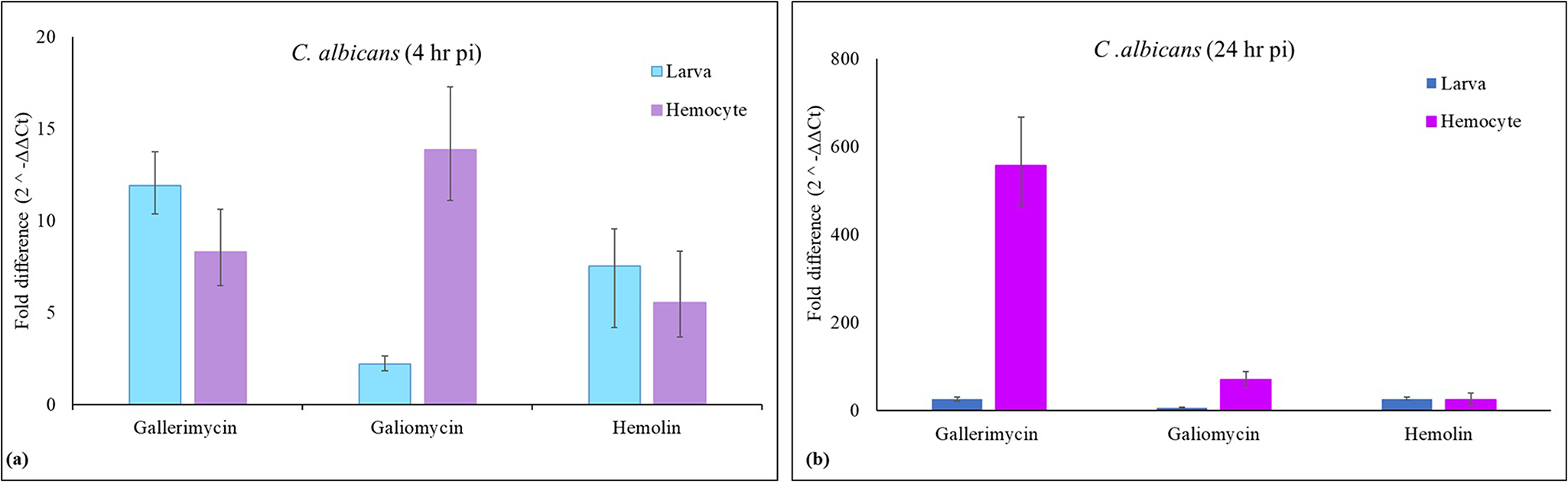
To investigate the effects of Z. jujuba fruit on the humoral immune response of G. mellonella, a lethal dose of C. albicans, silica beads, and Z. jujuba fruit extract was administered to larvae. In addition, no significant differences in gene expression were observed among the naïve, null, and water-injected control groups (p > 0.05); therefore, naïve larvae were used as the reference control in all subsequent analyses. Following C. albicans injection into larvae, the expression levels of the gallerimycin, galiomycin, and hemolin genes were assessed in both whole larvae and haemocytes at 4 and 24 h post-injection. At 4 h post-injection, gallerimycin and hemolin expression levels in whole larvae notably increased (12-fold and 8-fold, respectively), whereas galiomycin exhibited only a 2-fold rise (fig. 1A). In haemocytes, gallerimycin, galiomycin, and hemolin expression increased 8-fold, 14-fold, and 6-fold, respectively. No significant variation was observed in the expression of gallerimycin and hemolin between larvae and haemocytes. However, the expression of galiomycin in haemocytes was about seven times more than that in the whole larva (p < 0.01). After 24 h, the expression levels of gallerimycin, galiomycin, and hemolin in whole larvae increased 2–3 times relative to the 4 h expressions, reaching 26-fold, 6-fold, and 27-fold, respectively (fig. 1B). These values significantly increased in haemocytes, with gallerimycin increasing by 557-fold, galiomycin increasing by 71-fold, and hemolin enhancing by 24-fold compared to the control group. When comparing the 4- and 24-h expression levels, the expression of gallerimycin at 24 h was 70 times higher than at 4 h (p < 0.01), while the expressions of galiomycin and hemolin varied by 4–5 times (p < 0.05). The dramatic increase in gallerimycin expression, particularly in haemocytes, suggested that it played a pivotal role in defending against fungal infections. The relatively smaller increases in galiomycin and hemolin suggest that their contributions, while significant, may be complementary to the primary role of gallerimycin.
The expression of gallerimycin, galiomycin, and hemolin in larvae and haemocytes following pathogen injection at (A) 4 h and (B) 24 h.

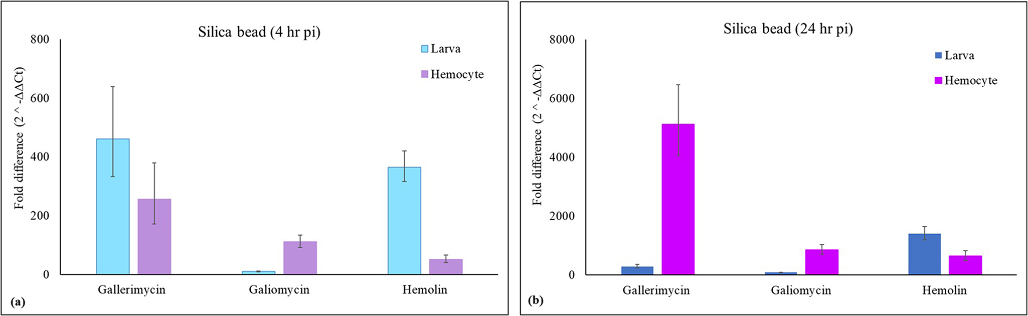
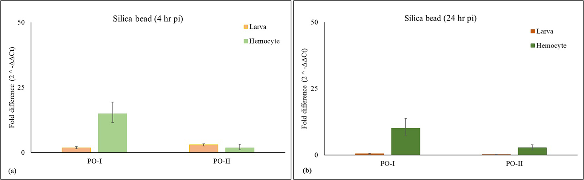
Significant variations in gene expression were detected between whole larvae and haemocytes following the injection of silica beads at both 4 and 24 h post-injection (fig. 2). At 4 h, gallerimycin expression significantly increased in whole larvae (460-fold) and haemocytes (255-fold) relative to control, although the difference between larvae and haemocytes was not significant. In contrast, galiomycin showed a modest rise compared to control (10-fold in whole larvae and 110-fold in haemocytes), with expression being significantly higher in haemocytes (p < 0.01). Hemolin, on the other hand, exhibited a robust response in whole larvae (364-fold) compared to haemocytes (51-fold), showing significant induction relative to control in both tissues but a significantly higher level in larvae (p < 0.01) (fig. 2A). Haemocytes exhibited elevated galiomycin expression, while whole larvae showed increased expressions of hemolin during the early immune response. After 24 h, the expression profiles exhibited a marked change in whole larvae; the expression of gallerimycin (293-fold) remained high compared to 4 h, while the expressions of galiomycin and hemolin showed significant increases of 95-fold and 1405-fold, respectively (p < 0.05), indicating a particularly strong and sustained activation of hemolin (fig. 2B). In haemocytes, the response intensified further, leading to an increase in the expression of gallerimycin (5125-fold; p < 0.001), galiomycin (851-fold; p < 0.01), and hemolin (642-fold; p < 0.01) with respect to control, highlighting the pivotal role of haemocytes in prolonged immune activation. In the late immune response, AMP expression was mostly elevated in haemocytes, whereas immune receptor protein expression increased throughout the whole larvae. These data reflect tissue-specific variations in the amplitude of immune responses and underscore the significant involvement of haemocytes in the synthesis of AMPs and immune signalling over time.
The expression of gallerimycin, galiomycin, and hemolin in larvae and haemocytes following silica bead injection at (A) 4 h and (B) 24 h.

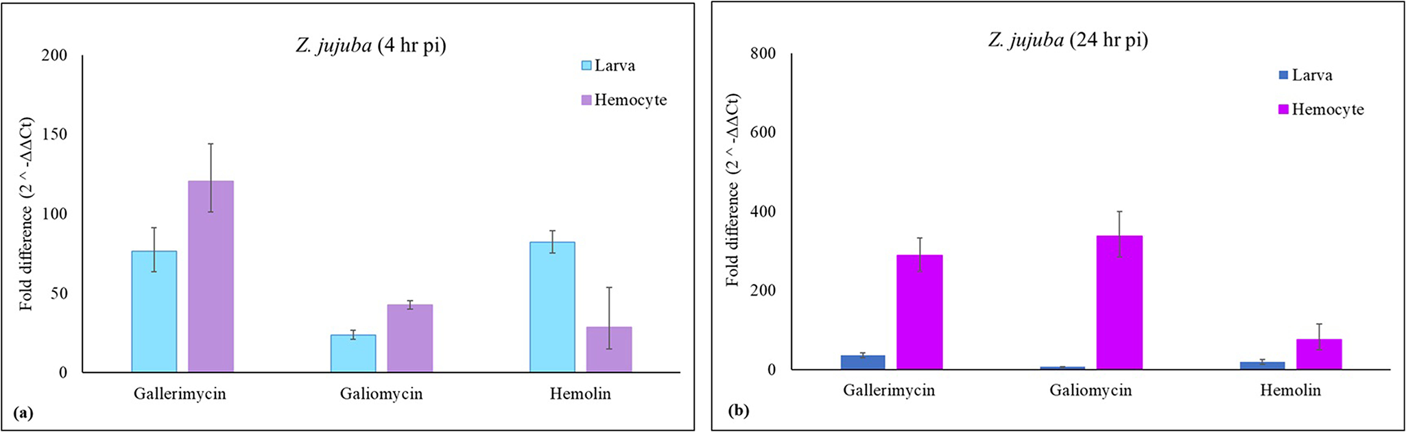
Following the injection of Z. jujuba extract into larvae, gene expression analysis revealed distinct temporal changes in gallerimycin, galiomycin, and hemolin expression in both whole larvae and haemocytes (fig. 3). At 4 h post-injection, the expression level of gallerimycin (76-fold) and hemolin (82-fold) in whole larvae was similar, although galiomycin expression (24-fold) was lower than the other two (fig. 3A), all being significantly higher compared to the control (p < 0.05). The level of gallerimycin expression (121-fold) was higher in haemocytes than the levels of galiomycin (43-fold) and hemolin (29-fold), which exhibited comparable expression levels, and all were significantly elevated compared to the control (p < 0.05). Whole larvae exhibited a slightly lower, but not significant, expression of gallerimycin compared to haemocytes (p > 0.05); whereas galiomycin expression was significantly lower in larvae (p < 0.05). On the other hand, hemolin expression was stronger in larvae compared to haemocytes (p < 0.01). The expression profile in whole larvae shifted after 24 h, showing a drop in gallerimycin (36-fold), galiomycin (7-fold), and hemolin (19-fold) compared to 4 h levels (p < 0.05, p < 0.01, and p < 0.001, respectively) (fig. 3B). In haemocytes, however, expression of gallerimycin (288-fold), galiomycin (338-fold), and hemolin (76-fold) further increased relative to 4 h levels (p < 0.001, p < 0.001, and p < 0.05, respectively), highlighting a sustained immune response, particularly within these immune-responsive cells. In general, the administration of silica beads significantly increased (p < 0.001) the expression levels in haemocytes and whole larvae at 4 and 24 h, compared to the results obtained after Z. jujuba extract. Therefore, Z. jujuba extract generated a noticeably weaker immune response than silica bead injection. However, compared to the pathogen injection results, the Z. jujuba extract elicited a more potent immune response, except for gallerimycin expression, which was higher in the pathogen injection (p < 0.01) than in the plant extract injection.
The expression of gallerimycin, galiomycin, and hemolin in larvae and haemocytes following Z. jujuba fruit extract injection at (A) 4 h and (B) 24 h.

PO gene expression
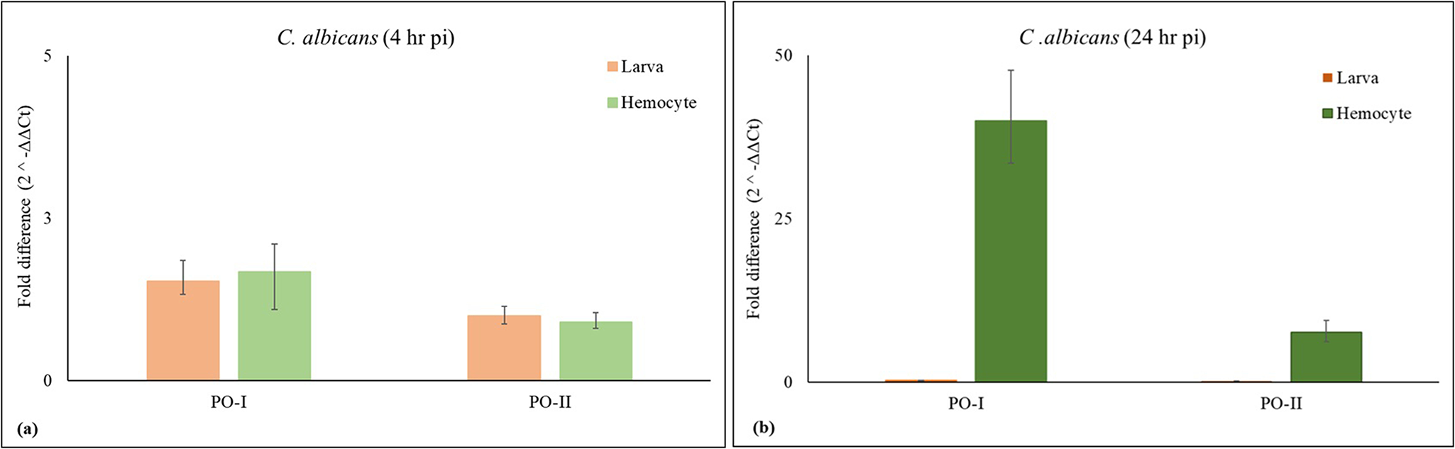
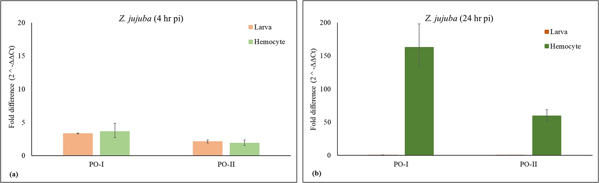
PO gene expressions were measured in both whole larvae and haemocytes at 4 and 24 h after C. albicans, silica bead, and plant extract injections in G. mellonella. Four hours post-injection with C. albicans, the expression levels of PO-I and PO-II in whole larvae and haemocytes were not significantly different from the control group (p > 0.05), except for PO-II expression in haemocytes, which was downregulated by 3-fold (p < 0.05) (fig. 4A). After 24 hours of injection, the expression of PO-I and PO-II in whole larvae was downregulated 5-fold (p < 0.05), while in haemocytes their expressions increased 40-fold (p < 0.01) and 8-fold (p < 0.05), respectively (fig. 4B). After 4 h post-injection of silica beads, PO-I and PO-II expression levels in whole larvae increased by 2 to 3-fold (p < 0.05). In haemocytes, PO-I expression went up 15-fold (p < 0.01), while PO-II expression increased by 2-fold (p < 0.05) (fig. 5A). After 24 h, PO-I and PO-II expression in whole larvae was downregulated 2-fold and 6-fold, respectively (p < 0.05). In haemocytes, however, PO-I and PO-II expression levels increased by 10-fold and 3-fold, respectively (p < 0.05) (fig. 5B). In both larvae and haemocytes, 4 h after injection of plant extract, PO-I expression increased 3-fold and PO-II increased 2-fold (p < 0.05) (fig. 6A). Both PO-I and PO-II expression were downregulated in whole larvae at the end of 24 h, but in haemocytes they were significantly upregulated by 163-fold and 60-fold, respectively (p < 0.001 and p < 0.05, respectively) (fig. 6B).
The expression of PO-I and PO-II in larvae and haemocytes following pathogen injection at (A) 4 h and (B) 24 h.

The expression of PO-I and PO-II in larvae and haemocytes following silica bead injection at (A) 4 h and (B) 24 h.

The expression of PO-I and PO-II in larvae and haemocytes following Z. jujuba fruit extract injection at (A) 4 h and (B) 24 h.

The immune response to C. albicans was delayed, with a significant increase in PO genes, particularly in haemocytes at 24 h, suggesting a localised defence against fungal growth despite systemic suppression in whole larvae. In contrast, silica beads triggered a rapid immune response at 4 h, particularly in haemocytes, with sustained activity at 24 h, reflecting their role in encapsulation. The plant extract induced a moderate immune activation at 4 h, but there was a dramatic increase in haemocytes at 24 h, potentially indicating increased immune priming or prolonged haemocyte activity. These findings indicate a time-dependent immune activation that is mostly localised in haemocytes. Moreover, after C. albicans, silica beads, and plant extract injections, PO-I expression in haemocytes was observed to be higher than PO-II expression. Therefore, PO-I is more dominant than PO-II in this context, and PO-I plays a more prominent role in the immune response, particularly in haemocytes.
PO activity
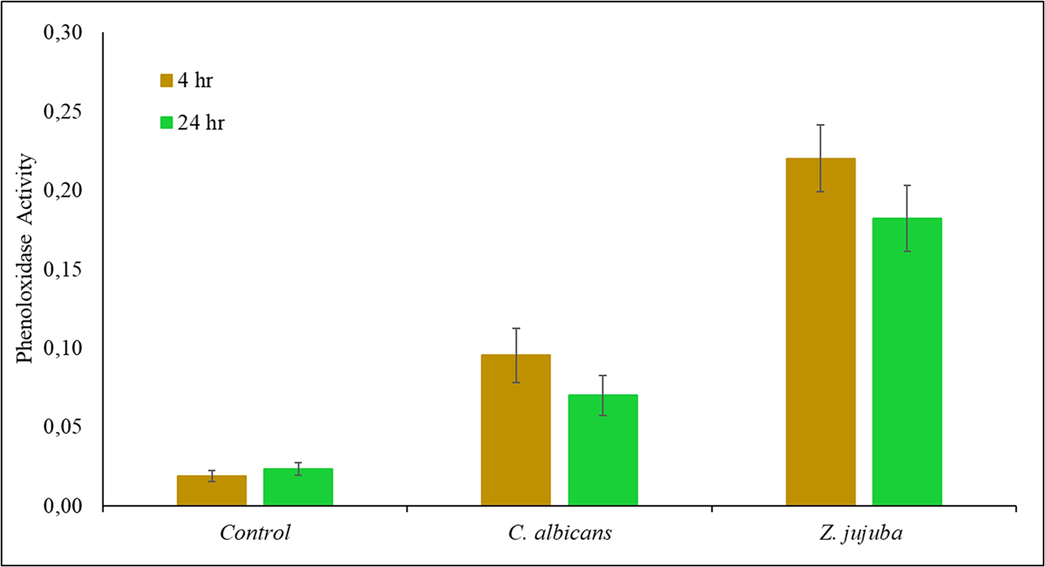
PO enzyme activity in G. mellonella larvae was determined following the injection of C. albicans and plant extracts at 4 and 24 h post-injection. In the untreated larvae (control), PO activity remained unchanged across both time points (fig. 7). Following pathogen injection, enzyme activity increased approximately 5-fold (p < 0.05) compared to the control at 4 h. At 24 h, enzyme activity remained 3-fold higher than the control (p < 0.05), although it showed a slight reduction compared to the 4-hour level. Similarly, following plant extract injection, enzyme activity was 12-fold greater than the control at 4 h (p < 0.01), with a slight decrease observed at 24 h compared to the activity of 4 h, which was non-significant, although at 24 h, activity remained 8-fold higher than the control (p < 0.01). However, no enzyme activity was detected after injection of silica bead at 4 and 24 h because of a high level of melanisation.
The phenoloxidase activity following pathogen and Z. jujuba fruit extract injection at 4 and 24 h. Enzyme activity was given as U/mg protein/min.

The results indicate that both pathogen and plant extract injections significantly increased PO activity compared to the control group. The initial rise in enzyme activity at 4 h for both injections reflects an immediate immune response. However, the magnitude of the response differed between the two challenges. The plant extract elicited a much stronger activation of PO (0,2201 ± 0,0212 U/mg protein/min) compared to C. albicans (0,0954 ± 0,0172 U/mg protein/min). Similarly, at 24 h, enzyme activity remained higher in the plant extract (0,1820 ± 0,0210 U/mg protein/min) than in the pathogen (0,0698 ± 0,0126 U/mg protein/min). This indicates that Z. jujuba extract may induce both immediate and sustained PO activation. The observed decline in enzyme activity at 24 h with respect to 4 h in both injections might reflect the consumption of active enzyme during the immune response. This is because, upon disruption of the physical barrier, the melanisation process, mediated by PO, is activated throughout the system, including the cuticle.
PO-I and PO-II expression levels in whole larvae and haemocytes after 4 h of C. albicans injection remained similar to controls, supporting the hypothesis that pre-existing PO stores were activated rather than newly synthesised. At 24 h, the gene expression levels of PO-I (40-fold) and PO-II (8-fold) in haemocytes were significantly elevated, reflecting a delayed synthesis phase, likely compensating for the depletion of active PO enzymes, as indicated by the decline in enzyme activity. After 4 h of plant extract injection, moderate (2–3-fold) increases in PO-I and PO-II expression in both larvae and haemocytes aligned with the higher enzyme activity. This suggests that the extract not only activates existing PO stores but may also contribute to new synthesis or early onset of gene expression possibly due to rapid consumption of existing PO enzymes. At 24 h, the substantial upregulation of PO-I (163-fold) and PO-II (60-fold) in haemocytes corresponds with sustained high PO activity (18 U). This implies a more robust and prolonged immune stimulation by Z. jujuba compared to C. albicans. As a result, the outcomes of both PO activity and gene expression are consistent. The plant extract promotes PO expression and activity during early humoral responses more effectively than pathogen treatments.
Molecular docking of hemolin to PO-I and PAP1 proteins
The HADDOCK docking results of hemolin with PO-I and PAP1 proteins were analysed to determine the most biologically plausible and energetically stable binding cluster. 0A summary of the results is provided in table 2. To identify the best cluster, several criteria were considered, including HADDOCK score, root mean square deviation (RMSD), cluster size, and buried surface area (BSA). Cluster 3 was identified as the optimal binding mode for the hemolin and PO-I, based on several key parameters: it exhibited the lowest HADDOCK score (−38.418), a moderate RMSD (1.871 Å), the largest cluster size (Nstruc: 7), and the largest BSA of 3141.142 Å2. Additionally, it demonstrated one of the most favourable interaction energies, indicating a strong and stable binding interface. These criteria collectively supported the selection of Cluster 3 as the most biologically and structurally relevant model for further analysis (Supplementary File 1). For PAP1 and hemolin, Cluster 7 was chosen based on the lowest HADDOCK score (−183.98), favourable RMSD (1.05 Å), and substantial BSA (3775.03 Å2), despite its smaller cluster size (Nstruc = 5). Cluster 4, as an alternative, with the lowest RMSD (0.60 Å), the largest cluster size (Nstruc = 7), and comparable BSA (3775.66 Å2), had a higher HADDOCK score (−214.96), suggesting relatively lower stability. Therefore, Cluster 7 was selected due to its balanced stability, consistency, and interaction interface (Supplementary File 2).
The four most optimal clusters from the docking study. Clusters utilised in the subsequent analysis are shown in bold. Comprehensive data and extended results are available in the supplementary files

Hemolin and PO-I: The protein–protein docking analysis of hemolin (Chain A) and PO-I (Chain B) revealed key electrostatic and hydrophobic interactions stabilising the binding interface (fig. 8). Two critical electrostatic interactions were observed with an attractive charge interaction between A:Lys62 and B:Asp464 (4.998 Å), and a pi–anion interaction between A:Glu109 and B:Tyr41 (4.932 Å). A total of 18 hydrogen bonds, including both conventional and carbon–hydrogen bonds, were identified, with distances ranging from 1.621 to 3.265 Å. Notable conventional bonds (e.g. A:Lys62–B:Ser16 at 1.621 Å and A:Glu109–B:Tyr41 at 1.628 Å) play a significant role in stabilising the complex. Longer-distance bonds (e.g. B:Arg51–A:Leu74 at 2.399 Å) and carbon–hydrogen bonds (e.g. A:Lys77–B:Thr466 at 3.265 Å) further contribute to the overall stability of the complex. Salt bridges such as A:Lys77–B:Glu17 (1.576 Å) and B:Lys40–A:Glu109 (1.589 Å) further reinforced the binding through strong electrostatic attraction. In addition, ten hydrophobic interactions were detected, primarily involving alkyl and pi–alkyl contacts. These included A:Ala18–B:Val300 (4.488 Å), A:Pro21–B:Leu383 (4.931 Å), and A:Lys77–B:Pro467 (4.846 Å), which contributed to the stability of complex by excluding water and promoting close contact between nonpolar residues. Longer-range interactions (e.g. A:Val83–B:Val170, 5.353 Å) provided flexibility, while shorter ones (e.g. B:Arg51–A:Pro73, 4.331 Å) enhanced local rigidity. Furthermore, the pi–alkyl interaction, such as B:His380–A:Pro21 (5.190 Å), provides an additional layer of stabilisation (Supplementary File 1).
The visualisation of protein–protein docking results of hemolin and PO-I proteins. Cyan: PO-I protein, yellow: hemolin protein, magenta: Ig domain of hemolin, and red: Ig domain of PO-I protein.

Hemolin and PAP1: The docking analysis of PAP1 (Chain A) and hemolin (Chain B) demonstrated key electrostatic and hydrophobic interactions that stabilise the protein–protein interface (fig. 9), a pi–anion interaction (B:Asp148–A:His186 at 3.629 Å), an attractive charge interaction (A:Arg149–B:Asp148 at 3.835 Å), and a pi–cation interaction (A:Arg149–B:Tyr243 at 3.861 Å), underscoring the contribution of charged and aromatic residues to interface stabilisation. Longer-range attractive charge interactions, such as A:Arg94–B:Glu157 (5.102 Å) and A:Arg241–B:Glu133 (5.546 Å), enhance stability. The hydrogen bonds were categorised into short- (1.5–3.5 Å), mid- (3.6–4.5 Å), and long-range (>4.5 Å) bonds. Short-range bonds, including a pi–anion bond (B:Asp148–A:His186 at 3.629 Å), an attractive charge interaction (A:Arg149–B:Asp148 at 3.835 Å), and a pi–cation bond (A:Arg149–B:Tyr243 at 3.861 Å), provided the strongest stabilising forces within the binding interface. The combined effects of hydrogen bonding and electrostatic attraction maintained the binding interface through salt bridges at distances between 1.561 Å and 2.934 Å. Hydrophobic contacts promote tight packaging and structural stabilisation by reducing solvent exposure. A short-range pi–sigma interaction (A:Leu95–B:Tyr155 at 3.702 Å), mid-range alkyl interactions (e.g. B:Arg143–A:Leu182 at 4.410 Å), and long-range pi–alkyl interactions (e.g. A:Tyr226–B:Lys138 at 5.460 Å) support hydrophobic core formation and enhance interface stability (Supplementary File 2).
The visualisation of protein–protein docking results of hemolin and PAP1 proteins. Blue: PAP1 protein and yellow: hemolin protein.

Chemical characterisation of the extract
When the larvae were challenged with the extracts of Z. jujuba fruit extract, the expression levels of several immune-related (gallerimycin, galiomycin, hemolin, PO-I, and PO-II) were significantly upregulated. Similarly, the activity of the PO enzyme was also elevated following the injection of the plant extract. The extract was not standardised to a specific polyphenol or sugar content. Instead, its chemical composition was analysed using GC-MS. The results, including chromatograms, major peaks, and compound identities, are provided in Supplementary File 3 and have also been deposited in the Zenodo database (Zenodo DOI: 10.5281/zenodo.10459999). GC-MS analysis identified several major compounds: 9-octadecenamide (59.11%), 3-(2′-hydroxy-4′-methoxyphenyl) propanoic acid (19.23%), hexadecanamide (9.94%), tridecane (4.11%), palmitic acid (3.31%), cis, cis, cis-7,10,13-hexadecatrienal (2.59%), and dodecane (1.71%). Among these, phenolic compounds are of particular interest because they can act as substrates for PO, thereby enhancing enzymatic activity. For instance, 3-(2′-hydroxy-4′-methoxyphenyl) propanoic acid, identified in the extract, is a phenolic molecule capable of stimulating PO activity. These bioactive chemicals may play diverse roles in modulating immune responses in biological systems.
Discussion
Z. jujuba, widely used in traditional Chinese medicine, has been shown to modulate the immune system in various model organisms (Yu et al., Reference Yu, Xu, Lu, Zheng and Chen2016; Zhang et al., Reference Zhang, Liu, Wang, Liu, Zhang, Zhao and Cheng2017; Liu et al., Reference Liu, Zhang and Wang2021). However, its immunomodulatory potential in invertebrate models, particularly in G. mellonella, has not yet been explored. Our findings demonstrate that G. mellonella larvae exhibit significant immune responses to Z. jujuba fruit extract, C. albicans, and silica beads, as evidenced by changes in AMPs (gallerimycin and galiomycin), the pattern recognition receptor hemolin, and PO gene expression and enzyme activity.
Gallerimycin and galiomycin are defensin-like AMPs that have been shown to be upregulated in response to fungal infections (Lee et al., Reference Lee, Yun, Jang, Kim, Lee, Park, Ryu, Seo, Kim and Lee2004; Bergin et al., Reference Bergin, Murphy, Keenan, Clynes and Kavanagh2006; Langen et al., Reference Langen, Imani, Altincicek, Kieseritzky, Kogel and Vilcinskas2006; Dekkerová-Chupáčová et al., Reference Dekkerová-Chupáčová, Borghi, Morace and Bujdáková2018). In this study, gallerimycin expression increased in haemocytes, particularly after C. albicans and bead injections (557-fold and 5125-fold, respectively). These data confirm that gallerimycin plays a key role in systemic antifungal responses. In contrast, galiomycin exhibited more moderate upregulation (71-fold and 851-fold, respectively), compared to gallerimycin in both tissues, suggesting that galiomycin functions as a secondary response element against fungal and physical challenges. This observation aligns with previous studies reporting that galiomycin is moderately upregulated following infection (Mowlds et al., Reference Mowlds, Barron and Kavanagh2008).
The response to Z. jujuba extract showed a more balanced AMP expression, with strong early upregulation of both gallerimycin (76-fold in larvae and 121-fold in haemocytes) and galiomycin (24-fold in larvae and 43-fold in haemocytes) compared to C. albicans. These results suggest that the bioactive compounds in the extract, such as polyphenols and flavonoids, act as immune modulators rather than potent stress inducers. Notably, while gallerimycin expression was higher in pathogen-treated larvae at 24 h (577-fold), the extract induced a stronger early response, suggesting stimulus-specific immune dynamics. The insect immune system may perceive the fruit extract as an immune modulator, while recognising C. albicans and silica beads as threats, thus eliciting a prolonged and appropriately vigorous immune response. This highlights the larvae’s ability to modulate their immune responses based on stimulus severity and pathogenicity. Our results support previous studies indicating that the insect immune system can regulate its response based on the magnitude and pathogenicity of the challenge, enabling it to differentiate between potentially nonlethal infections and life-threatening infections to provide appropriate and proportionate protection (Fallon et al., Reference Fallon, Troy and Kavanagh2011; Sheehan et al., Reference Sheehan, Tully and Kavanagh2020; Genç et al., Reference Genç, Kaya, Günay and Çakaloğlu2024). Similarly, even low pathogen doses can markedly affect host immunity and promote resistance development, further supporting the dose- and severity-dependent nature of pathogen-host interactions (Qayyum et al., Reference Qayyum, Zhihang, Wakil, Ali, Ali, Ghazanfar, Yasin, Danping, Almoallim, Ansari and Tahir2024).
Interestingly, Z. jujuba extract treatment did not elicit the dominance of gallerimycin over galiomycin expression observed with pathogens and beads. While some previous studies showed higher galiomycin levels in larvae challenged with C. albicans (Dekkerová-Chupáčová et al., Reference Dekkerová-Chupáčová, Borghi, Morace and Bujdáková2018; Vertyporokh and Wojda, Reference Vertyporokh and Wojda2020), others reported higher gallerimycin expression in response to fungal pathogens (Reis et al., Reference Reis, de Jesus, de Souza, Alcântara, Rodrigues, Brito, Penna, Vieira, Silva, Penna, Machado, Mora-Montes and Baptista2023), which is in agreement with our results. On the other hand, previous research revealed that larvae pre-challenged with pathogens, laminarin, or physical stress (shaking or injury) showed higher levels of galiomycin than gallerimycin (Bergin et al., Reference Bergin, Murphy, Keenan, Clynes and Kavanagh2006; Mowlds et al., Reference Mowlds, Barron and Kavanagh2008). These studies indicate that insects appear to tailor their immune responses to the nature of challenge, producing a broader AMP profile against microbial threats. In contrast, mild physical stress triggers fewer AMPs as such stress is typically self-limiting unless infection occurs (Mowlds et al., Reference Mowlds, Barron and Kavanagh2008). The antioxidant properties of Z. jujuba extract may also play a role in shaping this AMP expression pattern by reducing oxidative stress, which is known to suppress galiomycin. Hence, galiomycin may function not only as an AMP but also as a stress-related protein. Our results demonstrate a dynamic, challenge-specific regulation of gallerimycin and galiomycin, with higher AMP expression in haemocytes than in larvae, supporting our previous finding that haemocytes are the primary site of AMP production (Kaya et al., Reference Kaya, Turgut Genç and Günay2025).
Hemolin, a PRP from the IgSF, exhibits broad microbial recognition capabilities (Yu and Kanost, Reference Yu and Kanost2002; Kanost et al., Reference Kanost, Jiang and Yu2004; Terenius, Reference Terenius2008; Shaik and Sehnal, Reference Shaik and Sehnal2009; Browne et al., Reference Browne, Surlis, Maher, Gallagher, Carolan, Clynes and Kavanagh2015; Genç et al., Reference Genç, Kaya, Günay and Çakaloğlu2024; Subhagan et al., Reference Subhagan, Pathrose and Chellappan2025). It recognises and binds to bacterial and fungal PAMPs, including lipopolysaccharide (LPS) in Gram-negative bacteria, lipoteichoic acid (LTA) in Gram-positive bacteria, and fungal β-1,3-glucan or α-1,3-glucan (Yu and Kanost, Reference Yu and Kanost2002; Pinto et al., Reference Pinto, Barreto-Bergter and Taborda2008; Stączek et al., Reference Stączek, Zdybicka-Barabas, Wojda, Wiater, Mak, Suder, Skrzypiec and Cytryńska2021). Moreover, hemolin interacts with haemocytes, regulating their aggregation and phagocytosis, and promotes the opsonisation of foreign substances for immune clearance (Lanz-Mendoza et al., Reference Lanz-Mendoza, Bettencourt, Fabbri and Faye1996; Bettencourt et al., Reference Bettencourt, Lanz-Mendoza, Lindquist and Faye1997; Jung et al., Reference Jung, Sajjadian and Kim2019). Our study revealed no significant difference in hemolin expression between larvae and haemocytes after pathogen infection; however, it was elevated in larvae compared to haemocytes following bead injection. Hemolin expression was also higher at 24 h compared to 4 h after both pathogen and bead injection. Previous studies have established the systemic expression of hemolin in the epidermis, mid-gut, Malpighian tubules, fat body, and silk gland, not just in haemocytes (Aye et al., Reference Aye, Shim, Rhee and Lee2008; Shaik and Sehnal, Reference Shaik and Sehnal2009; Jung et al., Reference Jung, Sajjadian and Kim2019; He et al., Reference He, Zhou, Cai, Liu, Zhao, Zhang, Wang and Zhang2022). Additionally, it displays tissue-specific peak timings, indicating its continuous presence in larvae (He et al., Reference He, Zhou, Cai, Liu, Zhao, Zhang, Wang and Zhang2022). Therefore, no variation in hemolin expression may have been detected following pathogen injection in larvae and haemocytes; however, the elevated hemolin expression observed after bead injection (in both larvae and haemocytes) suggests that hemolin expression is modulated according to the strength of the challenge or the duration of the response. Moreover, hemolin expression increased 13-fold in haemocytes and 4-fold in larvae after 24 h, indicating a shift towards a haemocyte-based response to pathogens. Similarly, while hemolin was initially higher in larvae at 4 h post-extract injection, it became higher in haemocytes by 24 h, confirming a time-dependent shift of hemolin expression to haemocytes. These findings highlight the dynamic, adaptive, and tissue-specific regulation of hemolin expression, emphasising its role as a critical mediator in the immune response to varying types of challenges. Previous studies indicated that the expression of hemolin is sensitive to different microbial invaders, and that its pattern recognition mechanism varies across microbes (Ladendorff and Kanost, Reference Ladendorff and Kanost1991; He et al., Reference He, Zhou, Cai, Liu, Zhao, Zhang, Wang and Zhang2022). Our results indicate that hemolin expression occurs not only in response to microbial invaders but also against nonmicrobial agents such as plant-derived compounds.
The silencing of hemolin by RNA interference (RNAi) resulted in mostly unaffected AMP expression in Hyalophora cecropia, but it was variably regulated in Antheraea pernyi following microbial invasion (Terenius et al., Reference Terenius, Bettencourt, Lee, Li, Söderhäll and Faye2007; He et al., Reference He, Zhou, Cai, Liu, Zhao, Zhang, Wang and Zhang2022). In our study, no consistent correlation was found between hemolin and AMP expression, suggesting independent regulatory pathways.
PO, a central enzyme in the melanisation, is synthesised as inactive zymogens (proPOs) and activated by proPO-activating proteases (PAPs). Our results demonstrated time-dependent and challenge-specific regulation of the PO gene expression and enzymatic activity in response to all tested stimuli. PO gene expression and enzyme activity showed variable increases compared to the control at 4 h, which were sustained through 24 h. In wild-type Drosophila, low PO activity was observed without challenge and increased after 4 h, which was similar to our results (Ligoxygakis et al., Reference Ligoxygakis, Pelte, Hoffmann and Reichhart2002; Scherfer et al., Reference Scherfer, Tang, Kambris, Lhocine, Hashimoto and Lemaitre2008; Vertyporokh and Wojda, Reference Vertyporokh and Wojda2020). The initial activation of existing PO enables an immediate response, while subsequent gene upregulation ensures the long-term maintenance of immune defence capabilities. Therefore, the amount of inactive PO in haemocytes must be maintained at a certain level and constitutively expressed; when the level decreases, PO gene expression can be triggered to restore it. Our findings corroborate previous reports that insect haemocytes and fat bodies constitutively produce prophenoloxidase subunits at high levels, and that bacterial infections vary in their regulation of the prophenoloxidase-1 subunit (Terenius et al., Reference Terenius, Popham and Shelby2009). The interplay between enzyme activity and gene expression underscores the larvae’s dynamic immune regulation. These findings provide insight into the functional adaptability of the PO system in G. mellonella, demonstrating how enzyme activity and gene expression are fine-tuned to respond to different immune challenges.
Following silica bead injection, encapsulation and melanisation responses were observed around beads in G. mellonella larvae, but not around C. albicans. The reduced expression of PO genes in response to bead injection compared to pathogen injection suggests a distinction in immune recognition, likely influenced by the inorganic nature of beads and the organic properties of pathogens. Notably, the expression of immune-related genes, including galiomycin, gallerimycin, and hemolin, was higher in bead-injected larvae than in pathogen-injected larvae at both 4 and 24 h post-injection. Although PO activity was anticipated to align with the observed gene expression patterns, its divergence suggests that larval immune responses may differentiate between organic and inorganic entities. This discrepancy might reflect the specificity of PO activity in targeting invaders with certain physical characteristics or biological origins.
PO also produces other molecules such as proteases, cytotoxic quinines, reactive oxygen, and nitrogen intermediates, which can damage host cells if their levels are not properly controlled (Zhao et al., Reference Zhao, Lu, Strand and Jiang2011; González‐Santoyo and Córdoba‐Aguilar, Reference González‐Santoyo and Córdoba‐Aguilar2012). Therefore, PO activity must be regulated in a coordinated manner by mechanisms that operate in a time- and stimulus-dependent way, as previously indicated and supported by our results (Cerenius and Söderhäll, Reference Cerenius and Söderhäll2004; Nappi and Christensen, Reference Nappi and Christensen2005; Kanost and Gorman, Reference Kanost and Gorman2008). Previous studies have noted that the inhibition of hemolin via RNAi reduces PO activity in insects (Terenius et al., Reference Terenius, Bettencourt, Lee, Li, Söderhäll and Faye2007; He et al., Reference He, Zhou, Cai, Liu, Zhao, Zhang, Wang and Zhang2022). Our results demonstrated that plant extract injection induced greater PO enzyme activity and gene expression compared to pathogen injection. Likewise, hemolin expression was significantly elevated following extract injection compared to pathogen injection. The observed elevation in hemolin expression after extract injection may have influenced PO enzyme activity, leading to its enhancement. To validate these findings, future studies should investigate PO activity in the absence of hemolin in larvae injected with Z. jujuba extract.
Previous research revealed a connection between hemolin and the prophenoloxidase cascade, showing the knockdown of hemolin and reduced PO activity after microbial infections (Terenius et al., Reference Terenius, Bettencourt, Lee, Li, Söderhäll and Faye2007; He et al., Reference He, Zhou, Cai, Liu, Zhao, Zhang, Wang and Zhang2022). Therefore, we conducted protein–protein docking studies to investigate the interaction between G. mellonella hemolin and PO-I, as well as that of PAP1. The interaction between hemolin and PO-I exhibits strong electrostatic and hydrophobic binding motifs, indicating that hemolin can directly stabilise PO-I and maintain its active conformation. This suggests that hemolin may function as a regulatory protein for PO-I, directly supporting the enzyme’s activation and functionality. The interaction between hemolin and PAP1 is characterised by robust electrostatic bonding (pi–anion, pi–cation, and attractive charge interactions) and a substantial BSA, suggesting that hemolin may stabilise PAP1 with high affinity. This stabilisation could potentially activate the PAP1, thereby facilitating the proteolytic cleavage steps necessary for PO activation. Thus, hemolin may indirectly enhance PO activity by ensuring the effective function of PAP1. These findings suggest that hemolin plays a pivotal role in the PO activation cascade by directly stabilising PO-I and indirectly regulating the process via PAP1. The possible dual functionality underscores the significance of hemolin as a versatile regulatory protein in the immunological defence of G. mellonella.
The GC-MS analysis of Z. jujuba fruit extract identified key bioactive compounds grouped into lipid derivatives, hydrocarbons, and phenolics, which have varying degrees of biological significance. Although direct data regarding the role of fatty acids and their derivatives (palmitic acid and cis, cis, cis-7,10,13-Hexadecatrienal) in PO activity are limited, they are considered essential for modulating cellular and humoral immunity in insects (Kaczmarek and Boguś, Reference Kaczmarek and Boguś2021; Kim and Stanley, Reference Kim and Stanley2021; Elhoseny et al., Reference Elhoseny, El-Didamony, Atwa, Althoqapy and Gouda2024). The phenolic compounds, including 3-(2′-hydroxy-4′-methoxyphenyl) propanoic acid, serve as substrates for PO, which oxidises phenolics and initiates the melanisation process, an important immunological response in insects (Chase et al., Reference Chase, Raina, Bruno and Sugumaran2000; Marieshwari et al., Reference Marieshwari, Bhuvaragavan, Sruthi, Mullainadhan and Janarthanan2023). Therefore, these compounds have the potential to influence the activity of PO and immune-response proteins, thereby enhancing the immune response. Although these bioactive compounds have not been extensively studied in insect systems, their known biochemical properties suggest potential applications in enhancing insect immunity. Further experimental validation in insect models is necessary to elucidate their precise mechanisms of action and broader implications in immune defence and adaptation.
In conclusion, these results indicate that Z. jujuba extract triggers a time-dependent immune response in G. mellonella, with haemocytes playing a central role in prolonged expression of immune-related genes. The immune activation induced by Z. jujuba suggests its potential as an immunostimulant in biological research. Further research should explore the molecular mechanisms underlying these responses and the specific components of Z. jujuba responsible for immune modulation.
Supplementary material
The supplementary material for this article can be found at https://doi.org/10.1017/S000748532510076X.
Acknowledgements
The work is an outcome of research project number FHD-2022-3534 financed by the Scientific Research Coordination Unit of Çanakkale Onsekiz Mart University, Türkiye. The author would like to thank COMUDAM (Çanakkale Onsekiz Mart University Experimental Research Application and Research Centre) for providing the opportunity to use the Multiskan GO Microplate Spectrophotometer.
Author contributions
T.T.G. and S.K.: Conceptualisation, Investigation, Project administration, Resources, Supervision, Data analysis, Writing – original draft, Writing-review and editing. M.G.: Investigation, Data analysis, Writing – review and editing. All authors have read and agreed to the published version of the manuscript.
Declaration of competing interest
The authors declare that they have no known competing financial interests or personal relationships that could have appeared to influence the work reported in this paper.
Data availability statement
The data that support the findings of this study are available from the corresponding author upon reasonable request.