





Introduction
Serious cardiac events in the paediatric population are uncommon but potentially catastrophic. Reference Gajewski and Saul1 Serious cardiac events encompass sudden unexpected heart dysfunction, usually due to severe arrhythmias or blockages in flow to vital organs. Reference Zeppenfeld, Tfelt-Hansen and De Riva2 The most common aetiologies in the paediatric population include arrhythmic causes, such as arrhythmic channelopathies, long QT syndrome, and Brugada’s syndrome, or non-arrhythmic causes, such as hypertrophic cardiomyopathy, dilated cardiomyopathy, myocarditis, congenital heart disease (CHD), premature atherosclerosis, and coronary artery anomalies. Reference Gajewski and Saul1,Reference Meyer, Stubbs and Fahrenbruch3 These conditions can precipitate sudden cardiac arrests or sudden cardiac deaths, with sudden cardiac arrest defined as the abrupt cessation of normal cardiac activity with hemodynamic collapse, and sudden cardiac death occurring within one hour of symptom onset if witnessed or within 24 hours if unwitnessed. Reference Zeppenfeld, Tfelt-Hansen and De Riva2 In clinical research, serious cardiac events are commonly operationalised to include life-threatening arrhythmias, myocarditis, pericarditis, sudden cardiac arrest, sudden cardiac death, cardiogenic shock, and acute heart failure.
Prior to the COVID-19 pandemic, reporting limitations for sudden death meant that studies only focused on young athletes. This is due to the requirement of pre-participation cardiovascular screening procedures such as exercise, which is known to increase the risk of sudden cardiac death in the setting of predisposing conditions. Reference Meyer, Stubbs and Fahrenbruch3 In fact, sudden cardiac death was the main aetiology of death in young athletes, Reference Raukar and Cooper4 with an incidence of approximately 1:63,682 athlete-years in a 20-year study by the National Collegiate Athletic Association. Reference Petek, Churchill and Moulson5 Myocarditis accounted for 5–22% of sudden cardiac death events in athletes under 35 and could be triggered by viral infections. Reference Raukar and Cooper4 Sudden cardiac death events in the paediatric population (<18 years) were rarer and are associated with heterogenous causes depending on the age group, with infants (0–2 years) arresting most commonly due to congenital causes, children (3–13 years) due to hypertrophic cardiomyopathy, congenital causes, or arrhythmias among others, and adolescents (14–17 years) additionally due to dilated cardiomyopathy and myocarditis. Reference Meyer, Stubbs and Fahrenbruch3 Myocarditis may present suddenly without preceding respiratory symptoms, leading to arrhythmias or sudden death. Reference Raukar and Cooper4
SARS-CoV-2 infection has both direct and indirect cardiac impacts in adults and children. Direct effects include the acceleration of myocarditis, arrhythmias, and microvascular thrombosis while indirect effects encompass treatment-related cardiotoxicities. Reference Kuck, Schlüter, Vogler, Heeger and Tilz6 Vaccination-related myocarditis and arrhythmias have been reported through which there has been an increase in the likelihood of sudden cardiac death or sudden cardiac arrests. Even though these kinds of incidents are quite uncommon, they have sparked public reluctance and highlighted the importance of continuous monitoring and tailored immunisation plans, particularly for those with underlying heart disease. Reference Cho, Kim and Lee7 Furthermore, children with CHD or other comorbidities may also experience more severe COVID-19 disease, with an increased risk of ICU hospitalisation and complications, including multisystem inflammatory syndrome, a deadly inflammatory disorder that affects the heart and other organs. Reference Safi, Khouri, Zareef and Arabi8,Reference Zareef, Younis, Bitar, Eid and Arabi9
By acting as a direct cardiac toxin and a trigger for systemic failure, COVID-19 has altered the epidemiology of sudden cardiac death. Autopsy-attributed cardiomyopathy and “other causes” increased during the pandemic, indicating that the virus aggravates preexisting cardiovascular problems. Importantly, sudden cardiac death incidence increased by 38% overall and 77% among Hispanics, coinciding with peaks in the viral infection. Compounding this was the breakdown of healthcare: survival rates dropped from 15.3% to 10% due to delayed emergency medical services responses, decreased bystander cardiopulmonary resuscitation, and fewer shockable rhythms. This was exacerbated by ethnic differences, as Hispanics had a greater infection rate and even less access to resuscitation. Reference Chugh, Sargsyan and Nakamura10
Despite the documentation of COVID-19’s cardiovascular effects and vaccine-related risks, critical gaps are present. These include a limited understanding of how cardiac outcomes differ by age group (paediatric vs. adult populations), differential vulnerability toward vaccines, as well as short-term and long-term clinical outcomes. Moreover, adult studies dominate current literature, but data on the paediatric population remain sparse. Current strategies to mitigate risks in children, for instance, are mainly based on emerging adult data and may not fully apply to younger populations or others with underlying cardiac disease. Thus, this narrative review will synthesise existing literature to guide tailored interventions and strategies to mitigate such outcomes. The focus will be on clinical outcomes (e.g., hospitalisation, ICU admission, arrhythmia, myocarditis, sudden cardiac arrests, and sudden cardiac deaths), distinct risk profiles, and the efficacy of safety methods. This review will assist in directing age-appropriate and condition-specific treatments to minimise vaccine-related damage to guarantee safer immunisation and prevention practices. The paediatric population in this review refers to patients less than 18 years old, such as infants, children, and adolescents. When young adults who are older than 18 years old or athlete cohorts are discussed, these are explicitly labelled as comparators because several sudden cardiac death datasets originate from screening-based athlete registries.
Methods
The MEDLINE and PubMed databases were used to do a comprehensive review of the literature, focusing mainly on research that was published between 2019 and 2025 conducted on patients during the COVID era (from the end of 2019–2023), supplemented by background information from the pre-COVID years, henceforth defined as prior to 2019. “COVID-19,” “SARS-CoV-2,” “sudden cardiac death,” “arrhythmias,” “multisystem inflammatory syndrome,” “myocarditis,” “pulmonary embolism,” “adult,” and “pediatric” were among the main keywords. An example of how the keywords and medical subject headings (MeSHs) were combined is as follows: (“Heart Arrest”[Mesh] OR “pediatric multisystem inflammatory disease, COVID-19 related” [Supplementary Concept] OR “adult multisystem inflammatory disease, COVID-19 related” [Supplementary Concept] OR “Arrhythmias, Cardiac”[Mesh] OR “Myocarditis”[Mesh] OR “Death, Sudden, Cardiac”[Mesh] OR “Pulmonary Embolism”[Mesh] OR “sudden cardiac death” OR “sudden cardiac event” OR “sudden cardiac arrest” OR “arrhythmia*” OR “heart rhythm disorder*” OR “myocarditis” OR “MIS-C” OR “MIS-A” OR “Pulmonary embolism”) AND (“COVID-19”[Mesh] OR “SARS-CoV-2”[Mesh] OR “COVID-19” OR “SARS-CoV-2”) with filters applied to the different age groups. Only peer-reviewed research conducted during the COVID-19 pandemic and written or translated into English was included, with a focus on the disease’s causes, prevalence, results, or impacts, and whether they were caused by infection or vaccine. The studies were divided into three groups according to the population they addressed: paediatric patients, adults (over 18), or mixed age cohorts (both). We defined the paediatric population as less than 18 years old, and studies of young and adult athletes were retained for contextual comparisons, since they are separate populations. The causes of sudden cardiac death, population characteristics, sample sizes, clinical outcomes, mortality risk factors, autopsy results, immunisation status, and survival statistics were among the details gathered from each study.
Results
Epidemiology
Compared to the pre-pandemic era, there was a significant increase in the rates of out-of-hospital sudden cardiac death in adults during the pandemic, from 39 to 54 per 100,000 cases. Serious cardiac events were higher during the peak of infections, such as during the second wave that took place from November 2020 until February 2021. This increase persisted throughout those two years, even during months that did not contain peaks in COVID-19 infections. Reference Chugh, Sargsyan and Nakamura10 These findings contrast with those of other authors, who did not discover any meaningful association between hospitalisations or COVID-19 infection and ventricular tachycardia ablations or sudden cardiac death. Reference Foltran, Guilbeau-Frugier and Marimpouy11 Similarly, some researchers’ comparison of the burden of sudden cardiac death and sudden cardiac arrest among international football players before and during the pandemic did not find a statistically significant difference. Reference Ukaj, Meyer and Egger12 Figure 1 depicts the findings of a meta-analysis on the prevalence of ventricular arrhythmias and sudden cardiac death.
The prevalence and mortality linked to ventricular arrhythmias and SCD in hospitalised COVID-19 patients. The data used in the figure were adapted from a systematic review and meta-analysis, which included studies published between 2020 and 2021. Reference Tan, Huang and Mei44 Confidence intervals, when reported, are 95%. PVC: premature ventricular complex; SCD: sudden cardiac death; VA: ventricular arrhythmia; VF: ventricular fibrillation; VT: ventricular tachycardia.

Various studies have explored risk factors for sudden cardiac arrest and sudden cardiac death. Some ethnicities are more associated with in-hospital cardiac arrest than others during the COVID-19 period. The non-Hispanic black and Hispanic ethnicities were independent predictors of in-hospital cardiac arrest. However, systemic disparities were seen in a study, where half of the study participants were Hispanic and non-Hispanic Black. Reference Acharya, Ranka and Sethi13 Other risk factors included healthcare delays, as pandemic restrictions back then have increased out-of-hospital cardiac arrests by approximately 200% due to the restricted access of ambulances and the avoidance of hospitals. Reference Kuck, Schlüter, Vogler, Heeger and Tilz6 The “stay-at-home” mandate delayed patient presentation to the hospital and hence delayed reperfusion, which was associated with different mechanical complications, including papillary muscle rupture and ventricular septal defects due to necrosis of the myocardium. Reference Damluji, Gangasani and Grines14
The burden of the impact of myocarditis is even higher: compared to patients with non-COVID myocarditis, those with COVID-associated myocarditis experienced a 4.8x higher mortality. COVID-19 patients also had higher rates of sudden cardiac arrest, acute kidney injury, and cardiogenic shock. The risk is even higher in Hispanic patients, most likely related to socioeconomic variables as well as limitations and barriers to healthcare. Comorbid conditions such as obesity, diabetes, and hypertension are the primary risk factors of myocarditis. Furthermore, one study shows that, despite some athletes having electrocardiogram changes following infection, no athlete had myocarditis associated with SARS-CoV-2. Reference Petek, Churchill and Moulson5 Various case reports have been described in adults, including a case of giant cell myoendocarditis, Reference Blagova, Ainetdinova and Sedov15 aortitis, Reference Boor, Srinivasan and Stevenson16 double vessel coronary thrombosis, Reference Jokšić-Mazinjanin, Marić and Đuričin17 coronary dissection, Reference Hogea, Suciu and Chinezu18 myocarditis, Reference Shaukat, Petrasko, Petrasko, Rynders and Pham19 and sudden cardiac death in the setting of haemodialysis and hydroxychloroquine. Reference Murt, Dincer and Karaca20 These are summarised in Table 1.
Summary of case reports on the adult population

Table 1. Long description
A table summarizing case reports on the adult population affected by COVID-19. The table has 7 columns: Study, Date and place, Patient, Presentation, Investigations, Key findings, and Course and outcome. The table contains 7 rows of data. Row 1: Study, Simultaneous occurrence of myocarditis and COVID-19; Date and place, March 2020, New York, USA; Patient, 46-year-old male, no past medical history; Presentation, Chest discomfort, palpitations, and fever; Investigations, ECG (sinus rhythm, ST-segment elevation in SCL, T-wave inversion), troponin I, C-reactive protein, D-dimer, COVID-19 PCR; Key findings, ECG showed sinus rhythm, ST-segment elevation in SCL, T-wave inversion, troponin I elevated, C-reactive protein elevated, D-dimer elevated, COVID-19 PCR positive; Course and outcome, The patient was treated with hydroxychloroquine and QDCA and was in a stable condition at 30 days follow-up. Row 2: Study, Pregnancy-associated acute myocardial infarction in a young woman with COVID-19; Date and place, April 2020, Romania; Patient, 35-year-old female, smoking history, no other medical history; Presentation, Sudden onset chest pain, cold sweating, nausea, and vomiting; Investigations, ECG (ST-segment elevation in leads II, III, aVF), troponin I, C-reactive protein, D-dimer, COVID-19 PCR; Key findings, ECG showed ST-segment elevation in leads II, III, aVF, troponin I elevated, C-reactive protein elevated, D-dimer elevated, COVID-19 PCR positive; Course and outcome, The patient was treated with primary PCI and was in a stable condition at discharge. Row 3: Study, Necrotizing plaque erosion and rupture in COVID-19; Date and place, April 2020, France; Patient, 77-year-old male, history of hypertension and dyslipidemia; Presentation, Chest pain, dyspnea, and diaphoresis; Investigations, ECG (ST-segment elevation in leads II, III, aVF), troponin I, C-reactive protein, D-dimer, COVID-19 PCR; Key findings, ECG showed ST-segment elevation in leads II, III, aVF, troponin I elevated, C-reactive protein elevated, D-dimer elevated, COVID-19 PCR positive; Course and outcome, The patient was treated with primary PCI and was in a stable condition at discharge. Row 4: Study, Indirect cause of death in a patient with primary prevention ICD for non-ischemic cardiomyopathy; Date and place, April 2020, South Korea; Patient, 67-year-old male, history of hypertension and diabetes; Presentation, Mild dyspnea on exertion, productive cough, and fever; Investigations, ECG (sinus rhythm, no ST-segment elevation), troponin I, C-reactive protein, D-dimer, COVID-19 PCR; Key findings, ECG showed sinus rhythm, no ST-segment elevation, troponin I elevated, C-reactive protein elevated, D-dimer elevated, COVID-19 PCR positive; Course and outcome, The patient was treated with hydroxychloroquine and was in a stable condition at discharge. Row 5: Study, Progressive chest pain and sudden cardiac death; Date and place, April 2020, Massachusetts, USA; Patient, 49-year-old male, history of hypertension and diabetes; Presentation, Dyspnea on exertion, chest pain, and palpitations; Investigations, ECG (sinus rhythm, no ST-segment elevation), troponin I, C-reactive protein, D-dimer, COVID-19 PCR; Key findings, ECG showed sinus rhythm, no ST-segment elevation, troponin I elevated, C-reactive protein elevated, D-dimer elevated, COVID-19 PCR positive; Course and outcome, The patient was treated with hydroxychloroquine and was in a stable condition at discharge. Row 6: Study, Sudden cardiac death in a patient undergoing haemodialysis and hydroxychloroquine; Date and place, April 2020, Istanbul, Turkey; Patient, 73-year-old male, history of end-stage renal disease and hypertension; Presentation, Chest pain, dyspnea, and diaphoresis; Investigations, ECG (sinus rhythm, no ST-segment elevation), troponin I, C-reactive protein, D-dimer, COVID-19 PCR; Key findings, ECG showed sinus rhythm, no ST-segment elevation, troponin I elevated, C-reactive protein elevated, D-dimer elevated, COVID-19 PCR positive; Course and outcome, The patient was treated with hydroxychloroquine and was in a stable condition at discharge. Row 7: Study, Sudden cardiac death in a patient undergoing haemodialysis and hydroxychloroquine; Date and place, April 2020, Istanbul, Turkey; Patient, 80-year-old female, with a history of hypertension and diabetes; Presentation, Two episodes of syncope and chest pain; Investigations, ECG (sinus rhythm, no ST-segment elevation), troponin I, C-reactive protein, D-dimer, COVID-19 PCR; Key findings, ECG showed sinus rhythm, no ST-segment elevation, troponin I elevated, C-reactive protein elevated, D-dimer elevated, COVID-19 PCR positive; Course and outcome, The patient was treated with hydroxychloroquine and was in a stable condition at discharge.
ADA = anterior descending artery; APS 1 = autoimmune polyendocrine syndrome type 1; BiV-ICD = biventricular implantable cardioverter-defibrillator; CXR = chest X-ray; DES = drug-eluting stent; EF = ejection fraction; EMS = emergency medical services; ESRD = end-stage renal disease; GDMT = guideline-directed Medical Therapy; HFrEF = heart failure with reduced ejection fraction; IABP = intra-aortic balloon pump; ICCU = intensive coronary care unit; IMH = intramural haematoma; LAD = left anterior descending; LBBB = left bundle branch block; LCA = left coronary artery; LGE = late gadolinium enhancement; LMWH = low molecular weight heparin; LVEF = left ventricular ejection fraction; MI = myocardial infarction; NYHA = New York heart association classification system; OHCA = out-of-hospital cardiac arrest; PCI = primary percutaneous coronary intervention; PEA = pulseless electrical activity; PVCs = premature ventricular complexes; RCA = right coronary artery; ROSC = return of spontaneous circulation; STEMI = ST elevation myocardial infarction; TTE = transthoracic echocardiogram; TTM = targeted temperature management; VSMCs = vascular smooth muscle cells.
1 NYHA Class II, indicating mild limitation of physical activity and comfortable at rest.
On the other hand, paediatric patients (<18 years) are not simply young adults (≥18 years). Thus, COVID-2019’s associated risk factors, mechanisms, and outcomes may be different. Sudden cardiac death in children with multisystem inflammatory syndrome in children is rare, but still possible. Sudden cardiac death can occur days after apparent recovery from the hyperinflammatory syndrome, even without coronary artery aneurysms. For instance, the case of a 12-year-old with multisystem inflammatory syndrome in children who presented in cardiogenic shock, with elevated N-terminal pro-B-type natriuretic peptide, premature ventricular complexes on electrocardiogram, and biventricular dysfunction on echocardiography portrays how the myocardium in multisystem inflammatory syndrome in children cases is vulnerable to inflammatory-induced arrhythmias, even after apparent recovery. Reference Venkatesha, Srinivas, Mohamedali and Chandrasekar21 Similarly, another case report of a 36-day-old infant who was asymptomatic developed sudden cardiac arrest as the initial presentation of COVID-19. Echocardiography showed diminished left ventricular function. In parallel, the infant had elevated cardiac biomarkers such as troponin and B-type natriuretic peptide and high inflammatory markers. This suggests that they had an undiagnosed myocarditis, after ruling out all other causes. Reference Pierre, Kondamudi, Adeyinka, Trisha, Patki and Rimareva22 There are additional case reports of children who developed cardiac outcomes. Reference Cui, Tian and Huang23–Reference Unuma, Tomomasa and Noma28 A summary is included in Table 2.
Summary of case reports on the paediatric population

Table 2. Long description
A table summarizing case reports on pediatric COVID-19 patients and their cardiac outcomes. The table has 6 columns and 7 rows, including the header row. The columns are labeled as follows: Author, Year, Country, Patient, Presenting symptoms, Imaging findings, Other findings. Row 1: Author, Year, Country, Patient, Presenting symptoms, Imaging findings, Other findings. Row 2: Bostofte, 2020, Denmark, 12-year-old male, Fever, headache, and vomiting. Tachycardia, hypotension, and elevated N-terminal pro-B-type natriuretic peptide. Premature ventricular complexes on electrocardiogram, biventricular dysfunction on echocardiography, N/A. Row 3: COVID-19, India, 2020, 1-month-old girl, Asymptomatic, N/A, Elevated troponin and B-type natriuretic peptide, high inflammatory markers. Diminished left ventricular function on echocardiography, N/A. Row 4: Case report, April, 2020, 1-month-old male, Fever, cough, and diarrhea. Hypotension, tachycardia, and elevated troponin. Echocardiography showed global hypokinesis, N/A. Row 5: Cardiac Autopsy, Limited data, 12-year-old male, Fever, headache, and vomiting. Tachycardia, hypotension, and elevated troponin. Echocardiography showed global hypokinesis, N/A. Row 6: 11-year-old male, Fever, cough, and headache. Hypotension, tachycardia, and elevated troponin. Echocardiography showed global hypokinesis, N/A. Row 7: 12-year-old female, Fever, cough, and headache. Hypotension, tachycardia, and elevated troponin. Echocardiography showed global hypokinesis, N/A.
AKI = acute kidney injury; AST: aspartate aminotransferase; ATLE = apparent life-threatening event; BCP-ALL = B-cell precursor acute lymphoblastic leukaemia; CK = creatine kinase; CK-MB = creatine kinase-myocardial band; CPR = cardiopulmonary resuscitation; CRP = C-reactive protein; CVVH = continuous venovenous hemofiltration; CXR = chest X-ray; ED = emergency department; ESR = erythrocyte sedimentation rate; FS = fractional shortening; H&E = haematoxylin and eosin; HLH = hemophagocytic lymphohistiocytosis; IHC = immunohistochemistry; IVIG = intravenous immunoglobulin; ICV = inferior caval vein; LCA = left coronary artery; LDH = lactate dehydrogenase; LFTs = liver function tests; LMT = left main trunk; LMWH = low-molecular weight heparin; LVEF = left ventricular ejection fraction; LZTR1 = leucine-zipper-like transcription regulator 1; MISC = multisystem inflammatory syndrome; NIVS = non-invasive ventilator support; NT-proBNP = N-terminal pro-B-type natriuretic peptide; PAC = premature atrial contraction; PaO2 = arterial oxygen partial pressure; pRBCs = packed red blood cells; PICU = paediatric ICU; PVC = premature ventricular complexes; PVT = pulseless ventricular tachycardia; ROSC = return of spontaneous circulation; TTE = transthoracic echocardiography; VA-ECMO = venoarterial extracorporeal membrane oxygenation; VSD = ventricular septal defect; WES = whole exome sequencing.
1 defined as an event that is frightening to a witness, involving a combination of symptoms such as breathing apnoea, changes in skin colour or muscle tone or signs of choking or gagging.
Finally, a special discussion about patients with CHD is necessary. Epidemiological data regarding this subgroup mostly focus on overall patient outcomes rather than stratifying outcomes by cause, leaving data regarding specific complications sparse. Nevertheless, patients with underlying CHD are at higher risk of severe complications of COVID-19 infection and resulting mortality, with the highest risk being in patients with cyanotic lesions. Reference Schwerzmann, Ruperti-Repilado and Baumgartner29,Reference Soleimani and Soleimani30 In the context of serious cardiac events, one study involving 36,690 children finds that paediatric patients with CHD are at significantly higher risk of tachyarrhythmias and heart block but not at increased risk for myocarditis, sudden cardiac arrest, or the need for extracorporeal membrane oxygenation. Reference Ghimire, Chou, Aljohani and Moon-Grady31
Mechanisms of sudden cardiac arrest and sudden cardiac death
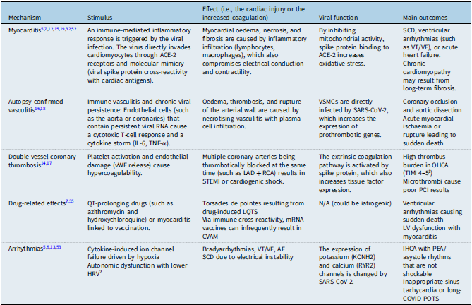
Different mechanisms could explain the development of sudden cardiac arrest and sudden cardiac death in adults with COVID-19, summarised in Table 3 and depicted in Figure 2. Myocarditis develops through the invasion of cardiomyocytes and vascular endothelium by SARS-CoV-2 via the angiotensin-converting enzyme-2 receptors, which enables viral entry and replication. Moreover, immune-mediated damage takes place through a hyperinflammatory response—a cytokine storm—that causes the infiltration of T-cells and activation of macrophages, triggering inflammation of the myocytes. Furthermore, endothelial injury increases ischaemia and thrombotic events, promoting cardiac injury. Reference Cannon, Bobba and Shekhar32 A case of SARS-CoV-2 infection-induced direct myocardial injury and inflammation was reported, which led to a significantly lower left ventricular ejection fraction (< 35%) and chronic myocardial damage, including myocarditis. Reference Shaukat, Petrasko, Petrasko, Rynders and Pham19 The incidence of ventricular arrhythmias and sudden cardiac death is greatly increased by this level of structural cardiac dysfunction, which produces a hazardous arrhythmogenic substrate. Primary preventive interventions, such as using an implanted cardioverter-defibrillator, are therefore required.
Mechanisms of sudden cardiac arrest. Image adapted from Servier Medical Art (https://smart.servier.com/), licensed under CC BY 4.0 (https://creativecommons.org/licenses/by/4.0/).

Summary of the mechanisms behind severe cardiac events

Table 3. Long description
A table with five rows and five columns. The columns are labeled Mechanism, Stimulus, Effect, Viral function, and Main outcomes. The rows detail different mechanisms: Myocarditis, Autopsy-confirmed vasculitis, Double-vessel coronary thrombosis, Drug-related effects, and Arrhythmias. Each row describes the stimulus, effect, viral function, and main outcomes for each mechanism. For example, Myocarditis is triggered by an immune-mediated inflammatory response to viral infection, leading to myocardial oedema, necrosis, and fibrosis, with outcomes including SCD and chronic cardiomyopathy. Autopsy-confirmed vasculitis involves immune vasculitis and chronic viral persistence, causing oedema, thrombosis, and arterial wall rupture, with outcomes like coronary occlusion and acute myocardial ischaemia. Double-vessel coronary thrombosis results from platelet activation and endothelial damage, leading to multiple coronary arteries being blocked, with high thrombus burden as a main outcome. Drug-related effects include QT-prolonging drugs causing torsades de pointes, with ventricular arrhythmias as a main outcome. Arrhythmias are driven by cytokine-induced ion channel failure, resulting in bradyarrhythmias and SCD.
AF = atrial fibrillation; ACE-2 = angiotensin-converting enzyme 2; CVAM = COVID-19 vaccine-associated myocarditis; HRV = heart rate variability; IHCA = in hospital cardiac arrest; IL-6 = interleukin 6; KCNH2 = potassium channel subfamily 2; LAD = left anterior descending artery; LQTS = long QT syndrome; LV = left ventricle; OHCA = out of hospital cardiac arrest; PCI = percutaneous coronary intervention; PEA = pulseless electrical activity; POTS = postural orthostatic tachycardia syndrome; RCA = right coronary artery; RYR2 = ryanodine receptor 2; SARS-CoV-2 = severe acute respiratory syndrome coronavirus 2; SCD = sudden cardiac death; STEMI = ST elevation myocardial infarction; TIMI = thrombolysis in myocardial infarction; TNF-α = tumour necrosis factor α; VF = ventricular fibrillation; VSMCs = vascular smooth muscle cells; VT = ventricular tachycardia; vWF = Von Willebrand factor.
1 grading system that refers to grades of coronary blood flow to assess perfusion in coronary arteries after interventions (e.g., thrombolysis, angioplasty). A TIMI thrombus grade of 4 corresponds to definite thrombus with its largest dimension bigger than or equal to two vessel diameters. A TIMI thrombus grade of 5 corresponds to total occlusion. In other words, no contrast can pass beyond it, requiring urgent revascularisation (Scarparo et al., 2022). Heart rate variability.
2 quantified using the root mean square of successive differences between normal heartbeats (RMSSD). It is a measure of parasympathetic nervous system activity. A lower RMSSD or reduced HRV was associated with SCD (Ranard et al., 2022).
Mechanisms are also revealed through pathological findings on autopsy, such as plasma cell-rich necrotising aortitis, which included diffuse circumferential inflammation of the aorta, along with IgG+ plasma cell infiltrates, neo-vascularisation, and medial necrosis. Furthermore, coronary arteritis mainly occurs on the left main coronary artery ostia, leading to stenosis of the vascular smooth muscle cells, which exacerbates acute myocardial ischaemia as well as contraction-band necrosis. Viral persistence is highlighted through the co-localisation of SARS-CoV-2 antigens with CD68+ macrophages in the aortic intima, and viral particles were seen in the aortic media through electron microscopy. Reference Boor, Srinivasan and Stevenson16
In summary, chronic immune activation takes place through the persistent SARS-CoV-2 antigens (approximately 4.5 months post-infection), which leads to the recruitment of T cells, B cells, and plasma cell differentiation in vascular tissue. Hence, ischaemia is vasculitis-mediated, along with inflammatory stenosis of the coronary and/or coeliac arteries, promoting coronary occlusion. Lastly, the binding of viral particles to angiotensin-converting enzyme-2 receptors disrupts vascular integrity, leading to vascular smooth muscle cell proliferation and oedema. Reference Boor, Srinivasan and Stevenson16
Other proposed mechanisms are related to the effect of SARS-CoV-2 on coagulation and inflammation in adults and include double-vessel coronary thrombosis. COVID-19 infection is associated with a hypercoagulable state, increasing vulnerability to atherosclerotic plaque rupture and creating a nidus for thrombus formation within coronary arteries. Reference Jokšić-Mazinjanin, Marić and Đuričin17 The virus can affect multiple vessels simultaneously, such as in the case of double-vessel coronary thrombosis. Some researchers describe the double formation of thrombi in the left anterior descending artery and right coronary artery, resulting in an ST-elevation myocardial infarction with an out-of-hospital cardiac arrests. Reference Jokšić-Mazinjanin, Marić and Đuričin17 This occurs even in patients without known risk factors. A D-dimer of less than 10 ng/mL is considered an independent predictor of 30-day survival in patients with out-of-hospital cardiac arrests. Reference Jokšić-Mazinjanin, Marić and Đuričin17
Another mechanism involves arrhythmias and QT prolongation from hydroxychloroquine and azithromycin, which were medications used early on in the pandemic but have since fallen out of use due to lack of efficacy. Reference Kuck, Schlüter, Vogler, Heeger and Tilz6,Reference Kamel, Monem, Sharaf, Magdy and Farid33,Reference Younis, Zareef, Al Hassan, Bitar, Eid and Arabi34 Hydroxychloroquine, originally used for autoimmune diseases and malaria, blocks the KCNH2-encoded hERG/Kv11.1 potassium channel in myocytes. Reference Cipriani, Zorzi and Ceccato35 In turn, ventricular repolarisation is delayed, which directly leads to QT prolongation. Other than gastrointestinal side effects, the risk of QT prolongation with hydroxychloroquine is even higher when used with other QT-prolonging medications or with patients who have pre-existing conditions. Reference Younis, Zareef, Al Hassan, Bitar, Eid and Arabi34 The same occurs with azithromycin. Furthermore, patients who have abnormal liver function tests had longer corrected QT (QTc) intervals, demonstrating involvement of the liver in altered drug metabolism and inflammation. QTc prolongation was also stable over 24 hours due to both tissue accumulation and the long half-lives. In this case, multiple daily electrocardiograms are not required to monitor arrhythmias. Reference Cipriani, Zorzi and Ceccato35 However, the drugs’ long half-life and dependence on hepatic and renal clearance increase the chances of toxicity, particularly in patients with impaired organ function. Reference Younis, Zareef, Al Hassan, Bitar, Eid and Arabi34 In addition, ventricular arrhythmias, including polymorphic ventricular tachycardia, ventricular tachycardia, and ventricular fibrillation, occurred more often in ICU patients infected with COVID-19. Rather than ventricular tachycardia or ventricular fibrillation, the majority of sudden cardiac arrests during COVID-19 were caused by non-shockable rhythms, like asystole or pulseless electrical activity. Reference Kuck, Schlüter, Vogler, Heeger and Tilz6
Ventricular arrhythmias are caused by ischaemia, microvascular thrombosis, inflammation, and viral invasion of myocytes or the conduction system. Inflammation also exacerbates electrical instability and ion channel malfunction. Ventricular arrhythmias are also facilitated by hyperadrenergic conditions (e.g., hypoxia and stress), metabolic disturbances (acidosis), electrolyte imbalances (hypokalaemia, hypomagnesemia), and renal or hepatic failure. Reference Kuck, Schlüter, Vogler, Heeger and Tilz6
Furthermore, due to its prothrombotic nature, the virus dramatically raises the frequency of pulmonary embolisms. Some reviewed studies have demonstrated that the prevalence of pulmonary embolisms in hospitalised patients ranges from approximately 12–32%, with considerably higher rates in ICU patients. Reference Abou Mansour, El Rassi, Sleem, Borghol and Arabi36 This increased risk is mediated by hypercoagulability, characterised by endothelial dysfunction, dysregulated immunological responses such as complement activation and neutrophil extracellular traps, as well as high von Willebrand factor. These may further trigger myocardial infarction or arrhythmias. In other words, some mechanisms may take place in parallel. ICU hospitalisation, serious sickness, and high D-dimer levels are important risk factors for PE. Moreover, emboli frequently damage the large pulmonary arteries, which can result in abrupt cardiovascular collapse. However, even for individuals who are not hospitalised, the risk of PE remains after infection, especially during the first 30 days. Reference Abou Mansour, El Rassi, Sleem, Borghol and Arabi36
Finally, patients with CHD, particularly in those with repaired defects, may be more prone to arrhythmia or heightened inflammatory response during COVID-19 infection due to underlying abnormalities in the conduction system and structurally within the myocardium itself, caused by past injury, surgical scarring, hypoxia or cyanosis, or cardiac remodelling. Reference Ghimire, Chou, Aljohani and Moon-Grady31,Reference Varney, Dong and Tsao37 This susceptibility is further increased by common residual complications, including valvular or shunt lesions, ventricular dysfunction, heart failure, arrhythmias, pulmonary vascular disease, cyanosis, renal or hepatic impairment, alongside an increased risk for infections and neurological complications. Reference Radke, Frenzel, Baumgartner and Diller38
Electrocardiographic changes
Adult patients with COVID-19 pneumonia demonstrate significant QTc prolongation compared to healthy controls, especially in severe disease, increasing risk for torsades de pointes. Conversely, these patients had a lower transmural dispersion of repolarisation, a measure of electrical heterogeneity across the ventricular wall during the heart’s recovery phase that is quantified as the T peak to end (Tp-e) interval or Tp-e/QTc ratio, ironically reducing the risk of torsades de pointes but still portending a higher chance of developing other arrhythmias like ventricular fibrillation and ventricular tachycardia. Furthermore, critically ill patients exhibited a higher index of cardiac electrophysiological imbalance, indicating prolonged ventricular repolarisation relative to depolarisation and a heightened susceptibility to arrhythmias. Reference Alareedh, Nafakhi, Shaghee and Nafakhi39 These findings contrast with other reports, which suggest that sudden cardiac arrests during COVID-19 are mostly due to non-shockable rhythms rather than ventricular arrhythmias. Reference Kuck, Schlüter, Vogler, Heeger and Tilz6
Conversely, in children, electrocardiogram changes included sinus bradycardia, transient ST-elevation-T negativity, and first-degree AV block, most of which had normalised over time and were not associated with echocardiographic changes. Reference Avcu, Arslan and Bal40,Reference Heching, Goyal and Harvey41 Some authors hypothesised that T-wave inversion, specifically in leads V4 and V5, which was seen in young athletes between the ages of 10 and 14 during the first year of the COVID-19 pandemic (2020), could indicate SARS-CoV-2 infection-induced myocarditis. Reference Šarčević and Tepavčević42 Compared to the pre-pandemic years of 2017–2019, there was a significant rise in the number of young athletes with electrocardiogram abnormalities requiring additional cardiac evaluation in 2020, raising the possibility that many of these paediatric patients may have had silent COVID-19 infections. A recorded example of a 13-year-old girl with normal pre-COVID electrocardiograms who experienced T-wave inversions in V4 and V5 after an asymptomatic but confirmed COVID-19 infection further demonstrates this notion. The fact that many athletes with abnormal electrocardiograms had family members with confirmed or suspected COVID-19 suggests that the virus may be transmitted through the household.
Myocarditis can happen even in asymptomatic instances, causing long-lasting myocardial damage. Electrocardiogram abnormalities, including aberrant ST segments or T-wave inversion, are known warning indicators of potential underlying heart disease, especially after vigorous exercise. Considering these concerns, the authors recommend that athletes who may be at higher risk undergo a thorough preparticipation assessment before resuming sports. Although echocardiograms were done on athletes with abnormal electrocardiograms and found to be normal, the study acknowledges a fundamental limitation: more definitive diagnostic techniques, such as cardiac MRI or biopsy, were not undertaken. Therefore, the proposed link between the observed electrocardiogram changes and myocarditis linked to SARS-CoV-2 infection is based only on temporal correlation, the documented case, and indirect familial exposure rather than direct histological or imaging evidence. Reference Šarčević and Tepavčević42
Biomarkers in COVID-19
In adults, COVID-19 is linked to various biomarkers. Among these is troponin, which predicts death and has a strong correlation with severity of infection. Patients with high-sensitivity cardiac troponin T greater than 14 ng/L had a 5.84-fold increased risk of dying. Furthermore, with a relative risk of 3.63, N-terminal pro-B-type natriuretic peptide levels are linked to adverse cardiac outcomes such as heart failure and arrhythmias. Reference Srour, Ismail, Zareef and Arabi43 This also holds for high D-dimer levels, a risk factor for thrombotic episodes, with low specificity and high sensitivity, and hence necessitating further diagnostic imaging, such as CT or pulmonary angiography. Similarly, inflammatory indicators like C-reactive protein or IL-6 are important in the pathophysiology of prothrombotic conditions, endothelial dysfunction, and immunological dysregulation, such as NETosis (neutrophil cell death). IL-1β, IL-6, and TNF-α cause damage to the endothelium glycocalyx, which results in a severe hypercoagulable state. Reference Abou Mansour, El Rassi, Sleem, Borghol and Arabi36
Furthermore, during their COVID-19 hospital stays, patients with high D-dimer levels or those in critical condition may benefit from anticoagulants (both preventative and therapeutic doses). For all hospitalised patients with COVID-19, prophylactic anticoagulation with low molecular weight heparin or unfractionated heparin is strongly advised by guidelines like the International Society on Thrombosis and Hemostasis, unless there are contraindications, such as in cases of bleeding risk. The authors did, however, address the disparity in other meta-analyses, some of which suggested that anticoagulants improved post-hospitalisation outcomes but did not significantly reduce outpatient mortality. Since biomarker-guided interventions have not been shown to directly improve outcomes beyond conventional risk-based techniques, more extensive research is necessary to improve the associated procedures. Reference Abou Mansour, El Rassi, Sleem, Borghol and Arabi36
As was previously mentioned, one of the main causes of cardiac problems in multisystem inflammatory syndrome in children is cytokine storm. Even in the absence of direct viral invasion, this is also marked by increased markers such as proBNP, ferritin, D-dimer, and C-reactive protein, which point to a post-infectious immune-mediated mechanism that causes diffuse cardiac inflammation and injury. Even after clinical recovery, which includes echocardiography normalisation, arrhythmia resolution, and biomarker improvement, the ensuing inflammatory myocardium becomes electrically unstable and forms an arrhythmogenic substrate. Reference Venkatesha, Srinivas, Mohamedali and Chandrasekar21
The impact of vaccination
Due to the vaccine’s recent and rapid development, numerous worries over its adverse effects have emerged during the pandemic alongside various case reports, Reference Tan, Huang and Mei44–Reference Valore, Junker and Heilmann48 summarised in Table 4. The complications experienced by the paediatric population are based on vaccination-related myocarditis or myopericarditis and infrequently due to the possibility of rhythm disturbances. Furthermore, these arrhythmias can be associated with the risk of developing sudden cardiac arrest or sudden cardiac death, which is why it is necessary to continue monitoring and plan immunisation in patients at high risk. Reference Cho, Kim and Lee7 Nevertheless, vaccination-related myocarditis was uncommon in a Korean national study (ages 12–30) with 1.08 per 100,000 patients experiencing this complication. It was particularly higher in males than in females and higher in males aged 12–17 years (5.29 per 100,000 patients), especially after mRNA vaccines. Reference Cho, Kim and Lee7 The data provided by the Centers for Disease Control and Prevention also reported the highest prevalence among males aged 12–29 years following the second dose of mRNA. Reference Abourjeili, Lattouf and Abou Mrad49
Summary of case reports regarding vaccine-related myocarditis

Table 4. Long description
A table summarizing case reports of vaccine-related myocarditis. The table has 8 rows and 7 columns. Column headers are Study, Date and location, Patient, Initial presentation, Time frame after vaccine, Interventions, Key findings, Outcome, and Conclusion. Row 1: Study: COVID-19 vaccine-induced myocarditis: A systematic literature review, Date and location: United States of America, 2021, Patient: 70-year-old female known to have systemic lupus erythematosus, Initial presentation: Dyspnoea, fever, and chills, Time frame after vaccine: Two days after receiving the second dose of the COVID-19 vaccine, Interventions: Intubation, ICU admission, vasopressors, inotropic support, and renal replacement therapy, Key findings: The patient was tachycardic, tachypneic, hypotensive, with an oxygen saturation of 79% with elevated D-dimer, troponin, and TTE yielded 2+ effusions, LVEF of 10% and diffuse left ventricular hypokinesis. She became oliguric with worsening renal function and elevated liver enzymes, Outcome: Death (day 8), Conclusion: Vaccine-induced myocarditis may be an adverse event following COVID-19 vaccination and requires early recognition to improve patient outcomes. Row 2: Study: Case report: Five patients with myocarditis following COVID-19 vaccination, Date and location: Japan, June 2021-January 2022, Patient: 16-year-old male, Initial presentation: Left anterior chest pain of 30 minutes duration, fever, and tooth pain, Time frame after vaccine: Three days after second vaccination with BNT162b2, Interventions: Colchicine, lopinavir, omeprazole, Key findings: ST elevation on ECG in leads I, II, III, aVL, aVF, V4-V6, pericardial effusion, and elevated troponin I, Outcome: Full recovery, Conclusion: ECG abnormalities and increased troponin I levels should alert the diagnosis of myocarditis following mRNA COVID-19 vaccination.
ECG = electrocardiogram; IV = intravenous; LVEF = left ventricular ejection fraction; NSAID = nonsteroidal anti-inflammatory drug; NT-proBNP = N-terminal pro–B-type natriuretic peptide; O2 = oxygen; CRP = C-reactive protein; TTE = transthoracic echocardiography.
Most vaccination-related myocarditis cases are clinically characterised by a self-limited course and moderate symptoms. The chest pain or discomfort, which has a prevalence of approximately 60%, is the most frequent manifestation, and treatment usually involves nonsteroidal anti-inflammatory drugs, colchicine, and the supportive therapy. Reference Cho, Kim and Lee7,Reference Abourjeili, Lattouf and Abou Mrad49 However, some outcomes may be serious. For instance, 19.8% of patients within the same population met the “poor outcome” criteria (such as fulminant myocarditis, ICU/extracorporeal membrane oxygenation requirement, transplant, or death) in the Korean dataset, along with a portion of the fatalities due to vaccination-related myocarditis being confirmed via an autopsy. Reference Cho, Kim and Lee7 Notably, other population-based studies failed to find a signal of the rise in sudden cardiac arrest, sudden cardiac death, and out-of-hospital cardiac arrest related to vaccination. These include Team USA athlete follow-up, an Australian out-of-hospital cardiac arrest study, and an Oregon mortality study. Reference Liko50–Reference Shah, Rizzo, Finnoff, Baggish and Adams52
Comparatively, multisystem inflammatory syndrome in children is a significant driver of paediatric COVID-19 cardiac risk with post-inflammatory myocardium possibly persisting to be electrically unstable or arrhythmogenic following clinical recovery. Reference Zareef, Younis, Bitar, Eid and Arabi9 In line with this mechanism, COVID-19 infection is a bigger risk factor than vaccination is for myocarditis, with a rate of 40 per 1 million patients within 28 days of infection. Reference Abourjeili, Lattouf and Abou Mrad49 Hence, vaccination is beneficial as a preventive measure. Furthermore, vaccinating patients starting 6 months of age is a key intervention in the health sector to prevent death and other complications. Reference Liko50
Discussion
According to this literature review, cardiac risk factors, mechanisms, and outcomes associated with COVID-19 differ between children and adults. In children, for instance, multisystem inflammatory syndrome in children-driven sudden cardiac death and silent myocarditis were important drivers with low vaccine risk, indicating that the benefits of vaccination outweigh the risks. In adults, vaccination reduced severe outcomes and the higher burden of infection, especially its prothrombotic effects.
Most studies highlighting public implications and prevention strategies stress the need to perform adequate imaging and electrocardiogram in patients who are at a high risk of cardiac-associated complications. Hospitalised COVID-19 patients who suffered from sudden cardiac death had smaller adrenal glands and considerably less heart rate variability as determined by root mean square of successive differences. These findings imply that decreased root mean square of successive differences and adrenal insufficiency could be useful indicators to detect adults who are more susceptible to stress cardiomyopathy and sudden cardiac death after contracting COVID-19. Thus, the authors draw attention to significant public health ramifications and recommend that people recuperating from severe COVID-19, particularly those displaying symptoms of physiological stress or adrenal dysfunction, have priority troponin testing and electrocardiogram screening. Reference Ranard, Megjhani, Terilli, Yarmohammadi, Ausiello and Park53
In a similar vein, some researchers noted the well-established significance of myocarditis as a cause of sudden cardiac death in athletes and found cardiac involvement, including myocarditis, among COVID-19 patients. Reference Raukar and Cooper4 They recommended athletes and those recovering from symptomatic COVID-19 to have a thorough cardiac evaluation, including an electrocardiogram and potentially an echocardiogram, prior to participating in strenuous physical activity again. Identifying those who are more vulnerable and preventing adverse cardiac events linked to post-COVID myocardial injury are the goals of this approach. Furthermore, biomarkers may be used for biomarker-guided therapies and risk classification of sudden cardiac deaths and thrombotic events in COVID-19 patients because their function is well understood. Reference Abou Mansour, El Rassi, Sleem, Borghol and Arabi36
As was established earlier, among COVID-19 patients receiving treatment with hydroxychloroquine, chloroquine, and/or azithromycin, a high incidence (14.15%) of drug-induced long QT syndrome was found in a systematic review. Reference Malaty, Kayes, Amarasekera, Kodsi, MacIntyre and Tan54 This increased risk of long QT syndrome significantly raises the risk of sudden cardiac death and torsades de pointes. As a result, the study emphasises the importance of public health policies that mandate rigorous electrocardiogram monitoring of the QTc in all hospitalised COVID-19 patients on these or other drugs that extend QTc. It is recommended to obtain a baseline electrocardiogram, perform routine follow-up electrocardiograms during treatment, and promptly address any severe QTc prolongation by stopping the offending medication or correcting electrolyte imbalances, Reference Malaty, Kayes, Amarasekera, Kodsi, MacIntyre and Tan54 as there may be a relationship between the severity of COVID-19 infection and the degree of QTc prolongation, a known risk factor for arrhythmias. Reference Alareedh, Nafakhi, Shaghee and Nafakhi39
It is also possible to use index of cardiac electrophysiological imbalance values for a risk stratification framework, which is determined by the TQc/QRS ratio and reflects the balance between ventricular depolarisation (QRS interval) and repolarisation (QT interval). For instance, the researchers provided a risk classification approach based on the combined interpretation of QTc, transmural dispersion of repolarisation, and index of cardiac electrophysiological imbalance in the adult population. Reference Alareedh, Nafakhi, Shaghee and Nafakhi39 Given the lower transmural dispersion of repolarisation, patients with mild-to-moderate and severe COVID-19 pneumonia may be at low risk for torsades de pointes. However, as evidenced by differences in index of cardiac electrophysiological imbalance, the risk for ventricular tachycardia or ventricular fibrillation persists. On the other hand, even though the transmural dispersion of repolarisation is lower, individuals in the critical COVID-19 group who have both a high index of cardiac electrophysiological imbalance and a considerably extended QTc may be more susceptible to torsades de pointes-mediated sudden cardiac death. These findings highlight the importance of careful electrocardiogram analysis in stratifying arrhythmia risk in COVID-19 patients across the spectrum of illness severity.
Given the possibility of cardiac consequences such as myocarditis and multisystem inflammatory syndrome in children, the disproportionate incidence of COVID-19 infections among minority children and those living in underprivileged areas raises severe concerns about their safety. Moreover, there is a crucial knowledge gap about the complete impact of the virus on this population since it is uncertain how much cardiac involvement there is in infected children who are not hospitalised or who have never been tested or identified. Reference Raukar and Cooper4
In addition, limitations in testing make it much more difficult to identify children who are at risk. Many infected infants may go undetected due to the PCR test sensitivity of approximately 70% and 5% to 40% false-negative rates, inconsistent antibody test sensitivity and specificity, and an unclear period of antibody presence, particularly in mild or asymptomatic infections. Some children are at risk for invisible consequences as a result of this underdiagnosis, which delays the prompt identification and monitoring of possible cardiac repercussions. Reference Raukar and Cooper4
Because cardiac injury can happen abruptly and severely, prevention and vigilance are crucial considering these difficulties, especially with relation to multisystem inflammatory syndrome in children. As the initial step in cardiac examination, an electrocardiogram is advised for children displaying symptoms during or after a COVID-19 infection, such as palpitations, exercise intolerance, or near syncope. Additional cardiac imaging, such as echocardiography and even cardiac MRI during recovery, may be necessary based on symptoms and preliminary electrocardiogram findings. Furthermore, clinicians who care for children participating in sports after infection must be properly trained in emergency action plans and the use of automated external defibrillators to ensure a timely response to cardiac emergencies because of the risk of arrhythmias linked to myocarditis. Reference Raukar and Cooper4
Furthermore, some findings suggest that the reported electrocardiogram abnormalities are more likely to be caused by benign electrical variability, such as physiological reactions to exercise, than by myocarditis. Reference Petek, Churchill and Moulson5 The study’s authors recommend against routine electrocardiogram screening during acute illness in athletes who do not show any symptoms because these changes are not indicative of true cardiac involvement and may lead to unnecessary testing or worry. Reference Petek, Churchill and Moulson5
Limitations
The interpretation and generalisability of recent studies examining sudden cardiac death and COVID-19-related myocarditis are limited by several significant factors. First, some authors point out that autopsy rate heterogeneity creates substantial bias. Reference De Gaspari, Fedeli and Saia55 Autopsy completion varied widely due to non-medical factors such as cultural or religious beliefs, national legal restrictions, and family concerns. This could have led to an incomplete or inaccurate cause of death attribution and concealed the true prevalence of myocarditis or other cardiac disorders because not all sudden cardiac death cases received an autopsy. The authors also note that these restrictions lead to a “lower-than-optimal percentage of autopsy or medical reports,” which diminishes the reliability and relevance of the conclusions drawn. Reference De Gaspari, Fedeli and Saia55
Second, a significant problem is underreporting among amateur and paediatric athletes. The FIFA registry, a crucial source of sudden cardiac death data among athletes, mostly depended on media reports (76%) and voluntary online submissions (5%), which may have left a sizable portion of cases unrecorded. Reference Ukaj, Meyer and Egger12 For unwitnessed incidents, such as those that take place during solo training (which are more frequent among young players and recreational players), this is particularly troublesome. Lowered participation in organised sports during COVID-19 lockdowns might have further lowered incident reporting, reduced exposure time, and complicated the estimation of actual incidence rates in these populations.
Finally, the absence of stratification by SARS-CoV-2 variant is a crucial gap in all the research. Analysis of whether some variants pose a higher risk for cardiac problems or sudden cardiac death is not possible because the examined studies did not directly distinguish data by variant (e.g., Delta vs. Omicron). The accuracy of existing risk assessments is limited by this missing data, which also makes it more difficult to properly understand how COVID-19-related cardiac risk is changing.
Future directions
To accurately determine the cause of sudden cardiac death, strict autopsy techniques are required, particularly when distinguishing COVID-19 or vaccine-related cardiac involvement from pre-existing cardiovascular conditions such as myocarditis or cardiomyopathy. In a study, a thorough autopsy analysis of 85 sudden cardiac death cases identified several cardiovascular conditions, including myocarditis in seven of them. Reference De Gaspari, Fedeli and Saia55 Importantly, the findings showed that the incidence of myocarditis-related sudden cardiac death was not much raised by the COVID-19 pandemic or vaccination initiatives, highlighting the significance of autopsy data in supporting evidence-based findings. Reference De Gaspari, Fedeli and Saia55 Finally, to monitor the occurrence and effects of VRM in children and adolescents, paediatric-specific registries must be created. The establishment of such registries would facilitate the development of targeted clinical guidelines for paediatric vaccine safety, maintain ongoing surveillance, and enable the early identification of at-risk groups. Reference De Gaspari, Fedeli and Saia55
Conclusion
In conclusion, the existing paediatric and population-level data indicate that a SARS-CoV-2 infection is linked to the increased burden of myocarditis and severe cardiac outcomes compared to vaccination, whereas vaccination-related myocarditis is rare. This is illustrated by the reported incidences of myocarditis of 40 per 1,000,000 for the first 28 days post-infection, compared to 1.08 per 100,000 post-vaccination (with even greater numbers in adolescent males). Reference Cho, Kim and Lee7,Reference Abourjeili, Lattouf and Abou Mrad49 Notably, population-based studies did not highlight the consistent signal of elevated sudden cardiac arrest/sudden cardiac death/out-of-hospital cardiac arrests following vaccination. In contrast, other datasets support that worse outcomes have stronger associations with complications related to infections than to those related to vaccination. Reference Liko50–Reference Shah, Rizzo, Finnoff, Baggish and Adams52 On the other hand, multisystem inflammatory syndrome in children is a serious SARS-CoV-2-associated hyperinflammatory syndrome in the paediatric population that often involves the cardiovascular system and can cause severe cardiac dysfunction. Reference Zareef, Younis, Bitar, Eid and Arabi9,Reference Joji, Subhash and Nisha Nair25 Results from a meta-analysis show that among affected children, 60% presented with shock, approximately 76% required ICU admission, and 66% had cardiac symptoms. Reference Santos, Gonçalves and Silva56 Important gaps remain. Current surveillance systems require strengthening through developing standardised sudden cardiac death registries. Longer-term follow-up as well as paediatric-specific research are required to inform age-specific recommendations in risk assessment, screening, and post-infection or post-vaccination care and to avoid reliance on adult or athlete-derived cohorts.
Data availability statement
Data sharing does not apply to this article as no new data were created or analysed in this study.
Acknowledgements
None.
Author contributions
K.T. and D.S. contributed to the conceptualisation of the project, writing, editing, and formulating the tables. D.S. constructed the figures. M.A. conceptualised and guided the project.
Financial support
This research received no specific grant from any funding agency, commercial, or not-for-profit sectors.
Competing interests
None.
Use of LLM models
The authors acknowledge the use of large language models for language editing (grammar) and clarity. All content was reviewed and approved by the authors.




 Open access

