Case report
Primary cardiac tumours during foetal life and infancy are a rare finding described with an incidence of 0.14%.Reference Isaac 1 Most of the foetal und neonatal cardiac tumours are benign; haemodynamic relevance and clinical symptoms will depend on size, number, and localisation of the tumour. During foetal life, pericardial and pleural effusion, cardiac dysfunction or hydrops, and arrhythmia or conduction disturbances might develop, and postnatally inflow or outflow obstruction will determine the clinical symptoms.Reference Isaac 1 , Reference Pucci, Botta and Sina 2 Rhabdomyoma and fibroma are the most common primary heart tumours in infancy. Myxomas are more frequent in older children predominantly arising at atrial level.Reference Isaac 1 , Reference Geipel, Krapp, Germer, Becker and Gembruch 3 Until 2003, myxomas have never been described in a foetus,Reference Paladini, Tartaglione, Vassallo and Martinelli 4 and only a few reports describe myxoma in the neonatal period arising from the right atrial and right ventricular structures.Reference Zhou, Fan, Peng, Zhang, Fu and Wang 5 , Reference George, Przygogzki, Lonas and Di Russo 6
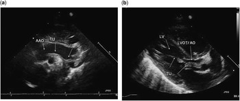
On the first day of life, the neonate was transferred to our unit because of the prescribed foetal ultrasound finding from the 25-week examination with a mild left ventricular outflow tract obstruction caused by a mass protruding across the aortic valve annulus in the presence of an otherwise normally developed left ventricle, aortic valve annulus, and aortic arch. On postnatal transthoracic echocardiography, an echogenic, pedunculated tumour was demonstrated arising at the anterolateral left ventricular wall next to the anterior papillary muscle extending across the left ventricular outflow tract and partially obstructing the structurally normal aortic valve (Fig 1a). There was no significant gradient across the outflow tract and only an insignificant aortic insufficiency. The tumour was loosely attached at the sinutubular junction with continuous extension into the aortic arch along its inferior curvature and isthmic region, with some adhesions superior and inferior to the origin of the left subclavian artery (Fig 1b). On colour flow mapping, there was some flow disturbance, and maximum blood flow velocity within the distal arch to the descending aorta was increased to 2.5 m/second. The infant was asymptomatic. Criteria for surgical removal at the age of 14 days were the risk for thromboembolic complications, further compromise of aortic valve function, and aortic arch obstruction. The intracavitary tumour was removed through an apical left ventriculotomy using cardiopulmonary bypass and hypothermic circulatory arrest. This approach allowed for detailed exposure and complete resection of the tumour, inserting nearby the anterolateral papillary muscle. The transaortic approach was used for removal of the transvalvular and intraluminal extension of the tumour within the aortic arch by careful suction. The postoperative period was uneventful.
( a ) Parasternal long-axis view with longitudinal extension of the left ventricular tumour across the left ventricular outflow tract. ( b ) Aortic arch view at the level of origin of the left subclavian artery: tumour extension within the aortic arch and adhesion around the origin of the left subclavian artery (arrow). AAO=ascending aorta; AO=aorta; LV=left ventricle; LVOT=left ventricular outflow tract; TU=tumour.

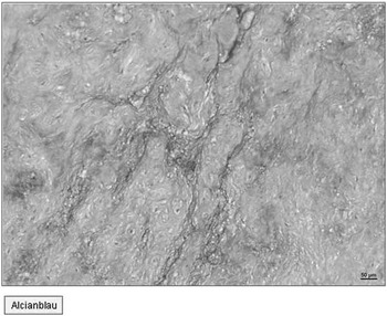
Histopathologic examination revealed ovoid cells with eosinophilic cytoplasma singly orientated and widely spaced in the myxoid stroma; elastic fibres and collagen bundles indicated fibrosis (Fig 2).
Histopathologic examination revealed the ovoid cells with eosinophilic cytoplasma singly orientated and widely spaced in the myxoid stroma; elastic fibres and collagen bundles indicate fibrosis (alcian blue staining).

Myxomas, as described in our neonatal patient and confirmed on histopathological examination, are extremely rare lesions, especially with regard to left ventricular wall origin. The exceptional tumour extension from the left ventricular cavity through the left ventricular outflow tract into the aortic arch has not been observed yet. Myxomas account for <1% of cardiac tumours in paediatric patients; they are more often found in older children and adult patients.Reference Isaac 1 The tumour appears as an echogenic, pedunculated, or broadly attached structure mainly localised in the left or right atrium. In children, a much more variable site of the origin of the myxoma has been considered, including the right ventricular wall or infundibulum.Reference George, Przygogzki, Lonas and Di Russo 6 , Reference Akyildiz, Tolgay, Öz, Yilmaz and Koc 7 Originating from the right or, extremely rare, left ventricular endocardium clinical symptoms of inflow or outflow obstruction have been described in adult patients.Reference Natale, Minardi, Casali, Pulignano and Musumeci 8 In this situation, protrusion of the mass through the aortic valve caused severe acute left ventricular outflow tract obstruction; the risk of sudden death has to be considered, and urgent removal of the mass is indicated.Reference Natale, Minardi, Casali, Pulignano and Musumeci 8
Tumour localisation and unusual tumour extension in our neonate might be explained by further extension of the tumour mass during pregnancy guided by the direction and streaming of blood flow. Postnatal echocardiography demonstrated extension of the echogenic mass within the aortic arch and some adhesions at the origin of the left subclavian artery – opposite to the entrance and predominant direction of streaming of the foetal arterial duct. An additional explanation for flow-related tumour extension might be that myxoma are thought to arise from the pluripotential mesenchymal cells dispersed in a myxoid stroma and have a soft, gelatinous consistency.Reference Isaac 1 , Reference Akyildiz, Tolgay, Öz, Yilmaz and Koc 7
Prenatal diagnosis of cardiac tumours have been described as early as 20 weeks of gestation mainly between 22 and 34 weeks; with further development during pregnancy the detection rate will increase. This observation has been made for rhabdomyomas and teratomas; fibroma or haemangioma have rarely been observed during infancy. Until 2003, myxoma have not been reported in a foetus and remain an exceptional finding on prenatal ultrasound.Reference Geipel, Krapp, Germer, Becker and Gembruch 3 , Reference Paladini, Tartaglione, Vassallo and Martinelli 4 Neonatal myxoma with severe symptoms of right atrial obstruction was reported in 1982.Reference Dianzumba and Char 9 George et al 2006 described a myxoma in a neonate arising from the infundibulum and partially obstructing the right ventricular outflow tract and found two other cases with right ventricular myxoma in the literature. Pathology studies in these cases were consistent with myxoma describing the stellate or ovoid cells with decreased or lack of mitotic activity within a myxoid matrix that undergoes fibrosis or calcification.Reference George, Przygogzki, Lonas and Di Russo 6 , Reference Pucci, Gagliadotto, Zanini, Pansini, Summa and Mollo 10 Histiogenesis of myxoma was described as “uncertain”. Immunohistochemical study indicated that they arise from the pluripotential mesenchymal cells;Reference Akyildiz, Tolgay, Öz, Yilmaz and Koc 7 another assumption is that the stroma cells derived from the endocardial neural tissue.Reference Pucci, Gagliadotto, Zanini, Pansini, Summa and Mollo 10 The principle diagnostic criteria of the eosinophilc cells – scattered stallate/spindle cells, singly or organised as nests or chords in a myxoid stroma were also found in our patient. Treatment of choice is complete resection, as haemodynamic compromise, arrhythmia, or embolic events might develop.Reference Zhou, Fan, Peng, Zhang, Fu and Wang 5 , Reference George, Przygogzki, Lonas and Di Russo 6 Risk for recurrence is only given after incomplete resection or recurrence at multiple sites.
Acknowledgement
None.
Financial Support
This research received no specific grant from any funding agency, commercial, or not-for-profit sectors.
Conflicts of Interest
None.