INTRODUCTION
Entamoeba moshkovskii is another species of Entamoeba which is morphologically similar to, but biochemically and genetically different from Entamoeba histolytica and Entamoeba dispar (Ali et al. Reference Ali, Hossein, Roy, Ayeh-Kumi, Petri, Haque and Clark2003; Beck et al. Reference Beck, Dogan, Maro, Sam, Shao and Houpt2008). It was initially thought to be a free-living environmental amoebae strain. However, the first E. histolytica-like strain isolated in 1961 from a resident of Laredo presented with diarrhoea, weight loss, and epigastric pain. Subsequently, molecular studies have confirmed that the E. histolytica Laredo strain is a strain of E. moshkovskii (Clark and Diamond, Reference Clark and Diamond1991). Since then, E. moshkovskii has been reported in humans worldwide (Ali et al. Reference Ali, Hossein, Roy, Ayeh-Kumi, Petri, Haque and Clark2003; Parija and Khairnar, Reference Parija and Khairnar2005; Tanyuksel et al. Reference Tanyuksel, Ulukanligil, Guclu, Araz, Koru and Petri2007; Beck et al. Reference Beck, Dogan, Maro, Sam, Shao and Houpt2008; Fotedar et al. Reference Fotedar, Stark, Marriott, Ellis and Harkness2008; Soumaya et al. Reference Soumaya, Karim, Nadia, Abdallah and Bouratbine2008; Farnaz et al. Reference Farnaz, Ebrahim, Ali, Ehsan and Bahram2011; Shimokawa et al. Reference Shimokawa, Kabir, Taniuchi, Mondal, Kobayashi, Ali, Sobuz, Senba, Houpt, Haque, Petri and Hamano2012).
The most recent study conducted by Shimokawa et al. (Reference Shimokawa, Kabir, Taniuchi, Mondal, Kobayashi, Ali, Sobuz, Senba, Houpt, Haque, Petri and Hamano2012) in Mirpur, Dhaka reported that diarrhoea occurred temporarily with the acquisition of a new E. moshkovskii infection among Bangladeshi children. Moreover, their study revealed that E. moshkovskii can cause diarrhoea, colitis and weight loss in mice. Similarly, other studies from Bangladesh, India, Turkey, Australia and Pakistan have reported E. moshkovskii as a sole potential enteropathogen in patients presenting with gastrointestinal symptoms and/or dysentery (Ali et al. Reference Ali, Hossein, Roy, Ayeh-Kumi, Petri, Haque and Clark2003; Parija and Khairnar, Reference Parija and Khairnar2005; Tanyuksel et al. Reference Tanyuksel, Ulukanligil, Guclu, Araz, Koru and Petri2007; Fotedar et al. Reference Fotedar, Stark, Marriott, Ellis and Harkness2008; Yakoob et al. Reference Yakoob, Abbas, Beg, Naz, Khan and Jafri2011).
E. moshkovskii is not distinguishable in its cystic or trophozoite forms from E. histolytica (the cause of invasive amoebiasis) or E. dispar (a common non-invasive parasite), and it is impossible to differentiate the 3 species based on traditional microscopic examination alone. However, polymerase chain reaction (PCR) tests have expedited the detection of E. moshkovskii. In Malaysia, many microscopically-based epidemiological studies have shown high prevalence of Entamoeba species infection. However, a previous molecular study that investigated the prevalence of E. histolytica and E. dispar did not consider the possible presence of E. moshkovskii (Noor Azian et al. Reference Noor Azian, Lokman Hakim and Maslawaty2006). Hence, to the best of our knowledge, this is the first identification of E. moshkovskii in Malaysia using single-round PCR assay through which the presence of E. moshkovskii was confirmed by sequencing.
MATERIALS AND METHODS
Fecal samples
A total of 500 fecal samples were collected over the period from June to December 2011 from Orang Asli communities in the states of Negeri Sembilan, Perak, and Pahang, in Malaysia. Approximately 5 grammes of each fecal sample were placed into a 15 ml centrifuge tube containing 3 ml of polyvinyl alcohol (PVA). The samples were examined by the trichrome staining technique. Additionally, the remaining part of the samples was kept unpreserved and stored at 4 °C upon arrival at the laboratory for further analysis by formalin-ether sedimentation and DNA extraction. Samples were considered microscopically positive if cysts and/or trophozoites of E. histolytica/E. dispar/E. moshkovskii complex were detected in at least 1 of the 2 techniques.
DNA samples
DNA isolated from axenically grown E. histolytica HM-1:IMSS, E. dispar SAW 760, and E. moshkovskii Laredo were used as positive controls in this study. All of these control DNA samples were a gift from Dr Graham Clark (London School of Hygiene and Tropical Medicine).
DNA extraction
DNA was extracted directly from fecal samples by using the QIAamp stool DNA extraction kit (QIAGEN, Hilden, Germany) according to the manufacturer's instructions. The extracted DNA was then stored at −20 °C until further use.
PCR amplification
The analysis was based on the amplification of the small subunit ribosomal RNA (SSU rRNA) gene of E. moshkovskii. Single-round PCR was performed according to an earlier protocol described by Hamzah et al. (Reference Hamzah, Petmitr, Mungthin, Leelayoova and Chavalitshewinkoon-Petmitr2006) based on the sequences of the SSU rRNA gene of E. moshkovskii (GenBank Accession no. AF149906). The sequence of the forward primer used EntaF (5′-ATG CAC GAG AGC GAA AGC AT-3′) was conserved in all 3 Entamoeba species, whereas the specific reverse primers, EhR (5′-GAT CTA GAA ACA ATG CTT CTC T-3′), EdR (5′-CAC CAC TTA CTA TCC CTA CC-3′), and for EmR (5′-TGA CCG GAG CCA GAG ACA T-3′) were specific for E. histolytica, E. dispar, and E. moshkovskii, respectively (Hamzah et al. Reference Hamzah, Petmitr, Mungthin, Leelayoova and Chavalitshewinkoon-Petmitr2006). The forward primer in combination with the appropriate reverse primer generates a 166-bp PCR product with E. histolytica DNA, a 752-bp PCR product with E. dispar DNA, and a 580-bp product with E. moshkovskii DNA.
The PCR amplification reaction was performed in a final volume of 50 μl in 0·2 ml PCR tubes by using an Eppendorf Pro-S thermal cycler (Hamburg, Germany). The reaction mixture contained 200 μ m of each deoxynucleoside triphosphate, 0·1 μ m of each forward and reverse primer, 6 mm MgCl2, 0·5 U of Taq polymerase (Nano Helix, South Korea), 1X Taq buffer (Nano Helix, South Korea) and 10 μl of extracted DNA samples. Amplification of each species-specific DNA fragment started with an initial denaturation at 94 °C for 3 min, followed by 30 cycles of 94 °C for 1 min, 58 °C for 1 min, and 72 °C for 1 min, with a final extension at 72 °C for 7 min. Amplified products were analysed by electrophoresis in 1·5% agarose gels and stained with GelRed (0·1 μl/ml: Biotium).
Sequencing of PCR product
The positive amplicons were then purified using the SolGent™ kit (South Korea) according to the manufacturer's instruction. All purified amplicons were sequenced in both directions using the same primer sets as in the respective PCR assay with an ABI 3730XL sequencer (Bioncer Corporation, South Korea). Sequence chromatograms were viewed using Sequence Scanner version 1.0 program (Applied Biosystem, USA). Forward and reverse sequences were edited, manually aligned, and the consensus sequence was created for each sample using the BioEdit Sequence Alignment Search Tool (BLAST) (Hall, Reference Hall1999) to the National Centre for Biotechnology Information (NCBI) reference sequences. The E. moshkovskii (GenBank Accession number AF149906) reference sequence was used in the analysis.
Ethical considerations
Ethical approval was obtained from the Ethics Committee of Universiti Kebangsaan Malaysia Medical Centre (UKMMC) (Reference number: UKM 1.5.3.5/244/FF-165-2011) and permission for field work was obtained from the Ministry of Rural and Regional Development Malaysia before starting the study.
RESULTS
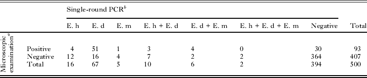
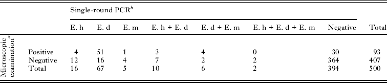
In total 500 fecal samples were screened by microscopy for the presence of either cysts and/or trophozoites of E. histolytica/E. dispar/E. moshkovskii complex. Overall, 93 (18·6%) fecal samples were positive for E. histolytica/E. dispar/E. moshkovskii complex. A single infection of E. histolytica/E. dispar/E. moshkovskii complex was detected in 20·4% (19/93) of the samples and 79·6% (74/93) were mixed infections of E. histolytica/E. dispar/E. moshkovskii complex with 2 or more other protozoan species.
All fecal samples (n=500) were subjected to single-round PCR for confirmation of the Entamoeba species. PCR products were detected in 106 (21·2%) samples. Of the 106 PCR-positive samples, single isolations of E. histolytica, E. dispar, and E. moshkovskii were reported in 16, 67 and 5 samples, respectively. Moreover, 10 samples were positive for E. histolytica and E. dispar and 6 samples were positive for E. moshkovskii and E. dispar, while E. moshkovskii was isolated in association with E. dispar and E. histolytica in only 2 samples (Table 1). Overall, E. moshkovskii isolates were recovered in 12·3% (13/106) of the samples. In addition, an amplicon of 580-bp specific for E. moshkovskii was successfully isolated from 5 samples that were positive for cysts and/or trophozoites by microscopy and 8 were isolated from microscopically negative samples.
Results of microscopic examination and PCR performed on 500 fecal samples
a Species cannot be distinguished.
b Species detected by PCR.
E.h, Entamoeba histolytica; E.d, Entamoeba dispar; E.m, Entamoeba moshkovskii.
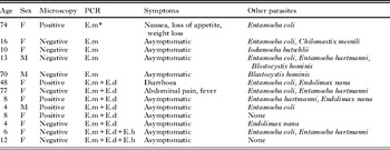
All 5 single isolation samples, as well as the reference strain E. moshkovskii Laredo produced the expected band at 580-bp. The sequences of all E. moshkovskii amplicons showed 99% to 100% similarity to E. moshkovskii sequences in GenBank (Accession number AF149906). The 13 positive samples were from 3 (23·1%) males and 10 (76·9%) females, aged between 4 and 77 years. Three E. moshkovskii-positive samples were from symptomatic individuals whereas the remaining 10 samples came from asymptomatic individuals (Table 2). Every PCR-negative sample was re-tested by PCR and each was again found to be PCR negative.
Clinical details of 13 subjects positive for Entamoeba moshkovskii by PCR

* E.h, Entamoeba histolytica; E.d, Entamoeba dispar; E.m, Entamoeba moshkovskii.
DISCUSSION
This study reports, for the first time, the presence of E. moshkovskii among Orang Asli communities in Malaysia. Findings of the present study demonstrated that 12·3% (13/106) of E. moshkovskii were detected in 106 PCR-positive products. A higher prevalence (24·7%) of E. moshkovskii was reported among a patient population in Australia (Fotedar et al. Reference Fotedar, Stark, Marriott, Ellis and Harkness2008). Similarly, a study by Ali et al. (Reference Ali, Hossein, Roy, Ayeh-Kumi, Petri, Haque and Clark2003) among pre-school children in Bangladesh found that the prevalence of E. moshkovskii was 21%. On the other hand, a study in a healthy population from Iran reported E. moshkovskii in 1·1% (1/88) of microscopically positive samples (Solaymani-Mohammadi et al. Reference Solaymani-Mohammadi, Rezaian, Babaei, Rajabpour, Meamar, Pourbabai and Petri2006).
The majority of E. moshkovskii isolations reported worldwide reported a mixed infection. In our study, the association of E. moshkovskii and E. dispar was found in 6 samples. Interestingly, this study also reported 2 combination cases of E. moshkvoskii, E. dispar, and E. histolytica. These results are in concordance with previous studies carried out in different parts of the world (Ali et al. Reference Ali, Hossein, Roy, Ayeh-Kumi, Petri, Haque and Clark2003; Parija and Khairnar, Reference Parija and Khairnar2005; Solaymani-Mohammadi et al. Reference Solaymani-Mohammadi, Rezaian, Babaei, Rajabpour, Meamar, Pourbabai and Petri2006; Hamzah et al. Reference Hamzah, Petmitr, Mungthin, Leelayoova and Chavalitshewinkoon-Petmitr2010). Likewise, a study by Fotedar et al. (Reference Fotedar, Stark, Marriott, Ellis and Harkness2008) reported 35% and 1·1% as mixed infections of E. moshkovskii with E. dispar and E. moshkovskii with E. histolytica, respectively. The mixed infections may be explained by the similar modes of human contamination by this entirely intestinal amoeba (Haque et al. Reference Haque, Ali, Clark and Petri1998).
Most of the studies support the commensal nature of E. moshkovskii (Scaglia et al. Reference Scaglia, Gatti, Strosselli, Grazioli and Villa1983; Beck et al. Reference Beck, Dogan, Maro, Sam, Shao and Houpt2008). However, a study in India reported an association of E. moshkovskii infection with dysentery (Parija and Khairnar, Reference Parija and Khairnar2005). Similarly, 2 mixed infection cases of E. moshkovskii and E. histolytica reported in Turkey presented with intestinal symptoms. However, it was unclear whether E. moshkovskii or E. histolytica caused the observed symptoms (Tanyuksel et al. Reference Tanyuksel, Ulukanligil, Guclu, Araz, Koru and Petri2007). A case-control study in Australia detected E. moshkovskii in 11 patients who presented with diarrhoea and other clinical symptoms of gastrointestinal disease, and no other pathogen including viruses and bacteria was isolated from their fecal specimens (Fotedar et al. Reference Fotedar, Stark, Marriott, Ellis and Harkness2008). Nonetheless, a previous study in Pakistan among 161 patients with chronic diarrhoea also reported that E. histolytica and E. moshkovskii were associated significantly with diarrhoea (Yakoob et al. Reference Yakoob, Abbas, Beg, Naz, Khan and Jafri2011). This is in accordance with another study from Iran where E. moshkovskii isolate was obtained from a dysenteric fecal sample in which common bacterial agents of dysentery was negative by routine stool culture (Farnaz et al. Reference Farnaz, Ebrahim, Ali, Ehsan and Bahram2011).
The current study found a single isolation of E. moshkovskii which was shown to be positive by both microscopic techniques and single-round PCR. This subject presented with gastrointestinal symptoms such as nausea, loss of appetite, and weight loss. Therefore, the present finding highlights a genuine infection rather than just passage or contamination sample with E. moshkovskii DNA. In addition, we also found 2 symptomatic cases with diarrhoea, abdominal pain, and fever, due to the combination of E. moshkovskii and E. dispar; 1 of them was also detected by microscopy. So, it can be postulated that E. moshkovskii may contribute to the development of diarrhoea and other gastrointestinal symptoms as E. dispar is well known to be a non-pathogenic species. These findings were in agreement with previous reports elsewhere (Fotedar et al. Reference Fotedar, Stark, Marriott, Ellis and Harkness2008; Yakoob et al. Reference Yakoob, Abbas, Beg, Naz, Khan and Jafri2011; Shimokawa et al. Reference Shimokawa, Kabir, Taniuchi, Mondal, Kobayashi, Ali, Sobuz, Senba, Houpt, Haque, Petri and Hamano2012) and provide further evidence in support of the hypothesis that E. moshkovskii could be a potential pathogenic amoeba. However, the cause-effect association could not be determined due to the limitation of the cross-sectional study design used in this study and no attempt was made to rule out other bacterial or viral causes of diarrhoea. However, more studies are necessary to define the exact role of E. moshkovskii in gastrointestinal disorders and the possible virulence of this organism.
We conclude that E. moshkovskii does exist in Malaysia. Thus, single-round PCR assay should be used for species-specific identification of E. histolytica/E. dispar/E. moshkovskii complex in epidemiological studies or for diagnostic purposes. Identification of this amoeba in fecal samples should not be dismissed as non-pathogenic protozoa especially in cases where other pathogenic protozoa, bacteria, and viruses are not detected. Further studies are needed to confirm the pathogenicity of E. moshkovskii infection and determine the epidemiology among Orang Asli communities in Malaysia.
ACKNOWLEDGEMENTS
We sincerely thank Dr Graham Clark (London School of Hygiene and Tropical Medicine) for providing us with lyophilized DNA of standard cultures of E. histolytica HM-1:IMSS, E. dispar SAW 760 and E. moshkovskii Laredo. We gratefully acknowledge the Ministry of Rural and Regional Development Malaysia for granting us permission to conduct this research. We also thank all the participants from Parit Gong village, Pasu village, Pian village, Bagan Balak village, Sungai Banun village, Desa Damai village, Sungai Raba village, and Pengkalan Permai village for their commitment and contribution in providing their fecal samples. Last but not least, our sincere thanks to Dr Edariah Abu Bakar for editing the manuscript.
FINANCIAL SUPPORT
This work was supported in part by the UKMMC Fundamental Research Grant (FF-165-2011) and Special Research University Grant (UKM-GUP-2011-316) from Universiti Kebangsaan Malaysia.