A vein of Galen malformation is a rare cause of cardiac insufficiency in neonates. Rarely, coarctation of the aorta has been described as a comorbid condition that may further compromise such an infant's haemodynamic state.
Either or both of these haemodynamically relevant lesions may require urgent therapy in the neonatal period. In general, surgical repair is considered the standard intervention for a primary coarctation. Vein of Galen malformations are generally treated by catheter-based endovascular embolisation.
This report describes a severely compromised term neonate, with a coexisting coarctation of the aorta and a vein of Galen malformation. The coarctation was first palliated by stent implantation as a temporising measure, followed by endovascular treatment of the vein of Galen malformation. After successful embolisation therapy, the stent was surgically explanted and the coarctation was successfully repaired.
Case report
A full-term neonate weighing 4.15 kilograms at birth became symptomatic with severe tachypnoea, dyspnoea, and mild hypoxaemia. The child had pulsations of the carotid arteries and a cardiac murmur.
Echocardiography showed a coarctation of the aorta with duct-dependent systemic circulation, and either a reverse flow in the aortic arch from the ductus arteriosus to the left carotid artery, or intermittently an antegrade flow with a late diastolic flow through the coarctation of the aorta. The diameter of the preductal part of the descending aorta was 3 millimetres compared with 12 millimetres in the ascending aorta. The intracardiac findings were normal.
A very large intracerebral arteriovenous malformation was detected by cranial ultrasound and confirmed by magnetic resonance imaging. These images confirmed the presence of a true vein of Galen malformation, with resultant hyperdynamic, high-output cardiac failure.
A vein of Galen malformation causing heart failure is generally treated in the neonatal period by repeated coil embolisation. However, in this case, the comorbid aortic coarctation also needed to be addressed in a semi-urgent manner. Medical treatment with prostaglandin infusion did maintain ductal patency, but this represented an additional aortic run-off lesion that compromised the infant's haemodynamics. We sought to address the coarctation while avoiding associated surgical morbidity. We chose stent placement as a means of temporising the aortic coarctation, and then pursued sequential embolisation of the vein of Galen aneurysm.
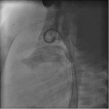
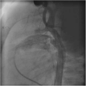
Cardiac catheterisation was performed via the femoral artery. Angiography – using a 4 French pigtail catheter – revealed a tubular hypoplasia of the descending aorta between the left subclavian artery and the ductus arteriosus, with a distinct preductal coarctation of the aorta (Fig 1). The ductus arteriosus was open, under infusion of prostaglandin E. The ascending aorta was enlarged, as well as both the carotid arteries. A 5 × 12-millimetre coronary stent (Liberté, Boston Scientific, Natick, Massachusetts, United States of America) was implanted through a 6 French sheath into the stenotic segment and was expanded up to 5.5 millimetres by a single inflation to 12 bars (Fig 2). The intervention was uneventful.
Lateral aortography shows the tubular hypoplasia and the juxtaductal coarctation of the aorta.

Aortic arch after implantation of a 5 × 12-millimetre stent. The stent covers the hypoplastic segment and the juxtaductal coarctation of the aorta.

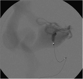
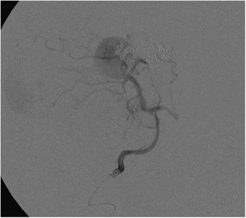
The symptoms of cardiac insufficiency did not improve following aortic stenting, and thus we proceeded with sequential embolisation of the vein of Galen malformation. On days 2 and 6 following stent placement, the initial embolisations were performed. Under general anaesthesia, femoral arterial and venous access was again obtained, and an arterial microcatheter was navigated into one of the main arterial feeding vessels of the malformation (Fig 3). Vessel location was derived from ultrasound and magnetic resonance imaging. A second microcatheter was navigated into the venous collector of the vein of Galen malformation via the transfemoral venous route. After visualising the fistulous connections, the microcatheter from the venous side was advanced into the feeding artery in a retrograde manner. At that point, both tips of the microcatheter crossed each other. Retrograde coiling of the arteriovenous fistula was then performed using microcoils. Multiple-staged embolisations were required in order to limit individual doses of contrast and radiation exposure. Following the initial two embolisation procedures, the infant's heart failure improved with ongoing anticongestive therapy. With ongoing heart failure, the infant was taken for a third course of embolisation 6 weeks later (Fig 4).
Contrast injection into the posterior cerebral artery – lateral view – showing the vein of Galen malformation.

After the third embolisation of the vein of Galen malformation – left vertebral artery injection; lateral view. The major part of the arteriovenous shunt had already been occluded, providing enough haemodynamic stability for surgical correction of coarctation of the aorta.

At the age of 12 weeks, arch reconstruction was performed via a median sternotomy using cardiopulmonary bypass and moderate hypothermia. The whole arch and descending aorta were dissected and mobilised. The procedure was done under selective antegrade cerebral perfusion into the brachiocephalic trunk. Externally, the stent was palpable just distal to the left subclavian artery, bridging the coarctation area through to the descending aorta.
Following cardioplegic arrest, the arch was opened and the stented area was removed completely. After removal of the stented area and extensive mobilisation, it was possible to connect the back wall of the native descending aorta with the distal part of the arch. In order to address the degree of arch hypoplasia, the anterior surface of the arch was enlarged with homograft material to achieve an equal-sized arch and descending aorta. Following separation from bypass, no gradient could be measured through the reconstructed area.
A fourth and final coil embolisation of the vein of Galen malformation was performed at 7 months of age. All embolisation procedures were performed by the senior author (F.B.). Upon follow-up at age 3, the child remains asymptomatic from a cardiovascular standpoint and is on no cardiac medications. He has a normal developmental status.
Discussion
Vein of Galen malformation is a rare cause of high-output cardiac failure in neonates. In some isolated cases, patients with vein of Galen malformation show signs of a coarctation of the aorta, either being associated with itReference Schievink, Mokri, Piepgras and Gittenberger-de Groot1–Reference Tomlinson, Piepgras, Nichols, Rüfenacht and Kaste3 or mimicking the disease.Reference Srinivas, Changela, Francis and Kumar4–Reference Doyle, Mastrobattista, Thapar and Lantin-Hermoso6
It has been assumed that an increased flow through the pulmonary artery into the aorta, as seen in patients with huge cranial arteriovenous malformations, is responsible for the development of an aortic coarctation or arch hypoplasia during foetal life.Reference Cumming7
Owing to their haemodynamic significance, both these disorders require attention in the neonatal period. Severe neonatal coarctation is typically addressed surgically;Reference Egan and Holzer8 vein of Galen malformations are typically treated by endovascular embolisation,Reference Lasjaunias, Chng, Sachet, Alvarez, Rodesch and Garcia-Monaco9 usually during the first few days of life.
A coarctation of the aorta impedes distal aortic flow, and this increased resistance augments flow superiorly through to the low resistance vein of Galen malformation.Reference Tomlinson, Piepgras, Nichols, Rüfenacht and Kaste3 When this pattern of flow is recognised, or there is an established diagnosis of a vein of Galen malformation, a coarctation must be suspected and definitively ruled in or out by careful echocardiographic examination of the aortic arch. In our patient, we identified the presence of a significant coarctation, and decided to pursue a catheter-based intervention. Owing to the fact that balloon dilatation of primary coarctation has shown suboptimal results, we opted for stent placement. We expected that several procedural embolisations of the vein of Galen malformation were likely to be necessary during the first few months of life, and we thought that stent placement would be better tolerated than operative intervention in this setting.
As repetitively reported, the prognosis of primary coarctation repair, without treatment of the vein of Galen malformation, is very poor.Reference Bagdure, Bartakian and Kaufman10 Thus, it is important to diagnose or to exclude a vein of Galen malformation in patients with symptomatic coarctation of the aorta. This combination of lesions is indicated by the presence of a severely enlarged ascending aorta, brachiocephalic trunk, and the right carotid artery in combination with a reversal of flow in the descending aorta from the ductus arteriosus to the left carotid artery. In this setting, there should be a strong suspicion for the presence of a cranial arteriovenous malformation, which can easily be diagnosed by ultrasound.
Conclusion
Coarctation of the aorta and vein of Galen malformation may occur simultaneously in some patients. In severely symptomatic patients with either of these diagnoses, the other has to be confirmed or excluded.
Vein of Galen malformations should be suspected in any patient with a coarctation and aortic arch dilatation; the presence of even transient reversal flow in the aortic arch greatly raises such suspicion.
We believe that therapeutic intervention for the vein of Galen malformation has priority over surgical coarctation repair. We propose that, in patients with suspected coincident coarctation of the aorta, cardiac catheterisation with the stent palliation of the coarctation has to be considered before vein of Galen malformation embolisation. Surgical coarctation repair can be performed after successful embolisation of the vein of Galen malformation.