Introduction
In vitro maturation (IVM) of oocytes is increasingly demanded in both human-assisted reproductive technology and animal in vitro embryo production (IVP). However, in vitro-matured oocytes have been shown to have lower fertilization and blastocyst formation rates than their in vivo-matured counterparts (Banwell & Thompson, Reference Banwell and Thompson2008). This low IVP efficiency using in vitro-matured oocytes may be related to a heterogeneous oocyte population at the beginning of IVM (Sirard et al., Reference Sirard, Richard, Blondin and Robert2006; Fair, Reference Fair2009). Thus, preselection of the immature oocytes that are most likely to develop is crucial for improving the IVP efficiency when using IVM oocytes.
Many morphological, cellular, and molecular predictors of oocyte developmental competence have been reported (Wang & Sun, Reference Wang and Sun2006; Bukowska et al., Reference Bukowska, Kempisty, Piotrowska, Walczak, Sniadek, Dziuban, Brussow, Jaskowski and Nowicki2012). Several intrinsic markers of oocyte quality are also known to be closely related to the onset of the nuclear maturation (germinal vesicle breakdown, GVBD), such as maturation-promoting factor (MPF) activity (Naito et al., Reference Naito, Daen and Toyoda1992; Christmann et al., Reference Christmann, Jung and Moor1994; Chesnel & Eppig, Reference Chesnel and Eppig1995; Ledda et al., Reference Ledda, Bogliolo, Leoni and Naitana2001; Catalá et al., Reference Catalá, Izquierdo, Uzbekova, Morató, Roura, Romaguera, Papillier and Paramio2011). However, although some reports refer to the relationship between the timing of completion of nuclear maturation (first polar body extrusion or reaching metaphase II: MII stage) and subsequent developmental competence of in vitro-matured oocytes in human and bovine (Dominko & First, Reference Dominko and First1997; Son et al., Reference Son, Lee and Lim2005), a direct comparison between GVBD timing and immature oocyte quality has never been reported.
Most procedures used to evaluate intrinsic predictors can have adverse effects on oocyte viability and/or subsequent developmental competence with varying degrees. Therefore, the objective of this study was to establish a non-invasive oocyte evaluation method based on GVBD timing for preselecting more developmental competent oocytes in a commonly used mouse oocyte model. First, we investigated the time course of changes in nuclear maturation and preimplantation developmental competence of in vitro-matured oocytes to estimate GVBD timing in high developmental competent oocytes. Second, we compared the preimplantation developmental competence of early and late GVBD oocytes. Because the O2 concentration during IVM may affect the nuclear kinetics (Zeilmaker et al., Reference Zeilmaker, Hulsmann, Wensinck and Verhamme1972; Mingoti et al., Reference Mingoti, Castro, Méo, Barretto and Garcia2011), all experiments were performed under two distinct oxygen concentrations generally used for mammalian oocyte culture: 5% CO2 in air (20% O2) or 5% CO2, 5% O2, and 90% N2 (5% O2).
Materials and methods
Experimental animals
Male and female ICR mice purchased from Japan SLC Inc. (Shizuoka, Japan) were housed in the animal housing facility at the Graduate School of Veterinary Medicine, Hokkaido University. Mice were kept under light- and temperature-controlled conditions (12-h light/12-h dark cycle; 23 ± 2°C) and given chow pellets and water ad libitum. All procedures described herein were conducted in accordance with the Hokkaido University guidelines for the care and use of animals (approved protocol: 17023).
Collection of oocytes
Immature (19 to 23 days old) female mice were treated intraperitoneally with 5 IU of equine chorionic gonadotropin (eCG; Aska Pharmaceutical. Co. Ltd, Tokyo, Japan) and euthanized 48 h later by cervical dislocation. Mouse ovaries were collected in a 35-mm plastic dish containing 2.5 ml of Leibovitz's L-15 medium (Gibco Life Technologies, Grand Island, NY, USA) supplemented with 4 mg/ml of bovine serum albumin (BSA; A-8022, Sigma-Aldrich, St. Louis, MO, USA) and 50 μg/ml of gentamicin sulfate (G-3632, Sigma-Aldrich). Cumulus–oocyte complexes (COCs) were released from the large antral follicles using a 25-G needle. Only oocytes with a diameter >70 μm (excluding the zona pellucida thickness) and completely enclosed by cumulus cells were selected. COCs with dark, shrunken, or irregularly shaped oocytes were discarded.
In vitro maturation (IVM)
IVM was performed as described previously (Adam et al., Reference Adam, Takahashi, Katagiri and Nagano2004) with a slight modification in the number of COCs cultured in one droplet. In short, approximately 10 COCs were transferred to a 50-μl droplet of Waymouth's MB 752/1 (Gibco Life Technologies) supplemented with 5% fetal calf serum (FCS; Gibco Life Technologies), 0.23 mM sodium pyruvate (P-4562, Sigma-Aldrich), 1 IU/ml of porcine follicle stimulating hormone (FSH; Antrin R, Kawasakimitaka Co. Ltd, Kawasaki, Japan), 10 ng/ml of human recombinant epidermal growth factor (hrEGF; Sigma-Aldrich), and 50 μg/ml of gentamicin sulfate; then, the entire droplet was covered with paraffin oil. COCs were incubated for varying times at 37°C in a humidified atmosphere of 5% CO2 in air (20% O2) or 5% CO2, 5% O2, and 90% N2 (5% O2).
In vitro fertilization (IVF)
Following IVM, oocytes surrounded by the cumulus cells were subjected to IVF by adapting the procedure of Toyoda (Reference Toyoda1971). In brief, spermatozoa collected from the caudal epididymis of matured (3 to 6 months old) male mice were preincubated for 2 h in a 0.4-ml droplet of Krebs–Ringer bicarbonate medium under paraffin oil at 37°C in a humidified atmosphere of 20% O2. Preincubated sperm was added to a 0.4-ml droplet of Krebs–Ringer bicarbonate medium containing approximately 10 COCs to give a final sperm concentration of 2.0 × 10⁵ cells/ml. COCs were coincubated with sperm for 5 h at 37°C in a humidified atmosphere of 20% O2.
In vitro culture (IVC) and evaluation of embryonic development
After IVF, presumptive zygotes freed from cumulus cells were transferred to a 25-μl droplet of potassium simplex optimized medium (KSOM) (Erbach et al., Reference Erbach, Lawitts, Papaioannou and Biggers1994) and cultured at 37°C in a humidified atmosphere of 5% O2 in air. Cleavage and development to the blastocyst stage were determined at 24 and 120 h after IVF, respectively. Cell numbers of the blastocysts obtained after 120 h of IVC were determined using an air-dry procedure described elsewhere (Takahashi & First, Reference Takahashi and First1992).
Experimental design
Experiment 1: Time course of changes in nuclear stage and preimplantation developmental competence of oocytes
To examine the time course of changes in nuclear stage, the nuclear status of the oocytes was first examined for every 2 h interval from 0 to 12 h of IVM in either 20% O2 or 5% O2. Thereafter, nuclear status was also examined at two additional time points; 7 h (the expected time of first oocyte reached MII stage) and 15 h (the time used for routine IVM work in our laboratory (Adam et al., Reference Adam, Takahashi, Katagiri and Nagano2004)) of IVM, to more fully characterize the time course of changes in nuclear stage of IVM oocytes. The nuclear status of oocytes was assessed by whole-mount preparation according to a previous report (Bedford, Reference Bedford and Daniel1971) with a slight modification. Briefly, COCs were transferred to a 50-μl droplet of Leibovitz's L-15 medium supplemented with 300 U/ml hyaluronidase (H-3506, Type 1-S from bovine testis; Sigma-Aldrich). The surrounding granulosa cells were removed by pipetting at room temperature (20 to 25°C) for 5 min. The cumulus-free oocytes were mounted on a glass slide and gently pressed down with coverslips supported by four beads of paraffin-vaseline wax until trapped. Oocytes were then fixed with 10% neutral formalin for over 6 h, stained with 1% (w/v) aceto-orcein, decolored with aceto-glycerol, and examined under a phase-contrast microscope. This experiment was repeated four times at each time point using 16 to 48 oocytes for each replicate.
To examine the time course of changes in preimplantation developmental competence, oocytes cultured for 10, 12, 15, and 17 h in IVM medium under 20% and 5% O2 were subjected to IVF and IVC, and cleavage and blastocyst formation rates were examined as described above. This experiment was repeated four times at each time point using 22 to 60 oocytes for each replicate.
Experiment 2: Relationship between GVBD timing and preimplantation developmental competence of oocytes
In vitro maturing oocytes were separated into two groups according to the presence (+) or absence (−) of a clear germinal vesicle (GV) (Fig. 1) examined at ×400 magnification under an inverted microscope 3 h and 4 h after the initiation of IVM under 20% and 5% O2, respectively. Both GV+ and GV− oocytes were separately transferred to new droplets in groups of approximately 10, cultured until 15 h from the initiation of IVM under distinct O2 tension, and then subjected to IVF and IVC; cleavage and blastocyst formation rates and the cell number of blastocysts were examined as described above. This experiment was repeated seven times using 21 to 56 oocytes for each replicate. Oocytes with an unknown GV status (without checking GV status) that were not transferred to new droplets were used as controls. In another five replicates (24 to 51 oocytes per replicate), cleavage rates and nuclear status of uncleaved oocytes were examined by whole-mount preparation 24 h after initiation of IVC. Cleaved and uncleaved oocytes with two pronuclei were considered fertilized.
Morphology of cumulus-enclosed mouse oocytes with (+) or without (−) intact germinal vesicle (GV). Representative images of cumulus-enclosed GV+ or GV− oocytes 3 h after the initiation of in vitro maturation (IVM) under 20% O2 at low magnification (A). High magnification images of GV+ (B) and GV− (C) oocytes. Arrowheads indicate GV. Bar = 100 μm (A), 50 μm (B, C).

Statistical analysis
Data are expressed as mean ± standard error of the mean (SEM). Percentage data were transformed using the arcsine transformation (arcsin √%) before analysis to normalize the variance. The effects of the maturation period and O2 concentration during IVM on the percentage of nuclear stage, cleavage, and blastocyst formation were analysed by two-way analysis of variance (ANOVA) followed by a Bonferroni post-hoc test. The effects of GVBD timing on cleavage and blastocyst formation rates and the cell number of blastocysts were analysed by one-way ANOVA followed by a Bonferroni post-hoc test. Differences in the frequency of fertilized, unfertilized, and fragmented GV+ and GV− oocytes were evaluated by chi-squared analysis with the Yates’ correction. Data analysis was performed using SPSS software (ver. 13.0, SPSS Inc., Chicago, IL, USA).
Results
Time course of changes in nuclear stage and preimplantation developmental competence of oocytes
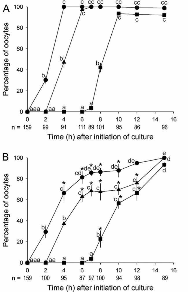
Under 20% O2, the first oocytes that underwent GVBD were found at 2 h after initiation of IVM, and the percentage of oocytes undergoing GVBD linearly increased to a maximum level after 4 h of IVM (Fig. 2 A). The percentage of oocytes that reached MI and MII stages was almost parallel to the time course of changes in GVBD with 2 h and 6 h delays, respectively. Under 5% O2, the percentage of oocytes undergoing GVBD increased in a biphasic manner; namely, approximately two-thirds of oocytes underwent GVBD during the first 4 h of IVM, gradually followed by the remaining third (Fig. 2 B). A similar biphasic pattern was also observed for oocytes that reached MI and MII stages with 2 h and 6 h delays, respectively.
Time course of changes in nuclear maturation of in vitro-matured mouse oocytes cultured under 20% O2 (A) and 5% O2 (B). Cumulative percentages of oocytes reached different stages of nuclear maturation [germinal vesicle breakdown (GVBD), black circle; metaphase I (MI), triangle; metaphase II (MII), square]. a,b,c,d,eDenotes statistical significance within the same nuclear stage among oocytes cultured under the same O2 concentration (P < 0.05). Asterisks (*) represent significantly lower values from oocytes at the same nuclear stage and time point cultured under 20% O2 (P < 0.05). This experiment was repeated four times. n represents the total number of oocytes examined at each nuclear stage at each time point.

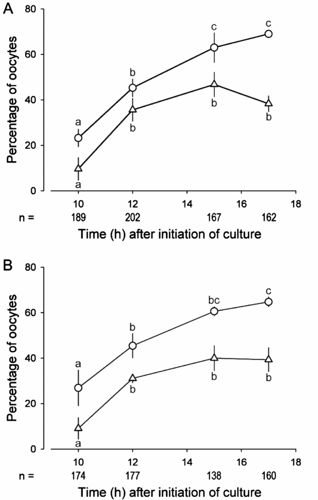
There were no significant differences between cleavage or blastocysts formation rates of oocytes cultured under 20% (Fig. 3 A) or 5% O2 (Fig. 3 B) at each time point. Cleavage rates tended to increase with prolonged periods of IVM, whereas blastocyst formation rates reached a maximum after 12 h, and these rates were not affected by prolonging the IVM period up to 17 h.
Time course of changes in preimplantation developmental competence of in vitro-matured mouse oocytes cultured under 20% O2 (A) and 5% O2 (B). Percentages of oocytes developed to 2-cell (white circle) and blastocyst (triangle) stages. a,b,cDenotes statistical significance within the same developmental stage among oocytes cultured under the same O2 concentration (P < 0.05). This experiment was repeated four times. n represents the total number of oocytes examined at each time point.

Relationship between GVBD timing and preimplantation developmental competence of oocytes
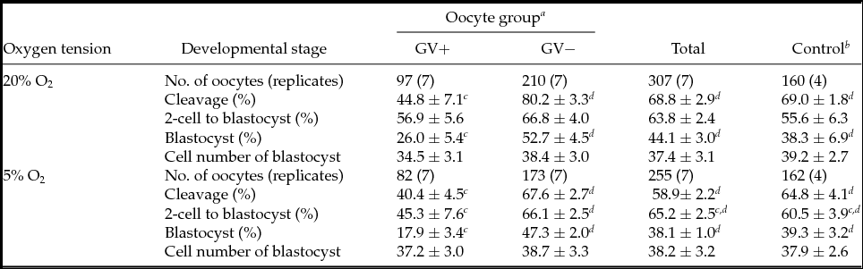
As shown in Table 1, GV− oocytes showed higher cleavage and blastocyst formation rates than GV+ oocytes, regardless of the O2 concentration during IVM (P < 0.05). The rate of blastocyst formation from the 2-cell stage in GV− oocytes cultured under 5% O2 was higher than that of GV+ oocytes (P < 0.05), but not when cultured under 20% O2. There was no difference between the cell number of blastocysts derived from GV+ and GV− oocytes, regardless of the O2 concentration during IVM. Mean percentages of cleavage, 2-cell to blastocyst, and blastocyst formation as well as the cell number of blastocysts in GV+ and GV− oocytes were not different from control oocytes, regardless of the O2 concentration during IVM.
Preimplantation developmental competence of early and late germinal vesicle (GV) breakdown mouse oocytes

a Oocytes with (GV+) or without (GV−) clear GV 3 and 4 h after initiation of in vitro maturation under 20% and 5% O2, respectively.
b Oocytes with an unknown GV status (without checking GV status) not transferred to new droplets.
c,d Values with different superscripts within the same oocyte developmental stage and O2 concentration differ significantly (P < 0.05).
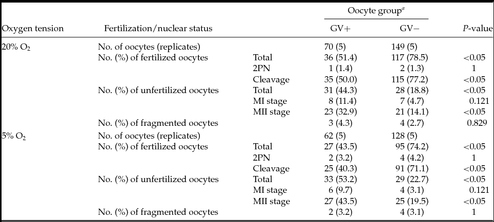
As shown in Table 2, the frequencies of GV+ and GV− fertilized oocytes stopped at the pronucleus stage, unfertilized oocytes stopped at the MI stage, and fragmented oocytes were not different, regardless of the O2 concentration during IVM. The frequency of GV+ unfertilized MII-stage oocytes was higher than that of GV− oocytes, regardless of the O2 concentration during IVM (P < 0.05).
Fertilization and nuclear status of early and late germinal vesicle (GV) breakdown in mouse oocytes 24 h after in vitro fertilization
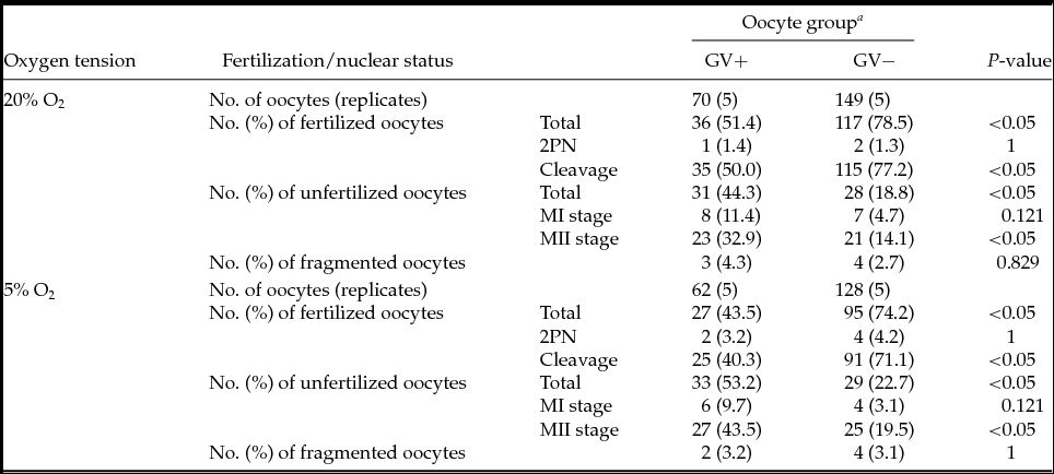
a Oocytes with (GV+) or without (GV−) clear GV 3 and 4 h after initiation of in vitro maturation under 20% and 5% O2, respectively.
2PN, two pronuclei formation; MI, metaphase I; MII, metaphase II.
Discussion
Although there were no comparative data on the nuclear progression of mouse oocytes cultured under 5% O2, the time course of changes observed in the nuclear stage under 20% O2 in this study fitted well with the previous studies (Donahue, Reference Donahue1968; Messinger & Albertini, Reference Messinger and Albertini1991; Liu et al., Reference Liu, Mal, Miao, Liu, Bao and Tan2005). Biphasic time course of changes in the percentage of oocytes that reached GVBD, MI, and MII stages under 5% O2 indicated that heterogeneous oocyte populations contained early and late nuclear maturation oocytes, even if they were obtained solely from large antral follicles of eCG-primed immature mice. Interestingly, two-thirds of the oocytes that underwent GVBD during the first 4 h of IVM under 5% O2 (early nuclear maturation group), showed similar nuclear kinetics to the oocytes cultured under 20% O2. Higher levels of intracellular reactive oxygen species in oocytes matured under 20% O2 compared 5% O2 was reported for bovine (Hashimoto et al., Reference Hashimoto, Minami, Takakura, Yamada, Imai and Kashima2000), and oxidative stress is known to stimulate meiotic resumption in mouse oocytes (LaRosa & Downs, Reference LaRosa and Downs2006).
Contrary to nuclear maturation, there were no detectable differences in cleavage or blastocyst formation rates for oocytes cultured under 20% or 5% O2 as reported previously (Eppig & Wigglesworth, Reference Eppig and Wigglesworth1995; Banwell et al., Reference Banwell, Lane, Russell, Kind and Thompson2007; Preis et al., Reference Preis, Seidel and Gardner2007). Furthermore, the blastocyst formation rate did not increase after 12 h of IVM even though one-third of oocytes cultured with 5% O2 underwent GVBD after that time. These results may indicate that early nuclear maturation oocytes seemed mainly to contribute to the blastocyst yield, and such high quality oocytes may acquire the developmental competence even under highly oxidative conditions.
To confirm this, we compared the preimplantation developmental competencies of early and late nuclear maturation oocytes selected based on the presence (+) or absence (−) of clear GV 3 h and 4 h after IVM initiation under 20% and 5% O2, respectively. These times were chosen because about two-thirds of oocytes were found to have undergone GVBD under the distinct O2 concentrations. GV− oocyte cleavage and blastocyst formation rates were almost double that of GV+ oocytes, regardless of the O2 concentration during IVM. This might be consistent with previous reports in humans and bovine; oocy- tes completing nuclear maturation early showed higher preimplantation developmental competence than those maturing later (Dominko & First, Reference Dominko and First1997; Son et al., Reference Son, Lee and Lim2005). However, although some late maturing bovine oocytes were improved, their developmental competence by prolongation of MII arrest before insemination (Dominko & First, Reference Dominko and First1997), a 3-h extension of IVM (18 h total) did not improve the developmental competence of GV+ oocytes in this study (data not shown). This finding may indicate that the low developmental competence of GV+ oocytes was not caused by a delay in cytoplasmic maturation. It should also be noted that no alteration in cleavage or blastocyst formation rates following the checking GVBD and subsequent transfer of in vitro maturing oocytes were observed.
Compared with GV− oocytes, although there were minor differences between the oocytes cultured under 20% and 5% O2, GV+ oocytes tended to remain unfertilized despite reaching MII or not form blastocysts despite successful fertilization/cleavage. Although the diameter of oocytes is reported to affect these parameters in mouse and bovine (Otoi et al., Reference Otoi, Yamamoto, Koyama, Tachikawa and Suzuki1997; Wu et al., Reference Wu, Liu, Zhou, Lan, Han, Miao and Tamm2007), the size (mean diameter excluding the zona pellucida) of GV+ and GV− oocytes were not different [GV+: 76.8 ± 1.6 µm (n = 75); GV−: 76.2 ± 1.2 µm (n = 150)]. Although the exact reasons for the difference in developmental competence of GV+ and GV− oocytes is unclear, it may be attributable to their different MPF activities. Because GVBD timing is precisely controlled by MPF activity, the appropriate sequential activation of MPF is needed for completion of nuclear maturation and successful fertilization, cleavage, and subsequent embryonic mitosis (Murray & Kirschner, Reference Murray and Kirschner1989; Kubiak et al., Reference Kubiak, Ciemerych, Hupalowska, Sikora-Polaczek and Polanski2008). Furthermore, lower MPF activity has been reported in the less developmental competent oocytes in several mammalian species (Naito et al., Reference Naito, Daen and Toyoda1992; Christmann et al., Reference Christmann, Jung and Moor1994; Ledda et al., Reference Ledda, Bogliolo, Leoni and Naitana2001; Catalá et al., Reference Catalá, Izquierdo, Uzbekova, Morató, Roura, Romaguera, Papillier and Paramio2011).
In conclusion, more developmental competent immature oocytes can be effectively preselected by choosing the early GVBD oocytes, regardless of the O2 concentration during IVM. Although further studies on implantation and postimplantation developmental competence are required, oocyte evaluation based on GVBD timing can be used as an efficient and non-invasive preselection method without the need for additional chemicals or equipment for high developmental competent oocytes.
Acknowledgements
This study was supported in part by a Grant-Aid for the 21st Century COE Program from the Ministry of Education, Culture, Sports, Science and Technology (MEXT) of Japan.