Introduction
Subglottic or tracheal stenosis is a well-recognised complication after prolonged endotracheal intubation in intensive care units. Stenosis occurs secondary to pressure-induced ischaemia and airway injury with subsequent scarring.Reference Siciliani, Rendina and Ibrahim1 Other, less frequent causes of stenosis include inflammatory, neoplastic, traumatic and idiopathic stenosis.Reference El-Fattah, Ebada, Amer, Abosamra and Tawfik2 Cricotracheal resection has a high success rateReference Siciliani, Rendina and Ibrahim1–Reference D'Andrilli, Maurizi, Andreetti, Ciccone, Ibrahim and Poggi3 and is considered the treatment of choice for severe (grade III and IV) stenosis according to the Myer–Cotton grading system.Reference Myer, O'Connor and Cotton4
Complications after cricotracheal resection are divided into anastomotic and non-anastomotic complications. Anastomotic complications include restenosis, granulation tissue formation, anastomotic dehiscence and fistulae of surrounding structures (oesophagus and innominate artery).Reference Auchincloss and Wright5 The incidence of restenosis after cricotracheal resection is between 3 and 9.5 per cent.Reference D'Andrilli, Maurizi, Andreetti, Ciccone, Ibrahim and Poggi3,Reference Morcillo, Wins, Gómez-Caro, Paradela, Molins and Tarrazona6–Reference Ashiku, Kuzucu, Grillo, Wright, Wain and Lo8
Restenosis has an insidious onset, and is the result of anastomotic problems including tension, ischaemia and/or subclinical separation leading to scar formation.Reference Tapias and Mathisen9 Management of restenosis after unsuccessful airway resection is challenging.Reference Ebada, Abd El-Fattah, Salem, Elkotb, Kamal and Tawfik10 Endoscopic dilatation, stenting and laser surgery may be performed, and satisfactory final results can be achieved with these manoeuvres.Reference Monnier and Monnier11 However, in the case of recurrent stenosis after the previously described procedures, repeat surgery should be considered. Re-intervention should be deferred for 6–12 months to allow the complete resolution of inflammation after the index surgery.Reference Tapias and Mathisen9
Revision cricotracheal resection is difficult given the unavoidable anastomotic tension associated with additional trachea resection. Furthermore, fibrosis after the primary surgery limits the mobility of the residual trachea.Reference Auchincloss and Wright5 Outcomes and success rates of revision surgical procedures are usually not as good as for primary surgical procedures.Reference Wright, Grillo, Wain, Wong, Donahue and Gaissert12,Reference Donahue13
This work aimed to review our experience over the last five years in revision cricotracheal resection performed for the management of recurrent stenosis following unsuccessful previous resection.
Patients and methods
Ethical standards
This retrospective study was conducted in the Otorhinolaryngology Department, Mansoura University Hospitals, Egypt, over a period of five years, from January 2015 to January 2020. The study was approved by the Mansoura Faculty of Medicine institutional research board (approval code: MFM-IRB: R.20.04.781).
Patients
A review of patients’ medical records revealed nine patients who underwent revision cricotracheal resection for the management of recurrent severe (grade III and IV) stenosis following previous failed cricotracheal resection.
Methodology
All nine patients underwent clinical evaluation, including a detailed history of previous airway surgical procedures and transnasal flexible laryngoscopy performed to assess the airway and true vocal fold mobility.
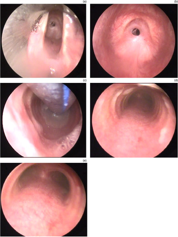
The grade and site of stenosis were assessed in more detail under general anaesthesia. The larynx was exposed using an anaesthetic laryngoscope with the blade inserted into the vallecula or by a suspension laryngoscope. Then, a rigid 4 mm diameter, 0° or 30° telescope was introduced, to offer a panoramic, clear view of the endolarynx, subglottis and trachea, all the way down to the carina (Figure 1). In grade IV stenosis, when the scope cannot pass the stenosis, the same assessment was performed by passing through the pre-existing tracheostoma. It is essential in the revision procedures to accurately assess the length of the remaining healthy trachea. If assessment reveals insufficient healthy trachea, a revision cricotracheal resection is contraindicated.
Endoscopic airway assessment. (a) The scope is introduced through the suspension laryngoscope to the level of the vocal folds. (b) The scope is introduced below the vocal folds. (c) Passing below the stenosis, the tracheostomy cannula is seen. (d) After cannula removal and examination of the lower trachea. (e) The scope reaches down to the carina.

The authors of this work adopted the surgical procedure described by Grillo and colleaguesReference Grillo, Donahue, Mathisen, Wain and Wright14,Reference Grillo, Mathisen and Wain15 and Monnier and colleagues.Reference Monnier, Lang and Savary16,Reference Monnier, Lang and Savary17 A cervical collar incision was planned with excision of the scar from the previous surgery and the stoma in patients with a pre-existing tracheostomy (n = 8) (Figure 2). The subplatysmal flaps were then elevated upwards to the hyoid bone and downwards to the sternum. Exposure of the laryngotracheal complex was achieved by separating the strap muscles in the midline. No attempt was made to identify the recurrent laryngeal nerves, because this would have put them at risk of injury. Tracheal dissection was performed close to the outer perichondrium to avoid injury to the recurrent laryngeal nerves. Circumferential dissection of the trachea was performed only at the level of the stenosis. Excessive dissection of the proximal and distal stumps was avoided in order to preserve the blood supply, so as to improve the healing of the anastomosis.
A patient with a previous failed cricotracheal resection. The incision for revision surgery should be planned to remove the stoma and scar tissue.

In revision cricotracheal resections, surgical dissection was challenging because of the scarring and fibrosis following the primary surgery. Identification of the landmarks was difficult, with a higher risk of injury to vital structures such as the oesophagus and the recurrent laryngeal nerves. However, dissection was performed carefully and meticulously, with no reported intra-operative complications.
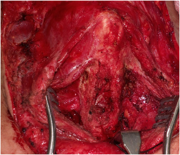
External airway deformity and scarring at the site of the recurrent stenosis was evident in all cases (Figure 3). Accordingly, resection of the stenosis was performed using the slicing technique: the trachea is opened transversely at the midpoint of the stenosis, and the trachea is then progressively sliced cranially and caudally until normal steady tracheal rings are reached. Re-anastomosis was performed between the proximal and distal stumps as cricotracheal anastomosis (n = 5) and thyrotracheal anastomosis (n = 4) according to the remaining stumps. After performing the anastomosis, Surgiflo flowable haemostatic matrix (Ethicon, Somerville, Massachusetts, USA) was applied over the whole operative field, including the line of airway anastomosis, with the purpose of providing adequate haemostasis and enhancing healing of the anastomosis. The authors of this paper believe that Surgiflo application may have a role in filling any possible micro-gaps in the anastomosis and, consequently, enhancing healing and decreasing the incidence of anastomotic complications.
Deformed tracheal segment.

Suprahyoid release is the standard procedure in our hospital, and it was conducted in all nine cases discussed here. In our institute, in primary surgical procedures, we perform a limited suprahyoid release when the resected segment is short (fewer than three rings). This entails cutting the suprahyoid muscles attached to the body of the hyoid (mylohyoid and geniohyoid) and the stylohyoid tendon attached to the lesser horn, with preservation of the digastric slings on either side. In cases requiring longer resections (more than three rings), full suprahyoid release is performed. The release is extended more laterally to both greater horns, with cutting of the digastric slings.
In this series, when suprahyoid release had been performed in the previous surgical procedures (n = 5), revision of the suprahyoid release was performed. This involved cutting the fibrosis at the site of the previous release (above the hyoid bone).
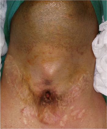
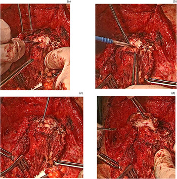
In one patient, because of previous suprahyoid release, there was excessive fibrosis, with almost complete adhesion and fusion of the body of the hyoid to the thyroid cartilage. This was associated with cosmetic deformity and a midline swelling in the neck, which was annoying for the patient (Figure 4). This patient had experienced significant choking and aspiration after the previous cricotracheal resection, which resulted in necrosis and dehiscence of the anastomosis, and finally restenosis and failure of the primary surgery. However, swallowing rehabilitation was performed by speech, language and swallowing pathologists in our hospital. The patient regained completely normal swallowing within three months. In the revision surgery, suprahyoid release was performed with excision of the body of the hyoid bone for cosmetic purposes (Figure 5). There was no reported post-operative dysphagia or choking.
(a) External neck swelling after previous suprahyoid release. (b) Post-operative photograph showing a marked decrease in the size of the swelling.

(a) Hyoid bone body is adherent to thyroid cartilage (the dissector is between the hyoid and thyroid). (b) Electrocautery is used to dissect hyoid bone from thyroid cartilage. (c) Further dissection was carried out. (d) The body of hyoid bone was removed after it was divided from the greater horns using heavy Mayo scissors.

A single-stage surgical procedure is preferred at our centre, and was performed in seven out of nine patients. This entails immediate extubation at the end of the procedure. Double-stage surgery entails performing a tracheostomy distal to the anastomosis. This procedure is preferred in infants or in patients who have severe co-morbidities, to secure the airway and protect against early complications after surgery (choking, aspiration and surgical emphysema). Decannulation is performed two or three weeks after complete healing of the anastomosis. Double-stage surgery was performed in two patients in the study.
The chin to chest suture is not preferred in our institute, and it was not used in any patient in the present study. Patients and caregivers were instructed to limit neck movements and to prevent neck extension during the early post-operative period (two to three weeks following surgery). A hard neck collar was fitted in a reversed manner in patients who were not compliant, to prevent extension.
After extubation, patients were sent to the intensive care unit, for 24-hour close observation and monitoring. On the next day, they were transferred to the ward to continue receiving their treatment and care. Drains were removed on the 7th post-operative day, and patients were then discharged home. Follow-up appointments were scheduled once weekly during the first month, then once monthly for the subsequent six months. The follow-up period ranged from 3 to 63 months (median of 38.5 months).
Results
Nine patients were included in this study (Table 1). Seven patients (77.8 per cent) were male and two patients (22.2 per cent) were female. The study included paediatric patients aged less than 18 years old (n = 6) and adults (n = 3).
Patients’ data

Stenosis was grade III in 44.4 per cent of patients and grade IV in 55.6 per cent. The stenosis was upper tracheal in 55.6 per cent (n = 5) and subglottic in 44.4 per cent (n = 4). Eight out of nine patients (88.9 per cent) were tracheostomised prior to the revision surgery. Single-stage surgery was carried out in 77.8 per cent of cases and double-stage surgery was conducted in 22.2 per cent.
All nine patients had restenosis after previous failed cricotracheal resections, performed in our centre (n = 3) or referred from other centres (n = 6). Data on the index surgical procedures (mainly the length of resected segment) were, therefore, not available in two patients. Furthermore, five patients had a history of multiple previous failed airway interventions other than the index cricotracheal resection, as follows: four patients had undergone multiple endoscopic procedures (dilatations and laser surgical procedures) before the index cricotracheal resection; and three patients had undergone costal cartilage laryngotracheoplasty before the index cricotracheal resection. In these latter three patients, the scarring and fibrosis in the revision cricotracheal resection made the surgery much more challenging after previous multiple open interventions (laryngotracheoplasty and cricotracheal resection).
For the three patients who were operated on in our hospital, the initial management of restenosis after the primary cricotracheal resection consisted of repeated endoscopic dilatations. One patient underwent one dilatation, and two patients underwent dilatations twice each. However, when dilatations failed to achieve a satisfactory airway, and stenosis recurred after dilatation to its original grade, revision surgery was considered.
The length of the resected airway segment in the index surgical procedures ranged from two to four rings (mean of 2.8 rings). When cricotracheal resection was performed in the first surgical procedure (n = 2), the resected cricoid arch was counted as one ring. In the revision procedure, the length of the resected segment ranged from one to three rings (mean of two rings). The mean total length of resection (first surgery plus revision) was five rings (range of four to six rings).
In the present study, no intra-operative complications were recorded. Regarding post-operative complications, minor complications occurred in two patients (surgical emphysema in one patient and temporary choking in the other). No major complications (such as anastomotic dehiscence, excessive granulation tissue formation or mortality) were reported. Restenosis occurred in one patient (11.1 per cent).
Successful decannulation was achieved in eight of the nine patients (88.9 per cent). In the follow-up period, those eight patients regained a normal lifestyle and activity, and no further airway procedures were indicated. In the remaining patient, restenosis was reported and decannulation was not possible. However, endoscopic dilatation was performed, and the patient will be considered for another revision surgery if the final outcome is not satisfactory.
Discussion
Revision cricotracheal resection after previous failed surgery is very challenging. More tracheal rings are removed and therefore anastomotic tension is unavoidable.Reference Monnier and Monnier18 It is not always possible to resect more rings. Some patients are not candidates for re-operation because of insufficient residual tracheal length.Reference Auchincloss and Wright5 In our centre, before embarking on a revision surgery, accurate assessment of the remaining healthy trachea is mandatory. This is achieved, first, by obtaining a detailed history of the previous surgery and the length of the previous resection. Moreover, the airway is endoscopically assessed under general anaesthesia before surgery, with careful measuring of the remaining airway and counting of the remaining tracheal rings.
MonnierReference Monnier and Monnier11 stated that up to five rings can be removed with laryngeal release procedures. Additionally, this author reported resection of up to eight rings in cricotracheal resections in paediatric patients. Similarly, Taylor et al.Reference Taylor, Collar, Wilson, Ohye and Green19 stated that up to half of the length of the trachea (8–10 rings) can be resected with full release manoeuvres. Wright et al.Reference Wright, Grillo, Wain, Wong, Donahue and Gaissert12 reported that adult patients can have up to 50 per cent of the trachea resected. In this study, it was possible to perform the revision cricotracheal resection with acceptable anastomotic tension by performing full suprahyoid release. The total length of the resected segment (index plus revision surgery) in this series ranged from four to six rings.
Anastomotic tension is unavoidable in revision cricotracheal resection. Excess tension leads to a higher incidence of complications. Previous reportsReference Wolf, Shapira, Talmi, Novikov, Kronenberg and Yellin20–Reference Nakache, Primov-Fever, Alon and Wolf22 concluded that repeat cricotracheal resections were associated with higher rates of complications and lower success rates. The largest series of laryngotracheal resections and reconstructions was published by Wright et al.,Reference Wright, Grillo, Wain, Wong, Donahue and Gaissert12 and it included 901 patients over a period of 28 years (1975–2003). They found that the incidence of anastomotic failure in revision procedures increased compared with initial surgery regardless of the length of trachea resected. In our previous studies,Reference El-Fattah, Ebada, Amer, Abosamra and Tawfik2,Reference El-Fattah, Kamal, Amer, Fouda, Elwahab and Tawfik23 the incidence of post-operative complications was higher in revision surgical procedures.
In order to decrease anastomotic tension, different laryngotracheal release manoeuvres have been described. Suprahyoid release was first described by Montgomery in 1974.Reference Montgomery24 This procedure is safe and well tolerated. Additionally, it can be performed through the same cervical approach. Mediastinal, hilar and pericardial release manoeuvres are more effective for lower tracheal or carinal resection, and a separate thoracic incision is required. In this series, full suprahyoid release was performed in all patients. Release manoeuvres should be performed liberally if excess anastomotic tension is expected, especially in revision cricotracheal resections.Reference Auchincloss and Wright5
The most common sequelae of suprahyoid release are related to dysphagia and aspiration.Reference Broussard and Mathisen25 It was assumed that preventing laryngeal elevation during swallowing might cause dysphagia. Yet, GrilloReference Grillo26 noted that dysphagia was significantly less often encountered after suprahyoid release in comparison with thyrohyoid release. In our institution, suprahyoid release is performed in a tailored fashion according to the length of the resected segment. Limited suprahyoid release (only the body of the hyoid) is performed in short resections (fewer than three rings), and full release (body and greater horn) is performed in long resections. Additionally, cutting of the greater horns is not carried out routinely in our centre. In this series, despite performing full suprahyoid release in all patients, only one patient developed temporary choking, and their condition improved spontaneously within a few days.
• Recurrent subglottic or upper tracheal stenosis after previous unsuccessful cricotracheal resection is challenging
• Revision cricotracheal resection can be successfully performed, provided there is sufficient length of healthy trachea remaining
• Adequate pre-operative evaluation, operative techniques and post-operative care are crucial for successful surgery
• Application of Surgiflo on the line of anastomosis enhances healing and improves the overall outcome
The authors of this study found that fibrosis and scarring made the revision surgical procedures much more challenging. Surgical dissection and the identification of landmarks and critical structures is difficult, and, consequently, the risk of injury to these structures is higher. However, meticulous surgical techniques were adopted to avoid intra-operative complications. Auchincloss and WrightReference Auchincloss and Wright5 stated that scarring after previous surgical procedures limits mobility of the trachea and leads to excess anastomotic tension. Accordingly, if repeat surgery is indicated, it should be deferred for 6–12 months after the primary surgery, to allow time for resolution of the peritracheal inflammation.Reference Auchincloss and Wright5
In our centre, in the last few years, we have started to use Surgiflo in cricotracheal resections conducted using either the primary or the revised procedure. Surgiflo is applied over the operative field and on the line of anastomosis. A study conducted by the authors of this work to assess the benefits of SurgifloReference Abd El-Fattah, Ebada and Tawfik27 revealed that our outcomes have improved significantly since adopting this procedure, specifically regarding the incidence of post-operative granulation tissue formation, anastomotic dehiscence, surgical emphysema, restenosis and overall success rates.
Surgiflo is an active flowable haemostatic matrix that contains gelatine and thrombin. It improves the clotting cascade to stop bleeding.Reference Price, Tackett and Patel28 It has been used in a wide range of head and neck surgical procedures, including laryngectomies, neck dissections and submandibular sialadenectomy.Reference Bannister and Ah-See29 Surgiflo consists of Surgifoam® absorbable gelatine powder, which is provided in a syringe and mixed either with saline or with human thrombin to create an injectable foam.
Gelatine provides a meshwork where the body's endogenous platelets, fibrin and thrombin may be entrapped, facilitating rapid clot formation and haemostasis.Reference Doria and Vaccino30 In a study performed to pathologically evaluate the role of haemostatic agents injected into the percutaneous tract after percutaneous nephrolithotomy, it was concluded that the haemostatic agents are effective at preventing urine leaks immediately post-operatively.Reference Lipkin, Mancini, Toncheva, Wang, Anderson-Evans and Simmons31 Surgiflo application causes significant histopathological alterations such as inflammation, adhesion and fibrosis. These changes may facilitate healing at the anastomotic site and consequently decrease the often-reported post-operative complications.
The authors believe in the importance of using a flowable haemostatic matrix like Surgiflo in tracheal resection anastomosis surgical procedures with the purpose of enhancing healing at the site of anastomosis, filling the proposed micro-gaps that may occur because of anastomosis dehiscence and consequently improving the overall outcomes.
Conclusion
Revision cricotracheal resection anastomosis is the definitive curative treatment for recurrent stenosis after previous unsuccessful resection. After initial failure, repeat surgery is indicated if dilatation fails to provide a satisfactory, stable airway. Revision cricotracheal resection has high success rates when careful pre-operative assessment and meticulous operative technique are performed. However, revision surgical procedures are contraindicated if there is insufficient healthy trachea, precluding primary anastomosis.
Competing interests
None declared