Muscular ventricular septal defects are common, and may accompany other congenital cardiac malformations.1 Their rate of spontaneous closure is said to vary from 24 to76%.2 The mechanism of closure is suggested to be variable, including growth and hypertrophy of tissue from the right ventricular side of the defect, formation of fibrous structures, and muscular encroachment or superimposed fibrosis.3, 4 As far as we are aware, however, formation of fibrous aneurysms has not thus far been shown to have any effect on closure or diminution in size of muscular defects. We present here six patients who were diagnosed with muscular ventricular septal defects in whom we discovered formation of fibrous aneurysms.
Material and methods
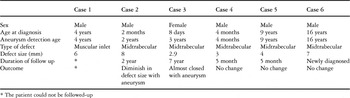
Among patients with muscular ventricular septal defect followed in our institute, we discovered six patients with aneurysmal formation, one being female and others male. Their age at the time of diagnosis ranged from eight days to 16 years. In one patient, the defect opened between the ventricular inlets, whilst in the others they occupied the middle part of the apical trabecular septum. Our fourth patient had an additional perimembranous defect. None of the others had any associated cardiac anomaly. The characteristic of the patients are summarized in the Table.
Table. Summary of the characteristic of patients.

Results
Of the patients, 3 were referred to our centre for evaluation of a cardiac murmur, while the diagnosis had already been made in the others. Pulmonary hypertension was not observed in any of the patients during the period of follow up. At initial diagnosis, the defects were measured echocardiographically to vary in size from 2.9 to 8 millimetres. We were unable to follow our first patient, whilst the sixth had already been diagnosed when seen in our centre. The period of follow up for the other patients was from five months to seven years. In two of the patients, we saw no evidence of aneurysmal formation at initial echocardiographic evaluation, the aneurysms being seen after 22 months after the initial study in the second patient, and four years in the third. Recent echocardiographic examination revealed an aneurysm which had decreased the size of defect from 8 millimetres to 4.5 millimetres in the second patient. In the third patient, the defect was nearly almost closed by the aneurysm, a minimal shunt being detected at its tip on colour Doppler echocardiographic examination. In this patient, the holosystolic murmur was no longer audible on physical examination. The pertinent echocardiographic images for all six patients are shown in Figures 1 through 6, respectively.

Figure 1. The defect opening between the muscular inlets measures 6 millimetres. Note the aneurysm. Abbreviations for this, and subsequent, figures: LV: left ventricle; RV: right ventricle; RA: right atrium; LA: left atrium; Arrow: aneurysm.

Figure 2. The defect in our second patients measures 4.5 millimetres. It is in the middle part of the apical trabecular septum.

Figure 3. The defect in our third patient, again in the middle part of the apical trabecular septum.

Figure 4. The defect in our fourth patient, again in the middle part of the apical trabecular septum, measures 3 millimetres.

Figure 5. The defect in our fifth patient, once more in the middle part of the apical trabecular septum, measures 4 millimetres.

Figure 6. In our final patient, defect in the middle part of the apical trabecular septum measures 7 millimetres.
Discussion
It is well recognised that aneurysmal transformation of the leaflets of the tricuspid valve, or adhesion of the septal leaflet to the margins of the defect, are a frequent cause for spontaneous closure, or diminution in size, of perimembranous ventricular septal defects. Prolapse of a leaflet of the aortic valve is also recognised as contributing to reduction in the size of defects, albeit usually at the cost of aortic regurgitation.5
With regard to muscular defects, several mechanisms have been suggested whereby they can become smaller or close spontaneously. These include growth and hypertrophy of tissue from the right ventricular side of the defect, formation of fibrous shelves, and muscular encroachment or superimposed fibrosis.3, 4 The mechanism is thought to involve endothelial roughening due to the high velocity jet through the defect, which leads to accumulation of platelets and production of fibrous tissue.6
As far as we are aware, however, the “classical” formation of aneurysms as seen in the setting of perimembranous defects has been reported only once when the ventricular septal defect is muscular.7
We suggested that turbulent flow of blood, and the other unknown factors, may be responsible for the formation of these fibrous aneurysm as seen in our patients with muscular defects. All the aneurysms, as far as we were able to judge, consisted of fibrous connective tissue and endothelial structures, without any underlying muscular layers. We presume that they developed as a result of the gradient in pressures between the ventricles. Since histopathology has not been available in any of our cases, our opinions of necessity remain speculative.