Introduction
Subglottic or tracheal stenosis is a well-recognised complication after prolonged endotracheal intubation in intensive care units. Stenosis occurs secondary to pressure-induced ischaemia and airway injury with subsequent scarring.1 Tracheal and/or cricotracheal resection was first reported in adults by ConleyReference Conley2 and in children by Gerwat and BryceReference Gerwat and Bryce3 In the 1990s, airway surgical procedures were further improved and refined by the work of Grillo et al.Reference Grillo, Mathisen and Wain4, Reference Grillo, Donahue, Mathisen, Wain and Wright5 and Monnier et al.Reference Monnier, Lang and Savary6, Reference Monnier, Lang and Savary7 Since then it has been performed with high success ratesReference El-Fattah, Ebada, Amer, Abosamra and Tawfik1, Reference Auchincloss and Wright8–Reference Mutrie, Eldaif, Rutledge, Force, Grist and Mansour10 for severe (grade III and IV) stenosis according to the Myer–Cotton grading system.Reference Myer, O’Connor and Cotton11
Dilatation and laser surgery are safe and well-tolerated procedures, but they typically lead to only short-term symptom relief. Recurrence is common, necessitating repeated procedures without a conclusive outcome.Reference Feinstein, Goel, Raghavan, Long, Chhetri and Berke12–Reference El-Fattah, Kamal, Amer, Fouda, Elwahab and Tawfik15 Tracheal and/or cricotracheal resection with end-to-end anastomosis is considered the treatment of choice.Reference Hentze, Schytte, Pilegaard and Klug16, Reference Monnier17 The main advantage of this procedure is that the stenotic pathologic segment of the airway is resected, with anastomosis between healthy proximal and distal stumps.Reference Piazza, Del Bon, Paderno, Grazioli, Mangili and Lombardi18
Patients with severe airway stenosis associated with neurological co-morbidities represent a major challenge. Pre-operative preparation, along with intra-operative anaesthetic and airway management, necessitates careful and tailored planning.Reference Rana, Guzman, Gourkanti, Chaudhry, Gratz, Trivedi and Pukenas19–Reference Ayoub, Smith, Cary, Dalton, Pinto and Ward22 Furthermore, intellectual and cognitive disabilities or neurodegenerative diseases may hinder co-operation with post-operative care.Reference Lange, Seer and Kopp23
Many studies demonstrated that neurological co-morbidities are associated with increased incidence of complications after tracheal resection or reconstruction, as well as poor overall outcomes.Reference Hentze, Schytte, Pilegaard and Klug16, Reference Bibas, Terra, Junior, Tamagno, Minamoto and Cardoso24–Reference Fiz, Filauro, Sampieri, Ioppi, Vallin and Fiz27 Accordingly, neurological conditions may be considered as a contra-indication for open airway surgical procedures, and a permanent tracheostomy may be preferred to alleviate the airway obstruction symptoms.Reference Abouyared, Szczupak, Barbarite, Sargi and Rosow28
The aim of the current study was to highlight our experience in the management of patients with severe subglottic and/or upper tracheal stenosis associated with neurological co-morbidities. Effective tips and tricks in the surgical technique that may enhance patient outcomes were also emphasised.
Patients and methods
Patients
This retrospective study was conducted in the Otorhinolaryngology Department, Faculty of Medicine, Mansoura University, Egypt, over the last five years (January 2020 to December 2024). The study included 33 patients who underwent tracheal and/or cricotracheal resection anastomosis for subglottic and/or upper tracheal stenosis, and all of these patients were also affected by neurological co-morbidities.
Informed written consents were obtained from all patients or patients’ caregivers, and the study was approved by the Mansoura Faculty of Medicine Institutional Research Board (IRB: R.25.02.3060).
Surgical techniques
All patients (n = 33) underwent clinical evaluation that involved a comprehensive history, as well as a trans-nasal flexible laryngoscopy to evaluate airway patency and vocal fold mobility. The levels and grades of stenosis were further evaluated under general anaesthesia. The Myer–Cotton system was applied for grading of stenosis.
The authors of the current work adopted the same surgical techniques that were described in their previous reports.Reference AM, Ebada, Amer, Abosamra and Tawfik29–Reference El‐Fattah, Ebada and Tawfik32 Suprahyoid release was the standard procedure and it was carried out in all patients (n = 33). Tailored suprahyoid release was performed according to the length of resection. A limited suprahyoid release was performed when the resected segment was short (less than three rings). This entails cutting the suprahyoid muscles attached to the body of the hyoid (mylohyoid and geniohyoid) and the stylohyoid tendon attached to the lesser horn, with preservation of the digastric slings on either side. On the other hand, in longer resections (more than three rings), full suprahyoid release is performed. The release is extended more laterally to both greater horns with cutting of the digastric slings.
Primary end-to-end anastomosis was performed. After completion of the anastomosis, Surgiflo® (Ethicon, Somerville, MA, USA) was spread over the entire surgical area, with particular emphasis on the airway anastomosis site, to ensure proper haemostasis and enhance the healing process as suggested by the authors in their previous work.Reference El‐Fattah, Ebada and Tawfik32
In addition, to decrease anastomotic tension, tracheo-hyoid de-tensioning stitches were performed in all surgical procedures. One or two 2/0 Vicryl stitches were performed, from the hyoid bone to the distal tracheal segment, one or two rings below the anastomosis.
A single-stage surgical procedure was the method of choice and was usually performed whenever possible. It involved immediate extubation on completion of the procedure. In contrast, double-stage surgical procedures involve performing a tracheotomy below the anastomosis with planned later decannulation. This option was typically selected for young children or patients with significant co-morbidities, such as respiratory conditions with increased risk of post-operative aspiration and chest infection. The double-stage approach helps secure the airway and minimises the risk of early post-operative complications, such as choking, aspiration and surgical emphysema. Decannulation was performed two to three weeks after adequate healing of the anastomosis.
The chin to chest suture is not preferred, and it was not applied to any patients in this work. Patients and their caregivers were advised to restrict neck movements and avoid neck extension during the initial post-operative period of two to three weeks.
Following extubation, patients were admitted to the intensive care unit for monitoring for a duration of 24 hours. On the next day, they were moved to the ward. Systemic antibiotics were prescribed along with a short course of corticosteroids, mucolytics and proton pump inhibitors. Drains were removed on the seventh day after surgery, after which the patients were discharged. Follow-up appointments were scheduled weekly for the first month and then monthly for the next six months. Additional appointments were scheduled in our institute’s neurology clinic tailored to each patient’s specific condition and needs.
Outcomes
The outcomes were evaluated in terms of both surgical and functional aspects. A successful surgical outcome after airway surgery was usually defined as decannulation without recurrent stenosis, and without the need for further interventions. Regarding functional outcomes, pre-and post-operative voice and swallowing scores were assessed by a speech therapist using the Voice Handicap Index,Reference Jacobson, Johnson, Grywalski, Silbergleit, Jacobson and Benninger33 the Eating Assessment Tool-10Reference Belafsky, Mouadeb, Rees, Pryor, Postma and Allen34 and the Penetration Aspiration ScaleReference Rosenbek, Robbins, Roecker, Coyle and Wood35 to screen for voice- and swallowing-related functions.
Statistical analysis
SPSS statistics version 22.0 (IBM, Armonk, NY, USA) for Windows was used for statistical analysis of the collected data. The Shapiro–Wilk test was used to check the normality of the data distribution. Normally distributed continuous variables were expressed as mean ± standard deviation, while categorical variables and the abnormally distributed continuous ones were expressed as median and inter-quartile range or number and percentage (as appropriate). The chi-square test was used for categorical data using the crosstabs’ function. In all analyses, p less than 0.05 was considered to indicate statistical significance.
Results
This study included 33 patients diagnosed with grade III or IV subglottic and/or upper tracheal stenosis associated with neurological co-morbidities. Table 1 shows the demographic data as well as the associated neurological co-morbidities among the study population.
Demographic and clinical data of the study population

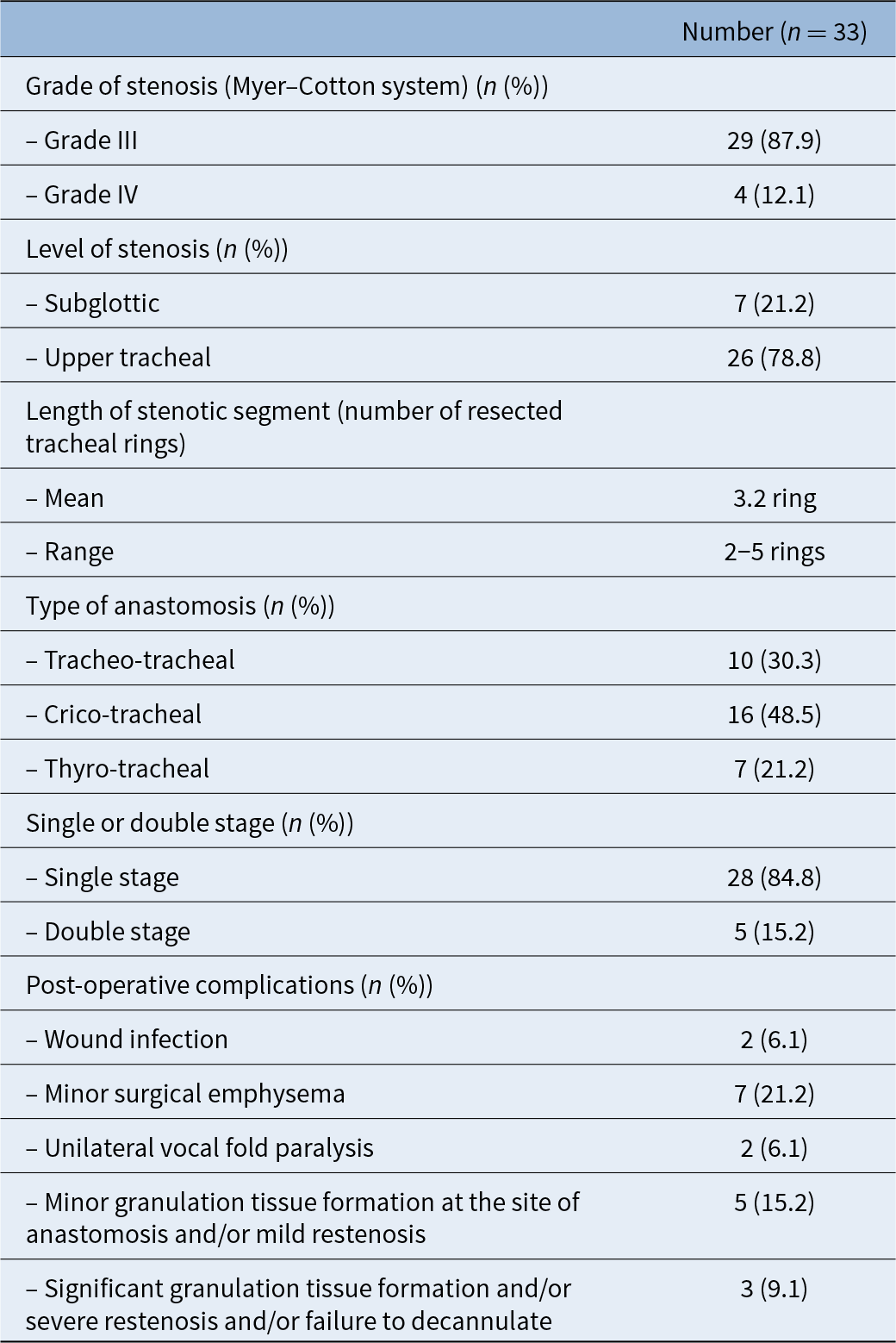
Prolonged endotracheal intubation in intensive care units was the cause of airway stenosis in all the study population (n = 33). Twenty-nine patients had grade III stenosis while four patients had grade IV (Table 2).
Intra-operative details and post-operative data of the study population

All patients underwent tracheal and/or cricotracheal resection with primary end-to-end anastomosis. The anastomosis was performed as cricotracheal (n = 16), trachea-tracheal (n = 10) and thyro-tracheal (n = 7) according to the level of stenosis and the remaining proximal and distal airway stumps. Single-stage surgery was performed in 28 patients (84.8 per cent), while double-stage surgery was performed in 5 patients (15.2 per cent).
Follow-up duration ranged from 6 to 66 months (mean 38.4 months). At the end of treatment, the overall surgical success rate (decannulation) was 30 of 33 (90.9 per cent). Three patients developed excessive granulation tissue that recurred despite multiple excisions and the topical application of mitomycin C. Ultimately, this led to severe restenosis, which was considered as treatment failure. On the other hand, minor degrees of granulation tissue and restenosis were treated with endoscopic procedures (laser- or coblation-assisted excision, topical application of mitomycin C and repeated dilation), with achievement of successful outcomes.
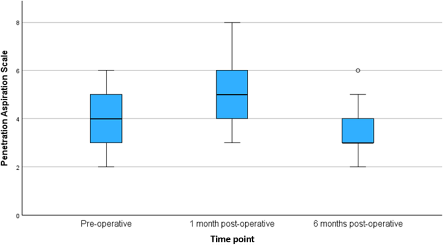
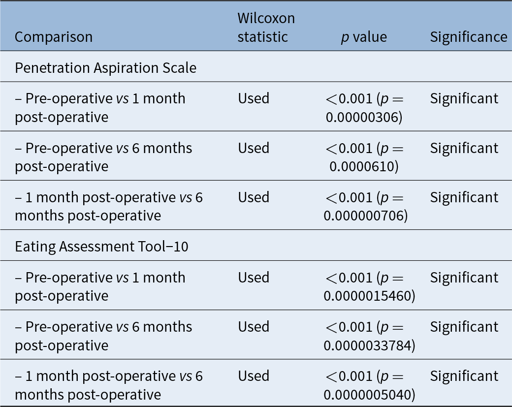
Regarding the functional outcomes, significant improvement of voice and swallowing was also noticed after surgery. The pre-operative Voice Handicap Index subscales were 22.49 ± 9.52 (functional), 20.38 ± 8.53 (physical), 21.74 ± 11.47 (emotional) and 64.62 ± 21.36 (total). After surgery and post-operative voice therapy, the Voice Handicap Index scores improved to 18.72 ± 8.35 (functional), 17.54 ± 7.18 (physical), 19.25 ± 9.40 (emotional) and 55.51 ± 21.43 (total). Swallowing initially worsened but significantly improved after six months of rehabilitation (p < 0.05) as shown in Figures 1 and 2 and Table 3. The neurological conditions of the patients remained stable and under continuous management by the neurology specialists. Notably, none of the patients experienced any deterioration in their pre-existing neurological conditions following surgery.
Box plot depicting improvement in the Eating Assessment Tool-10 (EAT-10) score.

Box plot depicting improvement in the Penetration Aspiration Scale score. * denotes statistical significance (p < 0.05).

Comparison between the pre-operative and post-operative Penetration Aspiration Scale and Eating Assessment Tool-10 scores among the study population

Discussion
To achieve optimal outcomes in airway surgery, it is crucial to have a highly specialised surgical team. In addition, when considering patients with neurological co-morbidities, a collaborative, multidisciplinary approach is essential, incorporating specialists from neurology, pulmonology and anaesthesiology. This collaborative effort is always considered in our tertiary referral centre to develop a personalised peri-operative plan that addresses the intricate relationship between neurological impairments and airway management, ultimately enhancing surgical results and ensuring patient safety.
Management of such patients is associated with several challenges. Regarding anaesthetic management, an individualised anaesthetic plan that minimises potential adverse effects on both the airway and neurological function is crucial. Patients with neurological impairments may have altered responses to anaesthetic agents.Reference Rana, Guzman, Gourkanti, Chaudhry, Gratz, Trivedi and Pukenas19 Conditions associated with spasticity and movement disorders like cerebral palsy or dystonia may affect positioning, airway management and recovery.Reference Ayoub, Smith, Cary, Dalton, Pinto and Ward22 Certain muscle relaxants and anaesthetic agents should be avoided in conditions like myasthenia gravis.Reference Radkowski, Oniszczuk, Opolska, Podlińska, Pawluczuk and Onichimowski36 In the current series all these conditions were addressed and managed by an experienced anaesthetic team who were familiar with these advanced surgical procedures.
Special attention was paid by the anaesthetic team to protect the airway during and after surgery, especially in patients with compromised airway reflexes due to their neurological condition. Poor cough reflex and secretion clearance can lead to post-operative aspiration pneumonia.Reference Mehanna and Jankovic20, Reference Thomson, Hall, Ambroggio, Stone, Srivastava and Shah21, Reference Hadjikoutis, Pickersgill, Dawson and Wiles37
Rehabilitation following surgery is a vital component of the patient’s management plan. Early involvement of speech and swallowing therapists can aid in the recovery of airway and swallowing functions, which may be affected by both the surgery and the underlying neurological condition. Additionally, long-term physical therapy, respiratory therapy and close neurological follow up are essential for adequate recovery.Reference Ha, Driver and Zopf38, Reference Patel, Keng, Masys, Amor, Patel and Keng39 In the current series, satisfactory outcomes were achieved by adopting adequate pre-operative planning, meticulous surgical techniques and careful post-operative care. The overall rate of decannulation in this study was 90.9 per cent, which is similar to the success rate of other series of tracheal and/or cricotracheal resection anastomosis.Reference Grillo, Mathisen and Wain4, Reference Marulli, Rizzardi, Bortolotti, Loy, Breda and Hamad9, Reference Wright, Grillo, Wain, Wong, Donahue and Gaissert40, Reference Macchiarini, Verhoye, Chapelier, Fadel and Dartevelle41
Suprahyoid release was first described by Montgomery in 1974.Reference Montgomery42 This procedure is both safe and well tolerated, and it can be performed using the same cervical incision. For lower tracheal or carinal resections, mediastinal, hilar and pericardial release techniques may be more effective, but these techniques necessitate a separate thoracic incision. The release manoeuvre should be adequately performed if excess anastomotic tension is expected, especially during revision tracheal and/or cricotracheal resection.Reference Monnier, Lang and Savary7 The authors of the current work perform the suprahyoid release in a tailored manner according to the length of resection. Limited suprahyoid release is performed in short resections and full release in long resections.
Surgiflo was used at the conclusion of the procedure, applied to the surgical area and the anastomotic line. In a previous study,Reference El‐Fattah, Ebada and Tawfik32 the authors of this work concluded that application of Surgiflo significantly improved the success rates and decreased the incidence of anastomotic complications such as granulation tissue, surgical emphysema and restenosis. The authors believed that application of Surgiflo might help fill any potential micro gaps in the anastomosis, thereby promoting healing and reducing the likelihood of anastomotic complications.
Trachea-hyoid detensioning stitches were applied to decrease the anastomotic tension, and consequently to decrease the incidence of anastomotic complications. Redmann et al.Reference Redmann, Rutter, de Alarcon, Hart, Manning and Smith43 described a similar procedure in which they placed internal suspension sutures using two 2-0 Polydioxanone (PDS) through the two tracheal rings just distal to the anastomosis and then around the hyoid bone. They demonstrated that applying these suspension sutures alleviates the need for post-operative chin to chest sutures. Atallah et al.Reference Atallah, Aldkhyyal and Castellanos44 described placement of a laryngosternopexy stitch between the thyroid lamina and the sternoclavicular ligament designed to take all of the tension off the anastomosis and to prevent inadvertent head extension.
Few studies have collected detailed information on functional outcomes after primary airway surgical procedures,Reference Timman, Schoemaker, WWL, Marres, Honings and Morshuis45–Reference Evermann, Schweiger, Kranebitter, Roesner, Aigner and Denk-Linnert48 although these outcomes should be incorporated into the definition of treatment success. These studies demonstrated that laryngotracheal resection and anastomosis is safe and provides excellent surgical and functional outcomes with an improved quality of life.
Nauta et al.Reference Nauta, Mitilian, Hanna, Mercier, Crutu and Fabre47 described the functional outcomes of 34 patients who underwent tracheal resection and anastomosis, utilising visual analogue scales for assessment. Their findings indicated a marked improvement in dyspnoea scores following surgery, compared with baseline measurements. However, dysphagia scores remained largely unchanged. Similarly, Timman et al.Reference Timman, Schoemaker, WWL, Marres, Honings and Morshuis45 evaluated the functional outcomes of 30 patients who underwent single-stage tracheal and/or cricotracheal resection anastomosis and reported significant improvement in breathing, voice and swallowing. In the current study, adequate functional outcomes were achieved in terms of voice and swallowing despite the neurological conditions of the patients. Additionally, none of the patients experienced deterioration of the pre-existing neurological conditions during or after airway surgery.
Despite the favourable outcomes associated with tracheal and/or cricotracheal resection in neurologically compromised patients, the current study has several limitations. It is a retrospective analysis involving a small sample size and a diverse range of neurological conditions. Additional larger studies are necessary to explore tracheal resection across various neurological disorders and to draw on different institutional experiences.
• It is well accepted that tracheal and/or cricotracheal resection with end-to-end anastomosis is the treatment of choice for severe airway stenosis
• Patients with severe airway stenosis associated with neurological co-morbidities represent a major challenge
• Many studies have demonstrated that neurological co-morbidities are associated with increased incidence of complications after tracheal resection or reconstruction, as well as poor overall outcomes
• Detailed pre-operative planning, meticulous and skilful operative techniques, as well as adequate post-operative care are essential to achieve optimum outcomes in this group of patients despite these challenges
• Certain refinements in the surgical technique, such as tailored suprahyoid release, tracheo-hyoid detensioning stitches and the application of Surgiflo®, are crucial to enhance healing, decrease anastomotic tension and improve the overall outcomes
Conclusion
Patients with severe subglottic and/or upper tracheal stenosis associated with neurological co-morbidities are challenging. However, careful pre-operative assessment, meticulous surgical techniques, post-operative care and multidisciplinary collaborative effort are essential for achieving optimum results. Certain refinements in the surgical technique such as tailored suprahyoid release, tracheo-hyoid detensioning stitches and the application of Surgiflo are crucial to enhance healing, decrease anastomotic tension and improve the overall outcomes.
Competing interests
None declared