Introduction
The mammalian ovary at birth consists of thousands of primordial follicles, which are considered the resting pool of follicles in the ovary. Throughout the female reproductive life span, a small number of primordial follicles are stimulated to grow, in a process referred to as follicular activation; whereas the vast majority (99.9%) becomes atretic toward ovulation (Skinner, Reference Skinner2005). Until recently, relatively little was known as to how these primordial follicles were activated and stimulated to develop into a more advanced stage. Moreover, it is known that in mammals, including primates, early follicular development goes through a complex process, in which the oocytes grow and their surrounding somatic cells proliferate and differentiate through preantral follicle stages (Gougeon, Reference Gougeon1996; Fortune, Reference Fortune2003). However, few long-term in vitro culture studies have achieved follicular activation in sheep (Muruvi et al., Reference Muruvi, Picton, Rodway and Joyce2005), human (Sadeu et al., Reference Sadeu, Cortvrindt, Ron-El, Kasterstein and Smitz2006) and goat (Rossetto et al., Reference Rossetto, Lima-Verde, Matos, Saraiva, Martins, Faustino, Araujo, Silva, Name, Sn, Campello, Figueiredo and Blume2009).
Leukemia inhibitory factor (LIF) is a glycoprotein with pleiotropic activity that exerts an important role on follicular growth, oocyte maturation and a wide variety of cell types including somatic and follicular cells (Demeestere et al., Reference Demeestere, Centner, Gervy, Englert and Delbaere2005). Shen & Leder (Reference Shen and Leder1992) suggested a specific role for LIF on mouse preimplantation development. Among its many activities, LIF can maintain embryonic stem cell monolayers in a pluripotent undifferentiated state. Recently, LIF was reported to inhibit myeloid leukemic cells and can enhance survival, migration, proliferation and meiosis resumption of primordial primitive cells in rodents (van den Hurk & Zhao, Reference Van Den Hurk and Zhao2005).
In rats, the level of LIF in the follicular fluid increases as ovarian follicles develop. There is evidence that LIF is responsible for oocyte development and maturation and preantral follicle viability in vitro (Haidari et al., Reference Haidari, Salehnia and Rezazadeh Valojerdi2008). Reports on the development of rat preantral follicles cultured with LIF showed them to promote the transition from primordial to primary follicles (Nilsson et al., Reference Nilsson, Kezele and Skinner2002; Haidari et al., Reference Haidari, Salehnia and Rezazadeh Valojerdi2006). Although LIF was identified as being involved in mammalian folliculogenesis, the role of LIF on preantral follicle activation is an issue that remains far from understood. Therefore, the aim of the present study was to investigate the influence of different concentrations of LIF on the activation of primordial follicles and further follicular survival after in vitro culture of caprine ovarian tissue.
Materials and methods
Unless mentioned otherwise, all chemicals used in the present study were purchased from Sigma Chemical Co.
Ovaries collection
Ovaries (n = 10) were collected from five non-pregnant mixed-breed adult goats (1–3-year-old). All animals were in the follicular phase of the estrous cycle with absence of corpora lutea. Immediately after being slaughter, ovaries were dissected from the surrounding connective tissue and washed once in 70% alcohol, followed by twice in minimum essential medium (MEM) supplemented with 100 μg/ml penicillin and 100 μg/ml streptomycin. Then, ovaries were transported to the laboratory at 4°C within 1 h.
Experimental design
In the laboratory, ovaries were stripped of surrounding tissues and the medulla and visible growing follicles were manually removed. Ovarian tissue samples from each ovarian pair were cut in to slices of 9 mm3 (approximate size 3 mm × 3 mm × 1 mm thickness) using a 26-G needle and a scalpel blade under sterile conditions. The tissue pieces were then either directly fixed for histological analysis (fresh control – FC) or placed in culture for 1 or 7 days. The tissues were transferred to 24-well culture Petri dishes containing 1 ml of culture medium. Culture was performed at 39°C in air with 5% CO2 in a humidified incubator and medium were incubated for 1 h prior to use. The basic culture medium (cultured control – CC) consisted of αMEM supplemented with ITS (10 μg/ml insulin, 5.5 μg/ml transferrin and 5 ng/ml selenium), 0.23 mM pyruvate, 2 mM glutamine, 2 mM hypoxanthine and 1.25 mg/ml bovine serum albumin. For the experimental conditions, basic medium was supplemented or not with LIF at different concentrations (1, 10, 50, 100 or 200 ng/ml). Each treatment was repeated five times and the culture medium was replaced every other day throughout the 7-day period.
Histological assessment of in vitro follicular growth
For the evaluation of follicular morphology (survival), all the ovarian pieces were fixed in Carnoy's solution for 12 h and then dehydrated in increasing concentrations of ethanol at 70, 80, 95 and 100%, before culture (FC) and after 1 or 7 days of culture. After paraplast embedding, caprine tissues fragments were sliced into 5 μm sections, mounted on glass slides and stained by periodic acid Schiff–haematoxylin. Follicle stage and viability were assessed microscopically on serial sections. Coded anonymized slides were examined on a microscopy (Nikon, Japan) under ×400 magnification (Silva et al., Reference Silva, Van Den Hurk, Costa, Andrade, Nunes, Ferreira, Lobo and Figueiredo2002). Each follicle was examined in every section and matched with the same follicle on adjacent sections to avoid double counting, thus ensuring that each follicle was only counted once, regardless of its size.
The developmental stages of follicles have been defined previously as primordial (one layer of flattened granulosa cells around the oocyte) or growing follicles (intermediate: one layer of flattened to cuboidal granulosa cells; primary: one layer of cuboidal granulosa cells, and secondary: two or more layers of cuboidal granulosa cells around the oocyte; Silva et al., Reference Silva, Ferreira, Costa, Santos, Carvalho, Rodrigues, Lucci, Báo and Figueiredo2004). Follicles were classified individually as histological normal when an intact oocyte was present and surrounded by a well organized of one or more layers of granulosa cells without the appearance of pyknotic nucleus. Degenerated follicles were defined as those with a retracted oocyte or a pyknotic nucleus, and/or surrounded by disorganized granulosa cells, which were detached from the basement membrane. Overall, 1950 follicles were evaluated for each treatment [30 follicles × 13 groups (1 or 7 days of culture) × five replicates]. To evaluate follicular activation, the percentages of healthy primordial follicles were calculated before FC and after culture in each medium with or without LIF.
Viability assessment of preantral follicles by fluorescence
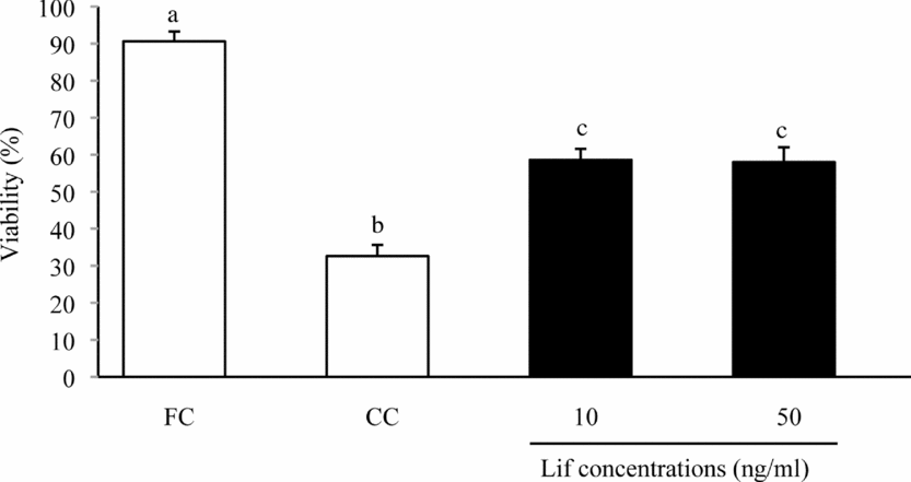
Based on the results of histological analysis, the viability of follicles cultured with LIF that provided the best outcome (10 and 50 ng/ml) was further analyzed using a more accurate method of assessment based on fluorescent probes. Goat preantral follicles were isolated using a mechanical method described by Lucci et al. (Reference Lucci, Amorim, Rodrigues, Figueiredo, Bao, Silva and Goncalves1999). Ovarian fragments were cultured for 7 days with CC, 10 or 50 ng/ml LIF. Briefly, using a tissue chopper (Mickle Laboratory Engineering Co.) adjusted to a sectioning interval of 75 μm, samples were sliced into small fragments, placed in MEM and suspended 40 times using a large Pasteur pipette (diameter of about 1600 μm) and resuspended subsequently 40 times with a small Pasteur pipette (diameter of about 600 μm) to dissociate preantral follicles from stroma. The obtained material was passed through 100 μm nylon mesh filters, resulting in a suspension containing preantral follicles smaller than 100 μm in diameter. This procedure was carried out within 10 min at room temperature.
Thereafter, the viability of preantral follicles were analyzed using a two-colour fluorescence cell assay based on the simultaneous determination of viable or degenerated cells by calcein-AM and ethidium homodimer-1 (Molecular Probes, Invitrogen), respectively. While the first probe detected intracellular esterase activity of viable cells, the second probe labelled nucleic acids of non-viable cells with plasmatic membrane disruption. Fluorescence analysis was performed by adding 4 μM calcein-AM and 2 μM ethidium homodimer-1 (LIVE/DEAD Viability kit: L, Molecular Probes, Invitrogen) to the suspension of isolated follicles, followed by incubation at 37°C for 15 min in a dark chamber. After being labelled, follicles were centrifuged at 100 g for 5 min, washed once and resuspended in αMEM. Then, labelled follicles were mounted on a glass microscope slide using 5 μl of anti-fading medium (DABCO, Sigma) to prevent photobleaching and finally examined using a DMLB fluorescence microscope (Leica). The emitted fluorescence signals of calcein-AM, and ethidium homodimer were collected at 488 and 568 μm, respectively. Oocytes and granulosa cells were considered viable if the cytoplasm was stained positively with calcein-AM (green) and chromatin was not labelled with ethidium homodimer (red); otherwise, they were classified as degenerated (Lopes et al., Reference Lopes, dos Santos, Celestino, Melo, Chaves, Campello, Silva, Bao, Jewgenow and de Figueiredo2009).
Statistical analysis
The mean percentages of primordial, viable (at all stages) obtained after 1 or 7 days of culture were subjected to analysis of variance (ANOVA) using the GLM procedure of SAS/STAT software. Data were tested for normal distribution using Shapiro–Wilk test and normalized when necessary. Dunnett's test was applied to compare LIF-treated groups with FC and CC groups. Duncan's test was used to compare differences among LIF concentrations and days of culture. Data from fluorescence analysis were subjected to chi-squared test. Differences were considered to be significant when p < 0.05 and data were expressed as mean ± standard error of means (SEM).
Results
Follicular activation and development
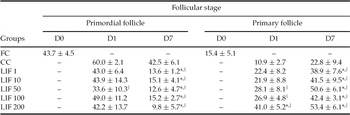
The percentages of primordial and primary follicles during culture with different concentrations of LIF are shown in Table 1. After 7 days of culture, the number of primordial follicles decreased (LIF from 9.8% to 15.2% vs. control groups FC 43.7% and CC 42.5%) and primary follicles increased (LIF from 38.9% to 53.4% vs. control groups FC 15.4% and CC 22.8%) when LIF was present in the culture (p < 0.05). In the presence of LIF, formation and differentiation (from flattened to cuboidal cells) of a new cellular layer of granulosa cells were observed, giving origin to primary follicle. These findings are evidence of preantral follicle activation induced by LIF.
Histological evaluation of in situ cultured fragments of goat ovarian cortex for 1 or 7 days using different concentrations of leukemia inhibitory factor (LIF) (ng/ml)

* Differs significantly from fresh control (FC).
† Differs significantly from cultured control (CC) in each day of culture (p < 0.05).
Data are shown as mean percentage ± standard error of means (S.E.M.) out of all preantral follicle stages (primordial and primary follicles). Data were pooled from five replicates. (D) Day of culture.
Follicular survival
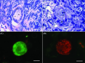
The preantral follicle survival was histologically evaluated after 1 or 7 days of culture in the presence or absence of LIF. An example of viable preantral follicle is depicted in Fig. 1A. The degenerated follicles presented a retracted oocyte, pyknotic nucleus, and/or cytoplasmic vacuolization (Fig. 1B). The viable preantral follicles were also labelled by calcein-AM green fluorescence (Fig. 1C) and the degenerated preantral follicles showed chromatin labelled by ethidium homodimer red fluorescence (Fig. 1D).
Viability assessment of caprine preantral follicles using histological and fluorescent analyses. (A) Histological section of preantral follicle cultured for 7 days showing the stroma and follicle integrity. (B) Histological section of preantral follicle cultured for 7 days showing cytoplasmic vacuolization, disorganized stroma cells and pycnotic nucleus. (C) An isolated viable preantral follicle labeled by calcein-AM (green fluorescence). (D) Degenerated preantral follicle showing chromatin labelled by ethidium homodimer (red fluorescence). gc: granulosa cells; n: nucleus; o: ooplasm; arrow: pycnotic nucleus. Bars = 50 μm.

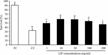
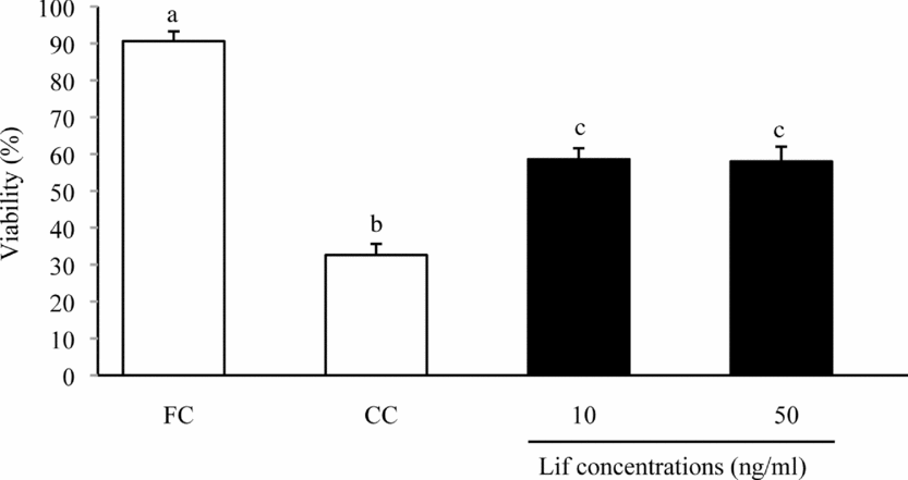
The preantral follicle survival did not differ among treatment and control groups after 24 h of culture. However, the survival of preantral follicles was improved when LIF was used in culture for 7 days (LIF treatments vs. CC groups; p < 0.05; Fig. 2), which was confirmed by fluorescence method (Fig. 3). Furthermore, the highest percentages of viable preantral follicles were obtained at concentrations between 10 and 50 ng/ml of LIF (from 58.6% to 58.0%), decreasing at 200 ng/ml (46.7%; p < 0.05). The survival of isolated preantral follicles before culture (FC) was above 90%, which demonstrated follicular health at the time of follicular isolation.
Histological analysis of the viability of preantral follicles (%) after 7 days of culture. The results are presented as the mean ± SEM of five independent cultures. a–dBars with no common letters are significantly different (p < 0.05).

Viability of preantral follicles analysed by a two-colour fluorescence cell using calcein-AM and ethidium homodimer-1. The best results observed in the histological analysis were repeated using probes to detected intracellular esterase activity of viable cells and to label nucleic acids of non-viable cells with plasmatic membrane disruption. The results are presented as the mean ± SEM of five independent cultures. a–cBars with no common letters are significantly different (p < 0.05).
Discussion
The effect of LIF on the activation of primordial and survival of preantral follicles was demonstrated in a 7-day culture system, using goat as a model. Our results revealed that: (1) LIF induced activation of primordial follicle, formation and differentiation of new cellular layer of granulosa cells, giving origin to primary follicle; and (2) LIF supported preantral follicle viability for 7 days in culture, based on histological and fluorescence analyses.
Slices of goat ovarian cortex were cultured in the absence and presence of different concentration of LIF to verify if LIF promotes primordial to primary follicle transition in ruminants. The percentages of primordial follicles decreased and primary follicles increased at all LIF concentration tested at 7 days of culture. The optimal LIF concentrations to maintain caprine preantral follicles viable and growing ranged from 10 to 50 ng/ml. In fetal rat ovaries, LIF induced premature follicular development and meiosis resumption when used at a concentration of 100 ng/ml (Lyrakou et al., Reference Lyrakou, Hulten and Hartshorne2002). Recently, Haidari et al. (Reference Haidari, Salehnia and Rezazadeh Valojerdi2008) reported that LIF at 50 ng/ml promoted follicular survival and preantral follicle development in vitro using rat follicles.
In the concentrations used in the present study, LIF induced increase in granulosa cell number and change from flattened to cuboidal shape, characterizing the transition to primary follicle. These results indicate that LIF can promote activation of primordial follicle and transition to primary follicle. Similarly, the dramatic decrease of primordial follicle and corresponding increase of primary follicle numbers after LIF treatment were observed in the rat ovary (Nilsson et al., Reference Nilsson, Kezele and Skinner2002; Haidari et al., Reference Haidari, Salehnia and Rezazadeh Valojerdi2008). In the rat, LIF also promoted the transition from primordial to primary follicles and supported their viability for 14 days in culture. However, there is little information on the role of LIF in promoting preantral follicle activation, development and viability in domestic animals.
LIF is expressed in rat preantral follicle (Nilsson et al., Reference Nilsson, Kezele and Skinner2002; Haidari et al., Reference Haidari, Salehnia and Rezazadeh Valojerdi2008) and in the ovarian stromal cells in human (Arici et al., Reference Arici, Oral, Bahtiyar, Engin, Seli and Jones1997). These last authors also demonstrated that LIF concentrations rise in periovulatory follicular fluid and regulate ovulation, estrogen production and early embryonic development. Despite being expressed in preantral follicles and promoting the development of primordial follicles, it appears that LIF has a role, but is not essential for activation and transition to primary follicles, considering that follicles reach ovulation in LIF knockout mice (Stewart et al., Reference Stewart, Kaspar, Brunet, Bhatt, Gadi, Kontgen and Abbondanzo1992). Therefore, there is strong evidence that the regulation of initial preantral follicle activation and growth is orchestrated by LIF and other signal factors [such as growth differentiation factor 9 (GDF9), kit ligand (KL), basic fibroblast growth factor 2 (bFGF2) and nerve growth factor (NGF)], having a compensatory action when one factor is missing in the system.
The expression and function of several transcription factors and hormones responsible for the regulation of follicle development is species specific and differences have been demonstrate in rodents, ruminants and primates (Shimasaki et al., Reference Shimasaki, Moore, Otsuka and Erickson2004; Young & McNeilly, Reference Young and McNeilly2010). For example, the tissue-type plasminogen activator (tPA) and urokinase-type plasminogen activator (uPA) activity is different between rodents and cattle during the periovulatory period (Dow et al., Reference Dow, Bakke, Cassar, Peters, Pursley and Smith2002). LIF and bone morphogenetic proteins (BMPs) are essential for mouse embryonic stem (ES) cells to maintain pluripotency (Smith et al., Reference Smith, Nichols, Robertson and Rathjen1992; Ying et al., Reference Ying, Nichols, Chambers and Smith2003); however, human ES requires activin/nodal (Vallier et al., Reference Vallier, Reynolds and Pedersen2004, Reference Vallier, Alexander and Pedersen2005; James et al., Reference James, Levine, Besser and Hemmati-Brivanlou2005). LIF and other factors [such as bFGF, BMP, GDF9, hepatocyte growth factor (HGF), insulin-like growth factor (IGF), IGF-binding protein (IGFBP), interleukin-1 (IL1), keratinocyte growth factor (KGF), luteinizing hormone (LH), stem cell factor/kit ligand (SCF), transforming growth factor beta (TGFβ) and tumour necrosis factor alpha (TNFα)] have species-specific patterns of expression and function (for review see Young & McNeilly, Reference Young and McNeilly2010). Therefore, the investigation of the LIF function in ovarian preantral follicle development is imperative in ruminants, which have a high economic importance and have been used as an experimental model for humans.
In conclusion, LIF induced activation of primordial follicles, differentiation of granulosa cells from flattened to cuboidal shape and maintenance of preantral follicle viability for 7 days in culture. To our knowledge, this is the first evidence that LIF is involved in the initial follicle development and viability in ruminant.
Acknowledgements
This work was supported by CNPq, FINEP and RENORBIO. We would like to thank contribution of Dr Maria Fátima Teixeira for help with the fluorescence analyses.