Introduction
Preimplantation genetic diagnosis (PGD) by fluorescence in situ hybridization (FISH) is one of the most common laboratory methods used to detect chromosomal abnormalities in human embryos (Munne et al., Reference Munne, Wells and Cohen2010). PGD by FISH includes three laboratory steps: embryo biopsy, blastomere fixation and FISH. Although improvements have been made to all three steps and these have increased the accuracy of the diagnoses, some samples still do not have signals after FISH, thus a second biopsy is needed for these embryos. Most failed (no result after FISH) FISH diagnoses are caused mainly by improper fixation, thus blastomere fixation becomes a crucial step in the FISH procedure (Xu et al., Reference Xu, Huang, Tiezheng, Zhongmig and Rosenwaks1998; Dozortev & McGinnis, Reference Dozortev and McGinnis2001).
Currently, two methods are being used for the fixation of biopsied blastomeres. One is the traditional method in which the blastomeres are cultured in a hypotonic solution for 5 min, and the operators blow air onto the blastomere such that the cell membrane can be broken during fixation (Tarkowski, Reference Tarkowski1966; Coonen et al., Reference Coonen, Dumoulin, Ramachers and Hopman1994). As a result, this method is difficult to learn and operators sometimes lose cells during fixation (Munne, Reference Munne, Wells and Cohen2010). The other method is a modified method developed by clinical embryologists (Xu et al., Reference Xu, Huang, Tiezheng, Zhongmig and Rosenwaks1998; Dozortev & McGinnis, Reference Dozortev and McGinnis2001; Velilla et al., Reference Velilla, Escudero and Munne2002), in which blastomeres are cultured in hypotonic solution shortly before fixation on a slide with Tween-20/HCI (Xu et al., Reference Xu, Huang, Tiezheng, Zhongmig and Rosenwaks1998; Dozortev & McGinnis, Reference Dozortev and McGinnis2001; Velilla et al., Reference Velilla, Escudero and Munne2002). This modified method is easy to learn, but the nuclei are not well dispersed after fixation, thus the FISH signals overlap in some samples and the data after FISH procedures are still not satisfactory (Velilla et al., Reference Velilla, Escudero and Munne2002), especially when five or more DNA probes are used.
In order to increase the efficiency of FISH technology for PGD in humans, it is therefore necessary to develop a simple, easy to learn and effective sample preparation method. In the present study, experiments were designed to develop this kind of method and evaluate its efficiency in human PGD.
Materials and methods
Embryo biopsy
For day 3 embryo biopsy, 4–10-cell stage embryos were placed in Sage biopsy solution (Cooper Surgical, CT, USA) and laser pulses generated by the ZILOS-tk™ laser system (Hamilton Thorn Bioscience Inc., MA, USA) were used to open a hole in the zona pellucida. One blastomere with an obvious nucleus was aspirated into a 35-μm biopsy pipette under a ×40 magnification objective. After biopsy, embryos were washed and cultured in Sage blastocyst medium with 10% serum protein substitute (SPS, Cooper Surgical), and the isolated blastomeres were fixed according to the methods mentioned below.
For blastocyst biopsy, a small hole on the zona pellucida was made with laser pulses on day 3, and blastocysts started to hatch from the hole on day 5 or day 6. A portion (< 5 cells) of hatched trophectoderm cells was aspirated into a 20-μm biopsy pipette and laser pulses were then applied to ‘cut’ cells between the cell connections. After biopsy, the blastocysts were frozen for future transfer, and the isolated cells were fixed according to the methods described below.
Blastomere fixation
Three solutions were used for blastomere fixation: hypotonic solution, Tween-20/HCI solution and fixative. Hypotonic solution is composed of 1% sodium citrate and 6% bovine serum albumin in distilled water. Tween-20/HCI solution is composed of 1% Tween-20 and 1% 0.01 N HCl in distilled water. Fixative is composed of 0.9 ml of methanol and 0.3 ml of acetic acid. All chemicals were purchased form Sigma (Sigma Chemical Co., St Louis, MO USA). Hypotonic solution and Tween-20/HCI solution were stored at 2–8°C for up to 6 months after preparation, while fixative was freshly made on the day of fixation.
Method I
Each blastomere was cultured in hypotonic solution for 5 min and then transferred to a drop (less than 1 μl) made with hypotonic solution on the glass slide. A drop of fixative was added to the hypotonic solution drop containing a blastomere immediately before it dried. During this period, humidity was applied by blowing air onto the blastomere so that the cell membrane could be broken. It is important to pay attention during this procedure as blastomeres/nuclei move around and may not be found easily. Blastomeres were fixed one by one until all blastomeres were fixed. The slides were kept at room temperature prior to the FISH procedure.
Method II
An equal number of hypotonic solution drops (10–20 μl) as blastomeres was made on a 60 mm culture dish and the drops were then covered with oil to avoid drying. On the bottom of the dish, each blastomere was marked with a number next to its drop. After embryo biopsy, all blastomeres were transferred individually from biopsy dishes to the hypotonic solution drops. The fixation procedures were started 5 min after the first blastomere was placed in the hypotonic solution. For fixation, each blastomere was transferred from the hypotonic solution drop to a glass slide under a stereomicroscope, and a small drop of Tween-20/HCI solution was dropped on the blastomere from a 0.16 mm pipette immediately before the hypotonic solution dried. A second drop of the Tween-20/HCI solution may be necessary if the cell membrane does not break. Finally, a few drops of fixative were added onto the slide to wash away the cytoplasm after the drop has completely dried.
After fixation, the presence of nuclei was verified using a ×10 or ×20 magnification objective under a phase contrast microscope. The slides were kept at −20°C for at least 30 min before the FISH procedure.
Data evaluation
The time was recorded from when the technician placed the first blastomere into the hypotonic solution until the complete fixation of all blastomeres. The average time (min) was calculated based on the total number of blastomeres to be fixed and the total time spent for the fixation, and then the data were compared between the two methods. The percentages of blastomeres with FISH signals were compared between the two methods. Differences between methods were analysed using Student's t-test. A P-value <0.05 was considered to be statistically significant.
Results
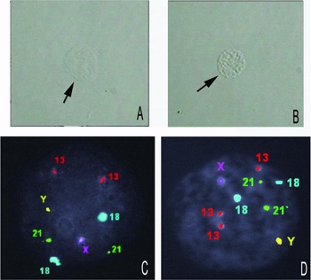
As shown in Table 1, 329 blastomeres from 30 patients were fixed by Method I, and the average time spent for fixation was 9.3 (8–10) min for each blastomere. By contrast, with Method II, 362 blastomeres from 32 patients were fixed, and the average time spent for the fixation was 3.2 (2–4) min for each blastomere. The nuclei fixed with Method II were well dispersed (Fig. 1 A,B), but most nuclei fixed with Method I were concentrated. After FISH, 86.9% of the nuclei had FISH signals when using Method I while 97.2% of the nuclei had FISH signals when using Method II (Fig. 1 C,D), a value that was significantly (P < 0.01) higher than that found with Method I.
Comparison of traditional method and modified method for preparations of blastomeres for FISH procedures in terms of time consumption and FISH signals

a,b Significantly different, P < 0.01.
Images of nuclei prepared with Method II for fluorescence in situ hybridization (FISH) from day 3 blastomeres. (A, B) Nuclear images taken with phase contrast microscope show that the nuclei (arrows) are well dispersed. (C, D) Chromosomal images after FISH show positive staining signals. Five DNA probes were used for the labelling of the chromosomes, and the staining was imaged using a fluorescence microscope. Nuclei in both (C) and (D) were labelled with five chromosomal probes (13, 18, 21 and XY).

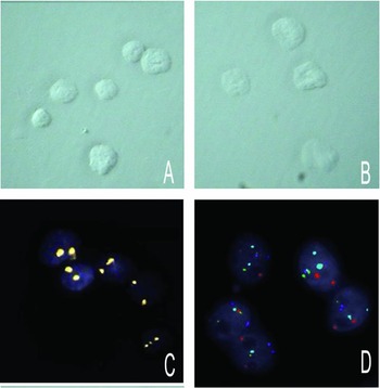
For fixation of cells biopsied from blastocysts, all nuclei were traceable during fixation and were distributed closely when fixed with Method II (Fig. 2 A,B). All 56 cells from 15 blastocysts (2–5 cells from each blastocyst) had FISH signals (Fig. 2 C,D). However, if they were fixed with Method I, some nuclei were washed away when fixative was added, and it was difficult to trace the cells due to multiple cells present on the slides, thus this method was not suitable for fixation of multiple cells biopsied from blastocysts.
Images of nuclei prepared with Method II for fluorescence in situ hybridization (FISH) from cells biopsied from blastocysts. (A, B) Nuclear images taken with phase contrast microscope show that each nucleus is well dispersed, and all nuclei are distributed very closely. (C, D) Chromosomal images after FISH show positive chromosomal staining signals. Nuclei in (C) were labelled with sex chromosome probes and nuclei in (D) were labelled with five chromosomal probes.

Discussion
PGD by FISH is one of the most common diagnostic approaches used in human infertility clinics for the detection of chromosomal abnormalities, such as aneuploidy and chromosome translocation. However, due to technical difficulties and limited chromosomes being examined, there is a possibility that this traditional cytogenetic diagnostic technology will be replaced by DNA microarray. Regardless, many in vitro fertilization (IVF) clinics still use FISH as a PGD for patients undergoing IVF, thus the professional application of this technology is important for patient service (Munne et al., Reference Munne, Wells and Cohen2010). In the present study, our results indicate that modifications in sample preparations for FISH procedures have some advantages over other methods.
First of all, we cultured all blastomeres in hypotonic solution for at least 5 min, a procedure similar to those methods reported previously (Tarkowski, Reference Tarkowski1966; Coonen et al., Reference Coonen, Dumoulin, Ramachers and Hopman1994). This treatment is crucial for dispersing the nuclei, and it can also prevent signals from overlapping after FISH. One modification of this method is to place all blastomeres in the hypotonic solution drops after biopsy, a difference from the traditional method in which blastomeres are processed one by one and operators wait for 5 min for each blastomere before fixation. By this modification, operators can save more than 50% of the time for fixation.
Secondly, we used Tween-20/HCI to break the cell membrane before adding fixative. Although in previous reports it has been stated that membrane breakage is not necessary for sample preparations during the FISH procedures (Dozortev & McGinnis, Reference Dozortev and McGinnis2001), we and other researchers have found that completely exposed nuclei were prerequisites for achieving better FISH signals (Munne et al., Reference Munne, Wells and Cohen2010). In this study, a small droplet of Tween-20/HCI solution was ‘dropped’ onto the blastomeres to break the membrane. This step is a quick and easy way to break the membrane and circumvents the need to apply humidity by blowing air onto the blastomere, an action not easy to apply. In addition, Tween-20/HCI solution is ‘dropped’ from a small pipette, and the volume of solution is limited, such that the nuclei do not move around after the membrane breaks. This factor is especially important for the fixation of multiple cells biopsied from blastocysts.
Finally, after fixation with Method II, the slides were stored at –20°C for at least 30 min, and the slides were also kept on an ice pad before the FISH procedures. Although we still do not know if this step is the main reason that more nuclei had a signal after FISH, our results showed that 97.2% of nuclei from the day 3 biopsy and 100% of nuclei from blastocyst biopsy had FISH signals, numbers that were significantly higher than those using the traditional method (Method I).
In the present study, we found that this new method is easy to use for cells biopsied from blastocysts. Usually, 3–5 cells are isolated from a blastocyst, and the cells are very small. For fixation, all cells must be kept very close together on the slide so that they are easy to track during fixation and the FISH procedures. When the new method was used, all nuclei were distributed very close together after addition of Tween-20/HCI. However, when the cells were fixed with Method I, some nuclei moved around after adding fixative and were very difficult to follow, a factor that may have contributed to the loss of nuclei in some cases. Therefore, the traditional method (Method I) is not suitable for the fixation of multiple cells biopsied from blastocysts. After FISH, we also found that more cells had signals if the isolated cells were fixed with Method II rather than with Method I. The data for multiple cell fixation presented in the present study are still limited, and further study is necessary to see the benefits of this modified method for the fixation of small cells isolated from blastocysts.
In conclusion, our study indicates that the modifications in sample preparations developed in the present study significantly reduce the time spent for fixation and increase the number of nuclei with FISH signals. With these modifications, operators require less time to fix blastomeres. Most importantly, the modifications increase the numbers of embryos with FISH signals, and therefore increase the efficiency of PGD, not only for a single cell biopsied from a day 3 embryo but also for multiple cells biopsied from blastocysts.