Introduction
Undernutrition during fetal development may alter emerging organ systems and/or homeostatic pathways, with consequences for health and survival over the life course.Reference Hanson and Gluckman1 Immune cell populations and lymphoid organs may be particularly vulnerable to early-life nutritional insults.Reference Palmer2 As a primary lymphoid organ, the thymus undergoes a critical period of growth and development early in gestation and attains its maximum relative size by the time of birth.Reference Kuper, van Bilsen, Cnossen, Houben, Garthoff and Wolterbeek3 Mid-gestation represents a second susceptible period, during which time immature thymocytes are subject to positive and negative selection.Reference Kuper, van Bilsen, Cnossen, Houben, Garthoff and Wolterbeek3 Altered thymic development during gestation may permanently affect phenotype. Epidemiological studies carried out in The Gambia and Bangladesh have demonstrated differences by season of birth, with those born during periods of nutritional deprivation having a smaller thymus and altered thymic function.Reference Collinson, Moore, Cole and Prentice4–Reference Fulford, Moore and Arifeen7 Similar work from the Philippines showed that children who were born small for gestational age had lower serum concentrations of thymopoietin, a thymic hormone, even during adolescence, than their peers.Reference McDade, Beck, Kuzawa and Adair8
Thymic function can be characterized by T cell receptor excision circles (TREC) or flow cytometry to identify recent thymic emigrants, both requiring freshly isolated peripheral blood mononuclear cells.Reference McFarland, Picker, Koup and Douek9 A thymic index determined by analysis of computed tomography scans or ultrasound images serves as a surrogate marker for thymic function.Reference McFarland, Picker, Koup and Douek9 The use of thymic hormones as functional markers has not been validated. However, hormone concentrations do correlate well with thymic size in the case of age-related thymic involution.Reference Brelinska10 Thymulin cannot be detected after thymectomy and is significantly reduced in immunodeficient patients or those with autoimmune disorders characterized by abnormalities in thymic epithelial cells.Reference Hadden11, Reference Safieh-Garabedian, Kendall, Khamashta and Hughes12 Thymulin is thought to be secreted exclusively by thymic epithelial cells in response to various cytokine and hormonal stimuli.Reference Reggiani, Schwerdt, Console, Roggero, Dardenne and Goya13 In its active state, bound to a zinc ion, the hormone induces surface protein expression on T cells, affecting their development, maturation, and differentiation.Reference Hadden11
The goal of the present study was to investigate nutrition-related correlates of thymulin, as a proxy for thymic size and function, among a birth cohort of rural Nepalese children aged 9–13 years, considering exposures during both fetal development and early infancy, as well as current health and nutritional status. As participants in our study were born to mothers randomly exposed weekly before conception, during pregnancy, and throughout lactation to an equivalent recommended dietary allowance of vitamin A, either preformed or as β-carotene, or a placebo,Reference West, Katz and Khatry14 we further considered treatment effect on thymulin concentration. Our a priori hypothesis for this analysis was that early-life exposures would be associated with thymulin concentrations in children aged 9–13 years, independent of current health and nutritional status.
Methods
This research was carried out in Sarlahi District, Nepal, which lies approximately 8 h by road southeast of Kathmandu, on Nepal’s border with India. It is typical of the Terai, or plains of southern Nepal, contiguous with the north-central Indian Gangetic flood plain. This district of roughly 500 sq km in area is home to a population of 636,000.15 Studies in this area have documented a high prevalence of stunting (height-for-age < −2 Z) and underweight (weight-for-age < −2 Z), 49.5% and 69.7%, respectively, and a high burden of infectious morbidity.Reference Wu, Katz and Mullany16 This represents a pattern of malnutrition consistently observed throughout the Terai.Reference Shrestha, Thorne-Lyman and Manohar17 Maternal undernutrition is also highly prevalent,Reference Christian, Katz and Wu18, Reference Jiang, Christian, Khatry, Wu and West19 contributing to an ∼40% incidence of low birth weight.Reference Christian, Khatry and Katz20
Data collection
From 1994 to 1997, we carried out a cluster-randomized, controlled trial to test the impact of low-dose vitamin A or β-carotene supplementation from preconception through lactation on maternal and infant mortality.Reference West, Katz and Khatry14 All married women living within a defined area (n = 44,646) were randomized by ward (n = 270 wards) to receive weekly doses of vitamin A (7000-μg retinol equivalents), β-carotene (42 mg), or a placebo. The vitamin A dose represented the equivalent of a recommended dietary allowance.21 Supplements were delivered to households each week. At these visits, field staff collected information on pregnancy and vital status, menses in the past week, and supplement compliance. Once a pregnancy was reported, we collected data on household demographics and socioeconomic status. In two visits over the course of the pregnancy, we took dietary and morbidity histories and assessed maternal weight (Model 727 digital floor scale; Seca, Hamburg, Germany), height (standing on foot plate with adjustable centimeter tape), mid-upper arm circumference (MUAC; to the nearest 0.1 cm at the midpoint of the left arm with an insertion tape), and hemoglobin (using a portable photometric hemoglobinometer; HemoCue Inc., Mission Viejo, CA, USA). Two additional visits were carried out at 3 and 6 months post partum. For a subsample of n = 2263 trial participants living in an accessible area equivalent to ∼10% of the field site (Supplementary Figure 1), we sought consent for additional assessments that were conducted at a study clinic in mid-pregnancy and at 3 months post partum, and at home just following the birth. We collected serum and breast milk from mothers in this subsample and took a heel prick blood sample from infants.
Mothers who participated in the supplementation trial and their children were recontacted in 2006–2008. We collected data on household socioeconomic status. Assessments of children included: (1) 7-day diet and morbidity histories; (2) measurements of height (Model 98.608 pocket stadiometer; Holtain Ltd., Crymych, UK), weight (Model 881 U scale; Seca), waist circumference and MUAC (Seca 200 measuring tape), and triceps and subscapular skinfold thicknesses (Model 98.610ND skinfold caliper; Holtain Ltd.) using standard protocols;Reference Gibson22 and (3) collection of venous blood in 10-ml sodium heparin-containing tubes. Whole blood samples were centrifuged at 1530× g for 10 min to separate plasma. Plasma samples were shipped in liquid nitrogen to the Johns Hopkins Bloomberg School of Public Health in Baltimore, Maryland for storage at −70°C.
Women may have had more than one pregnancy over the course of the trial. To avoid clustering within both wards and individuals, our present analysis considers only the first pregnancy during this time period and includes only children for whom we had a complete biospecimen archive from both the parent trial and cohort follow-up (Supplementary Figure 2).
Laboratory methods
All laboratory analysis was carried out at the Johns Hopkins Bloomberg School of Public Health in Baltimore, Maryland. Samples, standards, and controls were all run in duplicate. A quality control sample was run in duplicate on each plate. We measured thymulin using an enzyme-linked immunosorbent assay (ELISA) from ALPCO Diagnostics (Salem, NH, USA; Catalog # 30–9810). Intra-assay coefficient of variation (CV) averaged 11%. The inter-assay CV was 8% (n = 8). Interleukin-6 (IL-6) was measured by high-sensitivity ELISA (R&D Systems, Inc., Minneapolis, MN, USA; Catalog # HS600B), with mean intra- and inter-assay CVs of 7.2% and 9.6% (n = 8). C-reactive protein (CRP) was measured by a high-sensitivity chemiluminescent immunoassay on an Immunlite 1000 Analyzer (Siemens Medical Solutions Diagnostics, Tarrytown, NY, USA; Catalog # LKCRP1). Intra-assay CVs range averaged 12% and inter-assay precision was 15%. We used a commercial radial immunodiffusion assay to quantify α1-acid glycoprotein (AGP) concentrations (Kent Laboratories, Bellingham, WA, USA; Catalog # 123511), with a mean intra-assay CV of 2% and inter-assay precision of 3%.
Statistical analysis
We hypothesized that exposure to undernutrition during early life would have a permanent impact on thymic size, as indicated by lower circulating concentrations of thymulin during childhood, irrespective of current health and nutritional status. In the context of the randomized controlled trial to address widespread vitamin A deficiency, we further hypothesized that infants whose mothers were in the placebo group would have lower thymulin concentrations than those in the β-carotene or vitamin A groups.
Early-life exposure to undernutrition was characterized by maternal short stature (height < 145 cm vs. ≥145 cm) or thinness (MUAC < 21.5 cm vs. ≥21.5 cm) as measured during mid-pregnancy, a hungry season birth (post-harvest, from December to February; pre-monsoon, from March to May; monsoon, from June to August; pre-harvest, from September to November), delayed initiation of breastfeeding (no breastfeeding vs. breastfeeding in the first 3 days of life), and smaller infant size at 3 months of age, assessed by weight in kilogram, length in centimeter, and MUAC in centimeter as continuous measures. Supplement exposure was characterized by intent to treat with placebo versus either β-carotene or vitamin A. Our models further considered parity (primiparous vs. multiparous), and gestational age at delivery (calculated gestational age as the difference in time between the pregnancy outcome and the last prospectively reported date of a menstrual period from weekly surveillance). For childhood exposures, we considered inflammatory proteins (IL-6 in pg/ml; CRP in mg/l; AGP in mg/ml), season of interview (as described above), and anthropometric measures. Children were classified as having a low (< −2 Z-scores) body mass index (BMI) or low height-for-age based on sex-specific reference data for children aged 5–19 years.23 We considered the following as indicators of socioeconomic status: family size (>5 vs. ≤5 members); dwelling size (<3 vs. ≥3 rooms); asset ownership (one or more of eight possessions, e.g., radios, televisions, mobile telephones, etc.)
We conducted exploratory data analysis by reviewing frequencies and distributions. Variables with skewed distributions were natural log transformed and were reported, where applicable, as geometric means and 95% confidence intervals (CIs). We first explored whether children in our sample were representative of the original Nepal Nutrition Intervention Project-Sarlahi (NNIPS-2) study population, using analysis of variance and the χ 2 test for early-life exposures. We then assessed any differences between supplement allocation groups in terms of baseline variables, compliance, and characteristics of children at the time of follow-up. Between-group differences that were significant at P < 0.2 were flagged as potential factors for consideration in our modeling. Finally, given the established anti-inflammatory role of thymulin,Reference Lunin and Novoselova24 we explored associations between thymulin and inflammatory protein concentrations.
Modeling was carried out using linear regression. As the cluster-randomized design of the original trial may have resulted in factors other than supplement allocation clustering within wards, we used generalized estimating equations to correct standard errors.Reference Liang and Zeger25 We generated crude models for all early-life and childhood exposures. We then created separate “childhood” and “early life” models retaining exposures that were associated with thymulin concentrations at P < 0.2. We controlled for any socioeconomic status indicators that were associated with both the exposures of interest and the outcome variable at P < 0.2, but not on the hypothesized causal pathway. Finally, we used backward stepwise regression to generate a parsimonious multivariate model, from which inferences were drawn, retaining variables in the model that were statistically significant at P < 0.2. Known determinants of thymic size (sex and age) or correlates of thymulin concentrations (inflammatory markers) were maintained in all multivariate models. For child’s sex, we further tested for effect modification. For all final models (“childhood,” “early life,” and “combined”), we tested the normality of residuals and plotted residuals versus predicted values to assess for patterns. Graphs of independent variables against the dependent variable were reviewed, and influential points were excluded from models to assess whether this influenced interpretation.
We used Stata Statistical Software: Release 13 (Stata Corporation, College Station, TX, USA, 2009) for all analyses and considered P < 0.05 indicative of a statistically significant relationship.
Ethical considerations
The trial protocol was approved by the Joint Committee on Clinical Investigation of the Johns Hopkins School of Medicine in Baltimore, MD, USA, the Nepal Health Research Council of the Ministry of Health in Kathmandu, Nepal, and the Teratology Society in Bethesda, MD, USA. The Institutional Review Board at the Johns Hopkins Bloomberg School of Public Health, Baltimore, MD, USA and the Institute of Medicine at Tribhuvan University in Kathmandu, Nepal reviewed and approved the follow-up study.
Results
A total of 438 children were eligible for the present study based on the availability of complete clinical data from throughout pregnancy and early infancy, survival through childhood, and residence in the study area at the time of follow-up. Follow-up teams completed interviews with 95% of the children (n = 418) in 2006–2008; however, 88 children were excluded because they were not met for blood draw (n = 68) or because of insufficient plasma available for analysis (n = 20). Our final sample size for the present study was 290 children, representing 66% of the surviving and resident children born to mothers enrolled into the NNIPS-2 trial substudy. Given an α = 0.05, this sample size would enable us to detect a difference of roughly one third of a standard deviation between groups with 80% power. A comparison of children who consented to and were met for the blood draw versus those who did not provide a biospecimen or for whom insufficient plasma was available revealed differences. Comparing data at the time of the trial, mothers were of a similar age (24.2 ± 0.3 years vs. 24.0 ± 0.1 years for subsample and non-eligible, respectively; P = 0.4) and parity (2.3 ± 0.1 vs. 2.1 ± 0.05, respectively; P = 0.09), and children in the subsample were born into households of better socioeconomic status, as reflected by greater asset ownership (e.g., 36.5% vs. 26.9% had radios; P < 0.001; 35.2% vs. 17.2% had farmland totaling more than one hectare; P < 0.001). We also found minor differences across supplement allocation groups (Supplementary Table 1). Women in the β-carotene group were of higher socioeconomic status, as indicated, for example, by a higher proportion of Brahmin or Chhetri caste (P = 0.002) and greater access to electricity (P < 0.001).
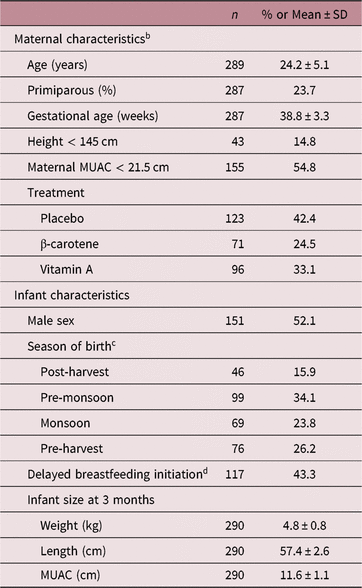
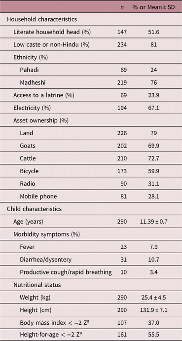
Characteristics of mothers and infants during the trial and of households and children at follow-up are presented in Tables 1 and 2, respectively. Maternal nutritional status was poor. Fifteen percent of mothers were shorter than 145 cm and greater than half (54.8%) were thin based on a MUAC < 21.5 cm. Our study sample was divided equally between boys and girls. Births were lower in the post-harvest season than pre-monsoon, but roughly equal in the monsoon and pre-harvest seasons. Breastfeeding was delayed (started >3 days after birth) for 43.3% of infants. Anthropometric data were available from early infancy for almost 90% of children. Infant anthropometric measures were normally distributed, with a mean (±SD) weight of 4.8 kg (±0.8), a length of 57.4 cm (±2.6), and a MUAC of 11.6 cm (±11.1). Based on socioeconomic status data collected at follow-up, only half of household heads were literate. Households were largely comprised of low caste or non-Hindu (81%) households, and roughly three quarters were of the Mahdeshi ethnic background. Access to electricity (67%) and the household’s ownership of selected assets suggested an upwardly mobile rural population. However, only one quarter of study households had access to an improved latrine. Over half (56%) of the children in our sample were stunted and the mean (±SD) BMI-for-age Z-score was −1.8 (±0.9). Twenty-six percent of children exhibited at least one morbidity symptom in the week prior to the interview, which were primarily gastrointestinal.
Maternal and infant characteristics during original trial (1994–1997) in rural Nepala

MUAC, mid-upper arm circumference.
a Mean timing of antenatal clinic visit was 18 weeks’ gestational age (range: 5–38 weeks); mean timing of neonatal home visit was 13 days of age (range: 0–117 days); infant anthropometric data missing for n = 42 children.
b Measurements taken at antenatal clinic visit (mean timing was 18 weeks’ gestational age; range 5–38 week).
c Defined as post-harvest (December–February), pre-monsoon (March–May), monsoon (June–August), or pre-harvest (September–November).
d Started >3 days after birth.
Household and individual characteristics of young adolescent children during follow-up (2006–2008) in rural Nepal

a Based on sex-specific reference data for children aged 5–19 years21.
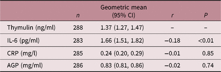
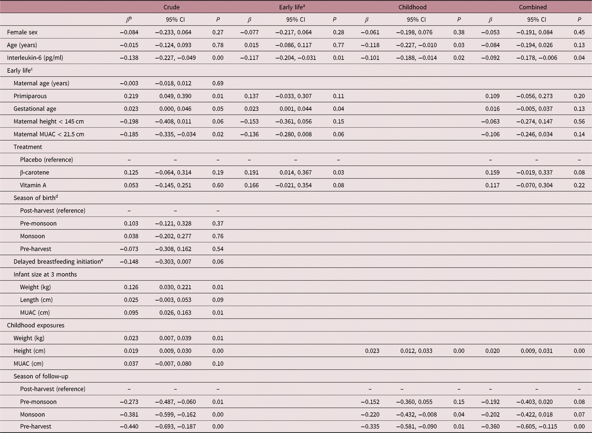
The geometric (95% CI) thymulin concentration was 1.37 ng/ml (1.27, 1.47). We found a weak negative correlation between thymulin and IL-6 concentrations (r = −0.179, P < 0.01), but no correlation with either CRP or AGP (Table 3). Unadjusted analyses revealed no significant difference between sexes (Table 4), nor any association with child’s age. Of the early-life exposures, thymulin concentrations in childhood only varied significantly by parity (P < 0.05) and low maternal MUAC (P = 0.02), with higher concentrations among first-born children and better nourished mothers. Concentrations also tended to be higher among children with a greater gestational age (P = 0.05), those with taller mothers (P = 0.06), and those who were breastfed during the first 3 days of life (P = 0.06). There were no apparent differences by season of birth. Relationships between thymulin and early anthropometry were consistent, with significantly higher concentrations in those with better nutritional status based on weight (P = 0.01) and MUAC (P = 0.01) at 3 months of age. Better nutritional status measures taken at follow-up were consistently associated with higher thymulin concentrations, although this was not statistically significant for child MUAC. Concentrations were strongly associated with season of measurement, with concentrations significantly lower in all other seasons compared to the post-harvest reference (P < 0.05).
Geometric mean serum immune marker concentrations and their correlations with serum thymulin in young adolescent children in rural Nepal (2006–2008)

All values were natural log-transformed.
IL-6, interleukin-6; CRP, C-reactive protein; AGP, α1-acid glycoprotein.
Differences in thymulin concentrations (ng/ml) among young adolescent children in rural Nepal (n = 290), by early-life and childhood health and nutrition exposures (2006–2008)

MUAC, mid-upper arm circumference.
a Multivariate models for early-life, childhood, and combined measures include all other variables in the column plus household electricity at follow-up.
b β coefficient (95% confidence interval) from simple or multiple linear regression analyses; multivariate models include all other variables plus household electricity at follow-up.
c Measurements taken at antenatal clinic visit (mean timing was 18 weeks’ gestational age; range 5–38 week).
d Defined as post-harvest (December–February), pre-monsoon (March–May), monsoon (June–August) or pre-harvest (September–November).
e Started >3 days after birth.
Of the variables significant in unadjusted analyses, only gestational age (P = 0.04) remained significant in the early-life multivariate model (Table 4). Parity and maternal nutritional status remained in the model but were not statistically significant. A relationship became apparent in the multivariate early-life model between maternal supplement allocation and childhood thymulin, with significantly higher concentrations in children born to β-carotene-supplemented mothers (P < 0.05). Concentrations also tended to be higher in children born to vitamin A-supplemented mothers; however, this did not attain statistical significance (P = 0.08). For the childhood model, both child height (P < 0.001) and season of measurement (P < 0.01) remained strongly associated with thymulin. All of the early-life relationships, with the exception of supplementation, were mediated in part by nutritional status at follow-up. Adding children’s height at 9–13 years of age to the model, in particular, decreased β coefficients by approximately one quarter.
Discussion
We hypothesized that exposures during the critical prenatal period of thymic development would have effects lasting into early adolescence. Unadjusted associations tended to support this hypothesis, although only gestational age and prenatal exposure to β-carotene remained statistically significant in our multivariate model. Marginally significant early-life exposures included parity, maternal stunting and wasting, and exposure to vitamin A. These observations were mediated, in part, by the strong contemporaneous influences of nutritional status and season. Of the known determinants of thymic size,Reference Pejcic-Karapetrovic, Kosec and Leposavic26–Reference Consolini, Legitimo, Calleri and Milani28 thymulin concentrations did tend to be lower in females and to decline with age. That these associations were not stronger may be due to a lack of sensitivity in our outcome measure or, for age, that this was a young study population with a narrow age range. Thymulin’s inhibitory effects on inflammationReference Henriques-Coelho, Oliveira and Moura29 were clearly evident in the strong negative association with serum thymulin.
Prior human studies have highlighted the importance of early-life exposures to thymic development,Reference Collinson, Moore, Cole and Prentice4–Reference Fulford, Moore and Arifeen7, Reference Moore, Prentice and Wagatsuma30 which may be driven by epigenetic changes,Reference Waterland, Kellermayer and Laritsky31 as well as by perturbations to the developing hypothalamic-pituitary-adrenal axis.Reference Reggiani, Schwerdt, Console, Roggero, Dardenne and Goya13 That only prenatal exposures were retained in our early-life model is supported by work by Fulford et al., which documented a direct association between fetal size and later thymic index, with the strongest association for fetal size measured at the earliest timepoints in pregnancy.Reference Fulford, Moore and Arifeen7 Greater gestational age, in particular, likely enabled further development of the thymus prior to birth. We found no relationship between season of birth and thymulin concentrations later in life. This is inconsistent with early research from The Gambia.Reference Collinson, Moore, Cole and Prentice4, Reference Ngom, Collinson, Pido-Lopez, Henson, Prentice and Aspinall5 It is possible that this association is less pronounced in South Asia.Reference Moore, Prentice and Wagatsuma30 Differences may also be due to age. Children in our study were aged 9–13 years, while prior research has been conducted during infancy.Reference Collinson, Moore, Cole and Prentice4, Reference Ngom, Collinson, Pido-Lopez, Henson, Prentice and Aspinall5 A study of thymic function in young adults similarly found no relationship with birth season.Reference Ngom, Solon, Moore, Morgan, Prentice and Aspinall32 We also cannot dismiss that associations were attenuated in our research by our reliance on thymulin, which is a less sensitive indicator of organ size and function than measurements of thymic index, TRECs, or recent thymic emigrants.Reference McFarland, Picker, Koup and Douek9 Our work is perhaps more comparable to that carried out in the Philippines, where researchers reported lower serum thymopoietin concentrations among adolescents who were born small for gestational age.Reference McDade, Beck, Kuzawa and Adair8 That study also failed to detect a relationship between birth weight and thymic hormone levels, except in interaction with the total duration of breastfeeding, an exposure that was not measured in our cohort.
The observed effect of early exposure to β-carotene, and marginally significantly effect of vitamin A, could be related to the role of vitamin A in development of the thymus. Mice with targeted mutations of the retinoic acid-synthesizing enzyme retinaldehyde dehydrogenase 2Reference Niederreither, Vermot, Le Roux, Schuhbaur, Chambon and Dolle33–Reference Ryckebusch, Bertrand and Mesbah35 or genetic manipulations of retinoic acid response element genes exhibit thymic aplasia or hypoplasia.Reference Mendelsohn, Lohnes and Decimo36, Reference Wendling, Dennefeld, Chambon and Mark37 However, the treatment effect was more pronounced in the β-carotene group and, further, it was strengthened in the multivariate model that included a marker of inflammation. We posit that the observed association involves early-life influences on the hypothalamic-pituitary-adrenal axis. Analyses from the original trial showed that supplementation with β-carotene reduced the prevalence of high fever during pregnancy and during the post partum period.Reference Christian, West and Khatry38 Fever, induced in animal models by lipopolysaccharide challenge, influences the developing neuroendocrine immune regulatory mechanisms.Reference Beloosesky, Maravi and Weiner39–Reference Spencer, Hyland, Sharkey and Pittman47 The neuroendocrine axis, in turn, regulates the production and secretion of thymulin.Reference Savino and Dardenne48
Effects on thymic size and function in early life are critical in that T cell development occurs largely in utero.Reference Sakaguchi, Miyara, Costantino and Hafler49 From mid-pregnancy, thymocytes are subject to both positive and negative selection, whereby those unable to bind major histocompatibility complex class I or class II, and those that are autoreactive, are deleted.Reference Murphy and Weaver50 An insult during this period of “thymic education” could theoretically limit the body’s T cell repertoire.Reference West51 The immune system’s developing regulatory mechanisms, notably the natural CD4+FOXP3+ TReg cell population, and its ability to differentiate between “self” and “non-self” may be affected as well. Indeed, some epidemiologic studies support a link between early-life undernutrition and heightened risk of autoimmune or allergic disease,Reference Phillips, Cooper and Fall52–Reference McDade, Kuzawa, Adair and Beck56 although others have refuted these findings.Reference Brix, Kyvik and Hegedus57, Reference Edwards and Cooper58 Future studies in this area should consider immunophenotyping, specifically considering differences in regulatory and/or γδ T cell populations.
Concurrent nutritional status is a well-known predictor of thymic size and function.Reference Prentice59, Reference Savino, Dardenne, Velloso and Dayse Silva-Barbosa60 Both protein-energy malnutritionReference Lyra, Madi, Maeda and Savino61, Reference Parent, Chevalier and Zalles62 and micronutrient deficienciesReference Kuvibidila, Dardenne, Savino and Lepault63–Reference Nodera, Yanagisawa and Wada66 can cause thymic atrophy. Morphological changes and decreased hormone production by thymic epithelial cells have also been shown in mouse models of undernutrition.Reference Mittal and Woodward67 We observed positive associations between thymulin concentrations and child height, weight, and MUAC. That only height was retained in our final model suggests that these associations may be partly explained by the correlation between organ and body size. Associations between weight, MUAC, and thymulin concentration may have been captured by the strong seasonal effect, which is generally attributed to seasonal patterns in infection and nutritional status. This seasonality is consistent with the literature.Reference Moore, Collinson and Fulford68,Reference Moore, Collinson and Prentice69 It should be noted that there is also seasonal variability in melatonin,Reference Kauppila, Kivela, Pakarinen and Vakkuri70 which regulates production of thymic hormones.Reference Molinero, Soutto, Benot, Hmadcha and Guerrero71
Our study contributes information on a thymic hormone, as a proxy for thymic size and function, from a population-based assessment among children in a low-income country setting. However, there were a number of limitations. Thymic hormone concentrations have not been validated as an indicator of size or function; however, concentrations are known to decline with age-related thymic involution.Reference Brelinska10, Reference Consolini, Legitimo, Calleri and Milani28 Our measurement of thymulin was further limited by our reliance on an ELISA method that does not differentiate inactive from the active, zinc-bound hormone. This may have had some influence on our results, particularly as children in our sample are at high risk of zinc deficiency.Reference Tielsch, Khatry and Stoltzfus72 Although our study was population-based, children were only eligible for participation if they and their mothers had consented to participate and provide biospecimens at a number of time points from pregnancy through later childhood. Reliance on this highly compliant group limited our sample size and resulted in a sample of somewhat better socioeconomic status than their peers, potentially affecting our ability to detect differences and our ability to extrapolate to the broader population. Some of our exposure measures were also limited, e.g., we did not have a measure of birth weight or detailed data on breastfeeding.
The findings reported here may have important relevance to later-life disease risk. Our understanding of the role of thymulin and other thymic hormones has evolved in recent years, with new work highlighting their importance in neuroendocrine regulation of the inflammatory response.Reference Reggiani, Schwerdt, Console, Roggero, Dardenne and Goya13, Reference Lunin and Novoselova24 Continued research in this cohort can provide important longitudinal information on immune dysregulation and, ultimately, help us to better understand its relevance for diseases such as metabolic syndrome, neurodegenerative conditions, cardiovascular disease, and cancer.Reference Brestoff and Artis73–Reference Ferguson76
Acknowledgments
We thank participating children and their families, as well as members of the Johns Hopkins and NNIPS study teams including Parul Christian, Joanne Katz, Steve LeClerq, Margia Arguello, and Veena Singh for their contributions to the study.
Financial Support
This analysis and follow-up studies were supported by grant GH614 (Global Control of Micronutrient Deficiency) and OPP141435 (Improving adolescent newlywed nutrition and health for pregnancy in Bangladesh and health across early-life stages in South Asia – Healthy Birth, Growth, and Development) from The Bill and Melinda Gates Foundation, Seattle, WA, USA; the original trial was supported by the US Agency for International Development, Washington, DC, under Cooperative Agreement No. DAN 0045 A 005094–00; additional assistance was received from the Sight and Life Global Nutrition Research Institute at Johns Hopkins University; and A.C.P. was supported by a pre-doctoral fellowship from the Procter & Gamble Company, Cincinnati, OH, USA.
Conflicts of Interest
None.
Ethical Standards
The authors assert that all procedures contributing to this work comply with the ethical standards of the national guidelines from the United States and Nepal on human experimentation and with the Helsinki Declaration of 1975, as revised in 2008, and has been approved by the institutional committees of the Johns Hopkins School of Medicine in Baltimore, MD, USA, the Nepal Health Research Council of the Ministry of Health in Kathmandu, Nepal, and the Teratology Society in Bethesda, MD, USA (trial protocol), as well as committees at the Johns Hopkins Bloomberg School of Public Health, Baltimore, MD, USA and the Institute of Medicine at Tribhuvan University in Kathmandu, Nepal (follow-up protocol and present analysis).
Supplementary materials
For supplementary material for this article, please visit https://doi.org/10.1017/S2040174419000485