Kawasaki disease is an acute febrile infantile disease causing ischaemic heart disease and was first described in 1967.Reference Kawasaki1 There is a patient population with a history of acute Kawasaki disease and cardiac sequelae occurring before 1967.Reference Burns, Shike, Gordon, Malhotra, Schoenwetter and Kawasaki2,Reference Gordon, Daniels and Kahn3 In most of those patients, coronary artery lesions caused by Kawasaki disease had been first recognised because of acute myocardial infarction or at autopsy for sudden death before.Reference Tsuda, Abe and Tamaki4 On the other hand, the prevalence of patients with a giant coronary aneurysm, which causes their ischaemic heart disease, in female is less than a half of that in male.Reference Tsuda, Hamaoka and Suzuki5 Therefore, female patients with coronary artery lesions caused by Kawasaki disease are a very small population. However, there are some problems that need to be resolved during the management of pregnancy and delivery in this population.Reference Gordon, Jimenez-Fernandez and Daniels6 We encountered two female patients who had delivered without any recognition of ischaemic heart disease caused by presumed Kawasaki disease.
Patient 1
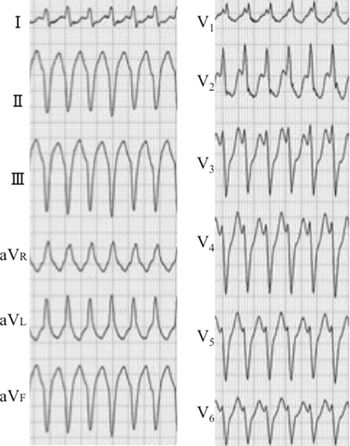
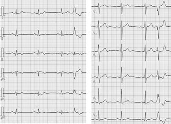
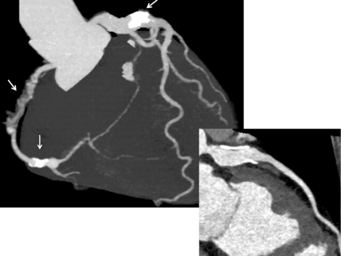
A 58-year-old female visited the hospital due to palpitation, while travelling. Her body height and body weight were 161 cm and 57 kg, respectively. A 12-lead electrocardiogram revealed wide QRS tachycardia with left axis deviation and a right bundle branch block morphology at 210 beats per minute (Fig 1). It was suspected to be ventricular tachycardia responding to verapamil, and she was restored to normal sinus rhythm after an intravenous administration of verapamil. An abnormal Q wave in lead III was detected (Fig 2). CT angiograms revealed segmental stenosis of the right coronary artery and a giant coronary aneurysm with calcification at the bifurcation of the left coronary artery (Fig 3). She was suspected to have coronary artery lesions caused by Kawasaki disease. She was referred to our hospital. Her blood pressure was 95/51 mmHg. Her low-density lipoprotein cholesterol level and brain natriuretic peptide level were 137 mg/dl and 50 pg/ml, respectively.
The 12-lead electrocardiogram of wide QRS tachycardia. An electrocardiogram revealed wide QRS tachycardia at a rate of 210 beats per minute. The electrocardiogram revealed an upper axis and right bundle branch block.

The 12-lead electrocardiogram at rest (Patient 1). Left axis deviation and an abnormal Q wave in lead III were detected.

CT coronary angiograms (Patient 1). The angiogram revealed segmental stenosis of the right coronary artery. Coronary artery calcification at segments 2, 4, and 5 was detected. A giant calcified coronary aneurysm at the bifurcation of the left coronary artery was found.

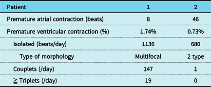
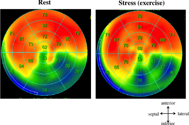
In the two-dimensional echocardiogram, the left ventricular diastolic dimension and left ventricular ejection fraction with the Simpson method were 59 mm and 45%, respectively. A giant calcified coronary aneurysm was detected at the bifurcation of the coronary artery, and hypokinesis of the inferior wall of the left ventricle was also detected. Multifocal premature ventricular contractions and non-sustained ventricular tachycardia were frequently observed on the 24-hours Holter electrocardiogram (Table 1). By the technetium-99m-tetrofosmin myocardial perfusion single photon emission computed tomography, hypoperfusion was detected in the inferior wall of the left ventricle, and the extent score was 34 (Fig 4). The summed rest score, summed stress score, and summed difference score were 20, 21, and 1, respectively, with normal being zero. These scores revealed severe myocardial involvement of the left ventricle. By the quantitative gated single photon emission computed tomography left ventricular ejection fraction was 48%. A beta-blocker and aspirin were prescribed. In the electrophysiologic study, delayed potentials were recorded at a basilar-mid inferior site of the left ventricle in the mapping during sinus rhythm, and three ventricular tachycardia with right bundle branch block morphology and upper axis were induced by extrastimulus delivered at the apex of the right ventricle. She underwent radiofrequency catheter ablation and an implantable defibrillator implantation.
The findings in the 24-hour Holter electrocardiogram

Technetium-99 m-tetrofosmin myocardial perfusion imaging (Patient 1). Hypoperfusion at the inferior wall of the left ventricle was detected at both rest and during exercise.

She was born in 1960. At the age of 3 years in her history of past illness, she was admitted for sepsis. Further, she remembered an episode of chest pain while sleeping, when she was in kindergarten. She was aware of premature ventricular contractions in her adolescent. She had delivered twice by vaginal delivery without any complication during her 20s. At the age of 48 years, she had undergone coil embolisation for pulmonary arteriovenous fistula in the department of respiratory medicine in another hospital. At that time, the left ventricular diastolic dimension was 58 mm, and it was slightly dilated. Premature ventricular contractions were also detected at that time. However, ischaemic heart disease with coronary artery lesions was not diagnosed at that time.
Patient 2
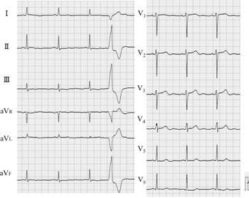
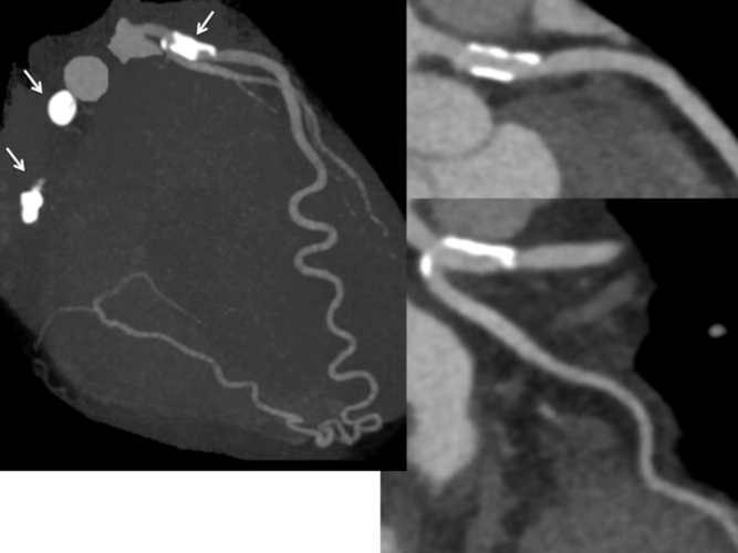
A 48-year-old female was accidentally found to have coronary artery calcification on CT, when she had pneumonia. She was transferred to our hospital because ischaemic heart disease was suspected. She was born in 1963. She had a history of “scarlet fever” before she was 12 months old. Premature ventricular contractions had been observed, when she delivered her first child at 31 years. She had delivered twice by a vaginal delivery without any complication. Her body height and body weight were 167 cm and 54 kg, respectively. Her blood pressure was 120/66 mmHg. Her low-density lipoprotein cholesterol level was 124 mg/dl. A-12-lead electrocardiogram revealed flat T wave in leads II, III, and V6 and poor progression of the r wave in leads V1, V2, and V3 (Fig 5). In treadmill test, ST-T depression in leads II. III. aVF V3, V4, V5, and V6 was detected, although she was asymptomatic. In the two-dimensional echocardiogram, her left ventricular diastolic dimension and ejection fraction were 48 mm and 71%, respectively. Mild mitral regurgitation was detected. The diameter of the proximal portion of the right coronary artery, left main trunk, and left anterior descending artery was 5.5, 5.0, and 3.4 mm, respectively. In her CT angiograms, the occlusion of the right coronary artery and the coronary artery calcification at segments 1, 2, 4, 6, and 11 were detected (Fig 6). The collateral arteries from the left anterior descending artery to the posterior descending artery were well developed. In the technetium-99m-tetrofosmin myocardial perfusion single photon emission computed tomography on exercise, localised mild hypoperfusion of the inferior wall of the left ventricle was detected. Her left ventricular end-diastolic volume and ejection fraction were 88 ml and 72%, respectively. Her coronary flow reserve of the left anterior descending artery with adenosine triphosphate by transthoracic echocardigraphy was 2.6. Isolated premature ventricular contractions, consisting of 680 beats (0.73%), were detected in a 24-hours Holter electrocardiogram (Table 1). Aspirin was recommended.
The 12-lead electrocardiogram at rest (Patient 2). A 12-lead electrocardiogram revealed flat T waves in leads II, III, and V6 and poor progression of the r wave in leads V1, V2, and V3.

CT coronary angiograms (Patient 2). (Left) An occlusion of the right coronary artery and coronary artery calcification at segments 1, 2, 4, 6, and 11 were detected. The collateral arteries from the left anterior descending artery to the posterior descending artery were well developed (Right upper). The angiogram revealed calcification at segment 6 of the left anterior descending artery (Right lower). The angiogram also revealed calcification at segment 11 of the left circumflex.

Discussion
Acute Kawasaki disease symptoms including fever in the post-inflammation period are self-limiting. There are probably many asymptomatic adult patients with coronary arterial lesions caused by Kawasaki disease who remain undiagnosed, because Kawasaki disease was not always known in the 1950s and 1960s. These two female patients first had diagnosis of coronary artery lesions at the age of 58 and 48 years, respectively. They had had a history of acute febrile disease in their childhood. One patient had sepsis, and the other “scarlet fever”. Because scarlet fever is likely to occur in children more than 2 years old, its diagnosis is questionable. These diseases are the differential diagnosis of Kawasaki disease and can often be misdiagnosed as Kawasaki disease. It was suspected that their episodes in children were acute Kawasaki disease. We think that they would have had coronary artery lesions caused by Kawasaki disease in their childhood.
Premature ventricular contractions were detected, while they were pregnant in their 20s or 30s. However, the coronary artery lesions caused by Kawasaki disease could not be diagnosed until fifties. As a result, they received no medications for ischaemic heart disease until they were middle-aged adults after childbearing. Recently, the use of transthoracic echocardiography has spread since the 1990s. The screening of coronary artery dilatation is needed in patients with multifocal premature ventricular contractions and abnormalities such as abnormal Q waves and negative T waves in electrocardiograms, although it is not always easy to detect the proximal portion of the coronary arteries in adults.Reference Tamaki, Tsuda, Igarashi, Tanaka and Fujieda7,Reference Suzuki and Kamiya8
Our female patients had giant coronary artery aneurysms with calcification at the proximal site of both coronary arteries. One was segmental stenosis of the right coronary artery, and the other was occlusion with calcified coronary aneurysm of the right coronary artery. Segmental stenosis, which is often found in patients with a history of Kawasaki disease, is considered typical of lesions caused by Kawasaki disease. It implies the development of several new small vessels, which represent recanalisation after coronary artery occlusion.Reference Tsuda, Tsujii and Hayama9,Reference Tsujii, Tsuda, Kanzaki, Ishiduka, Nakashima and Kurosaki10 Further, it is known that coronary artery calcification occur many years after severe acute Kawasaki disease vasculitis.Reference Tsuda, Tsujii, Kimura and Suzuki11 Calcified coronary arteries without dilatation imply the existence of coronary aneurysms in the acute phase and regression of aneurysms in the late period. Coronary artery calcification in the late period represents coronary artery involvement due to acute severe vasculitis. Coronary aneurysms are also likely to appear in the sites including segments 1, 2, 4, 5, 6, and 11, which were their coronary artery calcification existed. The characteristic distribution of coronary artery aneurysms and calcification is also an important finding that leads to suspecting a diagnosis of presumed Kawasaki disease.Reference Tsuda, Matsuo and Kurosaki12 These findings by CT angiograms in patients with a history of an unknown fever in childhood strongly suspect cardiac sequelae due to Kawasaki disease.
In the detection of their ischaemic heart disease, one had symptoms and the other was detected by accident. One patient was detected by the occurrence of ventricular tachycardia. It was suspected that the fatal ventricular arrhythmia was caused by myocardial fibrosis due to previous inferior myocardial infarction in her childhood. The myocardial involvement in patients after previous myocardial infarction in childhood can lead to sudden death in adolescents and adults.Reference Tsuda, Hirata, Matsuo, Abe, Sugiyama and Yamada13,Reference Yagi, Tsuda and Shimizu14 We reported that the cut-off value of the technetium-99m-tetrofosmin myocardial perfusion single photon emission computed tomography extent score for the appearance of non-sustained ventricular tachycardia was 17.Reference Abe, Tsuda, Sugiyama, Kiso and Yamada15 Such patients often have an increased left ventricular end-diastolic volume and low left ventricular ejection fraction with previous widespread infarct area.Reference Nakaoka, Tsuda, Morita and Kurosaki16 On the other hand, the other patient with occlusion of the right coronary artery had been asymptomatic for a long period of more than 40 years. She had well-developed collateral arteries from the left coronary artery to the right coronary artery. This finding is frequently detected in patients with coronary artery lesions caused by Kawasaki disease. It was considered that the degree of myocardial fibrosis in patients with preserved left ventricular ejection fraction was mild, even if a coronary artery was occluded.Reference Nakaoka, Tsuda, Morita and Kurosaki16
Pregnancy also induces a series of haemostatic changes, with an increase in the concentration of coagulation factors, fibrinogen, and platelet adhesiveness, as well as diminished fibrinolysis, which leads to hypercoagulability. The physiological adaptation for labour and the post-partum period requires four to six weeks. How to carry out antithrombotic therapy during pregnancy and how to perform the delivery should be considered. These problems had been touched at the 1980s and 1990s. Nevertheless, they delivered safely twice without any recognition of ischaemic heart disease and its treatment. A consensus on anticoagulant therapy in patients with coronary artery lesions caused by Kawasaki disease has not necessarily been decided at the present. Because the prevalence of myocardial infarction is very low in this population, it makes it difficult to determine any benefit from anti-coagulation therapy. It is suspected that an aggressive thrombotic therapy during pregnancy rather than that for non-pregnancy is not necessarily needed, although they might be lucky cases.Reference Gordon, Jimenez-Fernandez and Daniels6,Reference Tsuda, Kawamata and Neki17,18 Further, a conventional delivery in asymptomatic patients with a preserved left ventricular ejection fraction of more than 45% may be possible. However, we must take care of cardiac events including fatal ventricular arrhythmias in patients with a previous myocardial infarction.Reference Satoh, Sano and Suwa19
These patients had been almost asymptomatic for a long period of their lives until the detection of coronary artery disease. It may be a gender difference in the progression of coronary artery stenosis and the appearance of coronary artery calcification.Reference Tsujii, Tsuda, Kanzaki, Ishiduka, Nakashima and Kurosaki10,Reference Miura, Kobayashi and Kaneko20 In this Kawasaki disease population, the time of the occurrence of acute coronary syndrome in female patients may be later than that in male patients, as well as ischaemic heart disease due to atherosclerosis.
Conclusion
We encountered two female patients who had delivered without any recognition of ischaemic heart disease caused by presumed Kawasaki disease. They were lucky cases. Although they had premature ventricular contractions at the time of their delivery, no coronary artery lesions were diagnosed at that time.
Acknowledgement
The authors thank Mr. John Martin for his kind English language consultation.
Financial support
This research received no specific grant from any funding agency, commercial, or not-for-profit sectors.
Conflicts of interest
The authors state that they have no conflict of interest.
Ethical standards
All procedures performed in studies involving human participants were in accordance with the ethical standards of the institutional committee with the 1964 Helsinki declaration and its later amendments or comparable ethical standards.