Introduction
Fascioliasis is caused by two species of trematodes: Fasciola hepatica and Fasciola gigantica. Their ranges are governed by the distribution of suitable intermediate Lymnaeid snail hosts. Galba truncatula and other lymnaeid species of the Galba/Fossaria group are the intermediate hosts for F. hepatica, while snails of the Radix group are the intermediate hosts for F. gigantica – for example, Lymnaea (Radix) natalensis in Africa (Mas-Coma et al., Reference Mas-Coma, Valero and Bargues2009). Fasciola hepatica causes temperate fascioliasis and is commonly found in the Americas, Europe and Oceania. Fasciola gigantica is commonly found in areas of Africa and Asia; however, its range often overlaps with that of F. hepatica. Hybrids of F. hepatica and F. gigantica have been reported (Agatsuma et al., Reference Agatsuma, Arakawa, Iwagami, Honzako, Cahyaningsih, Kang and Hong2000).
Although there are increasing reports of zoonotic infections (Ashrafi et al., Reference Ashrafi, Bargues, O'Neill and Mas-Coma2014), fasciolosis is generally considered an agricultural problem and annual losses greater than US$2 billion and 600 million infected animals have been suggested (McManus & Dalton, Reference McManus and Dalton2006). Economic losses are due to mortality, poor carcass configuration, malnutrition, intercurrent disease, low weight gain, poor feed conversion efficiency, decreased milk yield and infertility (Sothoeun et al., Reference Sothoeun, Davun and Copeman2007).
There have been few published studies investigating fascioliasis in Cameroon. Ngole et al. (Reference Ngole, Ndamukong and Mbuh2003) described finding 80% of cattle being Fasciola positive through coprological testing and 13% of livers showing visible damage because of fascioliasis. More recently, a study in 2016 found 22.6% of livers affected by fascioliasis in Fako in the south-west of Cameroon (Kouam et al., Reference Kouam, Meningue and Fon2018). There have been more studies investigating fascioliasis in countries surrounding Cameroon, including Nigeria and Chad. A study in Jos, Nigeria, reported a prevalence of 12.7% of Fasciola in the various domestic livestock, including cattle, goats, sheep and dogs (Ekong et al., Reference Ekong, Juryit, Dika, Nguku and Musenero2012); however, an individual breakdown of each species was not provided. Studies focusing specifically on cattle have also been carried out. Opara (Reference Opara2005) found 47.6% of the cattle tested at three abattoirs in Uyo were Fasciola positive, and that cattle tested in the rainy season were more likely to be infected. Elelu et al. (Reference Elelu, Ambali, Colesm and Eisler2016) reported 74.9% of cattle to be infected with F. gigantica through faecal testing. In Chad, high infection rates have been found, including, for example, a prevalence of 68% in cattle (Jean-Richard et al., Reference Jean-Richard, Crump, Abicho, Naré, Greter, Hattendorf, Schelling and Zinsstag2014). The same study showed lower prevalence in sheep (23%) and goats (12%). Animals from near Lake Chad were more likely to be infected than animals that were further away from the lake, which most likely reflects the presence of the snail intermediate host.
In this study, we investigated the prevalence of Fasciola infections in cattle and sheep slaughtered at Etoudi slaughtering house in Yaoundé. Etoudi is one of the main abattoirs in Cameroon and animals from all ecological zones in Cameroon are transported there for slaughter.
Materials and methods
Study design
A study was undertaken in two separate sampling sessions at the Etoudi slaughtering house located in Yaoundé (fig. 1), the first occurred in November 2010 at the start of the dry season and the second in March 2011 at the end of the dry season. A simple random study was used to design the study (Thrusfield, Reference Thrusfield2007). Based on previous studies on prevalence of Fasciola infections in cattle and sheep, we used an estimated prevalence of 50% (Opara, Reference Opara2005) and 10% (Nwosu & Srivastava, Reference Nwosu and Srivastava1993), respectively. This gave a sample size of 384 and 138, respectively, to be sampled at 95% confidence and 5% precision. Before slaughter, the origin and breed of the animals were determined through discussion with the traders. Animals that were sampled at the abattoir came from nine locations within Cameroon (fig. 1): Bafia; Barfoussam; Batouri; Bertoua; Garoua Sambe; Maroua; Meiganga; Ngaoundal; Ngaoundere. In addition, cattle also came from the neighbouring country of Chad.
Map showing the locations within Cameroon where cattle (Batouri, Bertoua, Meiganga, Ngaoundal, Ngaoundere) and sheep (Bafia, Bafoussam, Garoua Sambe, Maroua, Ngaoundere) came from. Yaoundé, where the Etoudi slaughterhouse is situated, is underlined. Cattle that originated from Chad are not shown.

Sample processing
Over the two sampling sessions, a total of 767 cattle and 267 sheep were randomly inspected at Etoudi slaughterhouse for the presence of Fasciola species. At post-mortem, each liver, liver duct and the associated lymph nodes were examined for Fasciola infection. Any lesions seen at post-mortem were recorded on the data sheet. In all cases, where Fasciola were found at post-mortem in the liver or liver duct, total or partial condemnation of the liver was undertaken. The condemned liver was then transported to the Military Health Research Centre (CRESAR) laboratory in Yaoundé Global Viral Forecasting Johns Hopkins Cameroon (GVFI/JH) project laboratory for the separation, collection and storage of Fasciola with support from technical staff of Global Viral. In the laboratory, the infected liver was opened and put in saline water for 24 h. The saline water was then passed through a wire mesh and the liver flukes present were collected with the help of a fine brush. They were measured, photos taken and the parasites stored in 70% ethyl alcohol in 2 ml cryotubes at −80°C, for any future study.
Cost of liver
Livers with Fasciola infections were either fully condemned or the affected areas were trimmed. The amount of liver trimmed or the whole condemned liver was then weighed. The cost of 1 kg of fresh liver in the shop during the study period was 2100 FCFA (US$4.2) for cattle and 1000 FCFA (US$2) for sheep (the exchange rate at the time of the study was around 500 FCFA/US$). The total condemned liver was multiplied by the average price per kilo to obtain the value of the liver condemned. Figures for the number of cattle and sheep slaughtered in 2010 at the Etoudi slaughterhouse were obtained from the Centre Region Delegation of the Ministry of Livestock, Fisheries and Animal Industries. In 2010, 51,103 cattle and 2700 sheep were slaughtered.
Statistical analysis
Wilson's confidence intervals were calculated using WinPepi (http://www.brixtonhealth.com/pepi4windows.html). To compare age groups in this study, the cattle were split into two groups: group A included animals of four years old and under, while group B included animals of five years old and above. The sheep were classed in group A up to 23 months, and group B of 24 months or older. Fisher's tests were used to compare differences in prevalence between species and season; between the two age groups of cattle and sheep; and between the sexes of infected animals. For sex and age testing, the infection rates in the two groups were entered as a different stratum.
Results
A higher percentage of sheep (27%; 95% confidence interval (CI): 21.7–32.7) were infected with Fasciola parasites than cattle (18%; 95% CI: 15.3–20.9). The difference of infection between the two species of animals sampled was significant (P = 0.003)
Cattle
Over the two sampling periods, the overall prevalence of Fasciola infections in cattle was 18.0% (95% CI: 15.3–20.9). The prevalence in November 2010 was significantly higher (P < 0.001) at 24.9% (95% CI: 20.7–29.6) compared to March 2011 when it was 11.0% (95% CI: 8.0–14.6%). Older cattle (five years and older) with a prevalence of 22.6% (95% CI: 18.2–26.7) were significantly more likely (P = 0.005) to be infected than those under five years of age (14.3%; 95% CI: 11.2–18.0). Overall, the prevalence in female cattle was 23.2% (95% CI: 18.1–29.1), while it was 15.6% (95% CI: 12.6–19.0) in male cattle. Although we did find a significant difference (P = 0.012) between male and female animals, age confounded the relationship (P = 0.365).
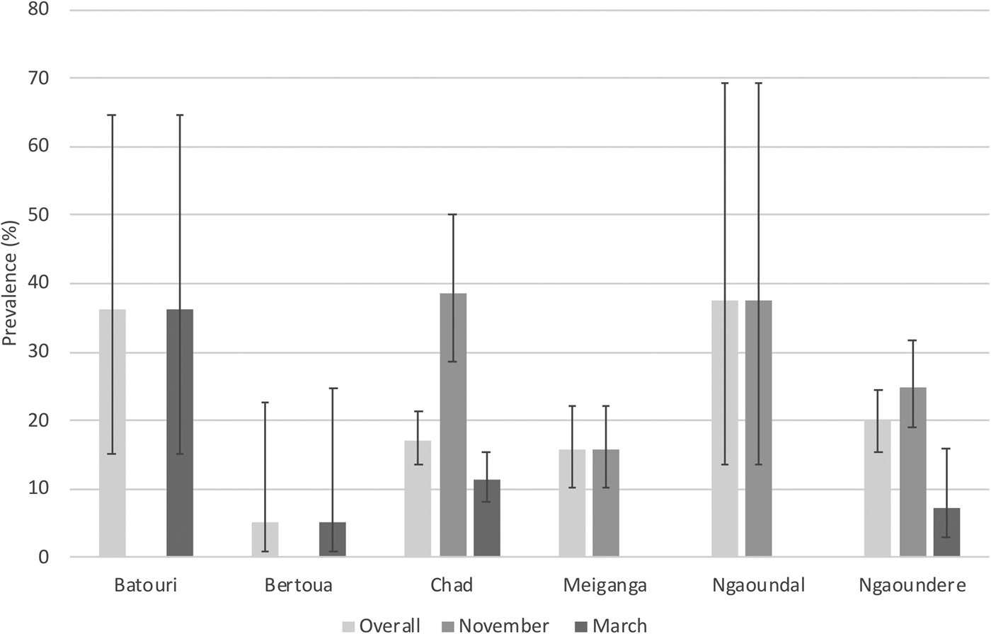
Overall, there were no differences in the prevalence in terms of the origin of the cattle (fig. 2). However, significant differences (P = 0.001) were found during the first sampling session. In November, cattle from Chad (38.7%; 95% CI: 27.6–50.6) were more likely (P = 0.003) to be infected than cattle from Meiganga (15.7%; 95% CI: 9.7–23.4). No differences were found in March 2011. Comparing the two sampling periods, cattle from Chad (P < 0.001) and Ngaoundere (P = 0.001) showed significantly higher prevalence in November when compared to March 2011.
Prevalence of Fasciola in cattle sampled at the Etoudi slaughterhouse in Yaoundé, Cameroon, by origin. Cattle were sampled in either November 2010 or March 2011. Presence of Fasciola was detected through examination of the liver post-slaughter. Error bars show 95% confidence intervals.

Overall, there were no significant differences between the three breeds of cattle that were sampled at the abattoir, these being Aku (16.9%; 95% CI: 11.3–23.8), Gaudali (20.3%; 95% CI: 15.5–25.9) and Mbororo (16.9%; 95% CI: 13.2–21.1). However, Gaudali were significantly (P = 0.002) more likely to be infected in November 2010 (24.9%; 95% CI: 18.8–31.8) than in March 2011 (7.7%; 95% CI: 2.5–17.1). Mbororo were significantly (P < 0.001) more likely to be infected in November 2010 (37.0%; 95% CI: 26.6–48.5) than in March 2011 (11.2%; 95% CI: 7.8–15.4). Mbororo were also significantly (P = 0.002) more likely to be infected in November 2010 (37.0%; 95% CI: 26.6–48.5) when compared to Aku (17.1%; 95% CI: 10.9–24.9).
Sheep
Over the two sampling periods, the overall prevalence of Fasciola infections in sheep was 27.0% (95% CI: 21.7–32.7). The prevalence in November 2010 was significantly (P = 0.004) lower at 18.8% (95% CI: 12.6–26.5) compared to March 2011 when it was 35.1% (95% CI: 27–43.8%). Overall, the prevalence (P = 0.355) in female sheep was 29.6% (95% CI: 22.2–37.8), while it was 22.4% (95% CI: 15.6–30.4) in male sheep. There were no significant differences in terms of the two age groups tested (P = 0.716) or sex (P = 0.335) and there was no confounding (P = 0.728) due to sex.
Sheep from three of the five sampled areas were Fasciola positive, these being Garoua Sambe (29.1%; 95% CI: 22.1–38.5), Maroua (63.3%; 95% CI: 45.6–78.1) and Ngaoundere (29.9%; 95% CI: 20.8–40.9). Sheep were found to be Fasciola positive at both sample points except for Ngaoundere, with no positives being found during the November sampling. No affected livers were found in sheep from Bafia and Bafoussam.
Two breeds of sheep were sampled during the study – Sahel and African dwarf. The prevalence of Fasciola infection in Sahel was 37% (95% CI: 31.8–45.6); no African dwarf sheep were found to be infected. Most of the African dwarf sheep came from Bafia and Bafoussam, where no infected animals were found. Twenty-three of the African dwarf sheep came from Ngaoundere, although infected Sahel also came from Ngaoundere – these were from the end of the dry-season sampling period, while African dwarf sheep were only presented at the abattoir at the start of the dry season.
Economic losses
From the 138 affected cattle livers, a total of 54 livers were fully condemned and 84 were partially condemned (affected areas were trimmed). The total weight of condemned or trimmed livers totalled 267.86 kg, with an average of 1.94 kg per infected animal for cattle. The total lost was calculated by multiplying the weight of liver condemned by the price per kg of liver. This totalled 562,495 FCA (US$1124.42). If the figures were representative of cattle slaughtered over a yearly period at the Etoudi slaughterhouse, then the yearly money lost through fascioliasis condemnation based on 51,103 cattle being slaughtered per year and 18% being affected was US$74,945.30. In addition to Etoudi, there are two other large abattoirs in Cameroon, situated in Douala and Obala. During 2010, there were 115,800 and 47,500 cattle slaughtered at these abattoirs, respectively. Applying the same parameters to these abattoirs, the amount of liver material lost would total US$169,836 (Douala) and US$69,665 (Obala).
All infected sheep livers were fully condemned, amounting to 57 kg in total, with an average of 0.79 kg per infected animal. As with cattle, the total lost was calculated by multiplying the weight of liver condemned by the price per kg of liver. This totalled 57,000 FCA (US$113.76). If the figures were representative of sheep slaughtered at Etoudi slaughterhouse over a yearly period, then the yearly money lost through fascioliasis condemnation based on 2700 sheep being slaughtered per year and 27% being affected was US$1151.82.
Discussion
Generally, the prevalence of liver fluke may be explained in terms of the origin of the animals, presence of intermediate snail hosts, treatment, systems of production, the ability of the meat inspector to diagnose the presence of liver fluke and the quality of the meat inspection facilities (Githigia et al., Reference Githigia, Kimoro, Mwangi and Gichange1995).
The findings of this study showed that fascioliasis is present in cattle and sheep slaughtered at the Etoudi SODEPA slaughterhouse, causing economic losses. The infection rate in cattle was found to be 18.0% (95% CI: 15.3–20.9), while sheep were significantly higher at 27.0% (95% CI: 21.7–32.7). However, as the animals were from different areas it is difficult to come to firm conclusions as the sheep might have come from areas where there are increased levels of infected pasture.
The findings in this study also reveal that in the early phase of dry season (November) the infection rate of 25% in cattle was higher than the end of the dry season (March) at 11%. Previous work in Nigeria has suggested that most acute cases of fascioliasis occur at the end of the dry season, with infections being picked up from the end of the wet season through to the middle of the dry season (Schillhorn van Veen, Reference Schillhorn van Veen1980). Similar results have been found by Umar et al. (Reference Umar, Nwosu and Philip2009) who found that infection peaked in the dry season and was lowest during the peak of the rains. It is unclear why we have found a higher infection rate at the beginning of the rainy season when compared to the late dry season. However, it might be that animals that were presented for slaughter near the end of the dry season had been treated with a flukicide that killed adult parasites. Due to the nature of the study, we did not have any records related to treatments with flukicides. Therefore, it is unclear if any of the tested animals had been treated. Recent work in Nigeria by Damwesh & Ardo (Reference Damwesh and Ardo2015) has suggested that animals should be treated with a flukicide before the dry season starts (September) and later in the dry season (January). This will reduce damage to the liver caused by migrating flukes and reduce infections in the late dry season when animals are concentrated around marshy areas where water is present and may, therefore, be more likely to become infected. Therefore, the farmers that owned the cattle used in this survey might have treated their animals in January, which may result in the lower prevalence rates seen in April in our study.
Although the above prevailed for the cattle under study, the opposite was true for the sheep, whereby we observed a lower infection rate during the start of the dry season (18.1%) than at the end of the dry season (35.1%). This falls into line with what has been suggested by Schillhorn van Veen (Reference Schillhorn van Veen1980) and Umar et al. (Reference Umar, Nwosu and Philip2009). It may be that sheep are less likely to have been treated with a flukicide, therefore explaining why infection levels peaked near the end of the dry season. During the current work, we found no African dwarf sheep to be infected; it is unclear if the lack of infection was due to resistance in this breed or a lack of infection pressure. Previous work has shown that under controlled experiments the African dwarf sheep is susceptible to infection (Ogunrinade, Reference Ogunrinade1984).
In relation to sex, females had higher infection rates (23.5%) than males (15.6%). However, this was confounded by the age of the cattle. There were very few young female cattle sent for slaughter (as they are most likely kept for milk). Therefore, it tends to be the older females that are sent for slaughter and, therefore, will have been more likely to have picked up an infection.
Analysis shows a significant difference in infection rates between the origins of the cattle under study. This may be due to the animal husbandry systems (extensive, nomadic) that are practiced in the geographic origins of the cattle. This is seen especially with cattle from the northern zones, which are inhabited by pastoralists who go in search of pasture and water in ecosystems with climatic conditions favourable to the development of the snail intermediate host. Hence, the movement of animals in search of fodder and water points presents conducive conditions favourable for infection (Githigia et al., Reference Githigia, Kimoro, Mwangi and Gichange1995). Equally, the wet and humid climate in the eastern region also provides optimal growth conditions for the survival of liver flukes and snails. This, coupled with the presence of Fulani cattle farmers who own large herds of cattle and move them in search of pasture, may also account for the high infection rates, especially in the cattle that came in from Bertoua and Batouri located in the eastern region of Cameroon. In terms of sheep, the three areas with positive cases are in the northern part of the country with very hot and dry weather conditions most of the year, while Bafoussam and Bafia, where no infections were detected, have cold and wet climatic conditions most of the year. It is unclear why Bafoussam and Bafia did not have any Fasciola-positive sheep, as the wet climates might suit the snail intermediate host. Bafoussam is situated at over 1500 m above sea level (asl), and it may be that this is not suitable for the intermediate host. Previous studies in Uganda have found a higher prevalence of F. hepatica below 1500 m asl compared to above (Howell et al., Reference Howell, Mugisha and Davies2012). However, Bafia at 467 m asl is not so high; therefore, it is unclear why no infections were found. A study that was carried out in 2012/2013 suggested a prevalence of 0.4% in cattle at the Bamenda abattoir, around 80 km from Bafoussam, which may suggest that there is a low prevalence within this area (Kelly et al., Reference Kelly, Mazeri and Hartley2019). Further work should be undertaken to investigate fascioliasis in these areas and to examine if the snail intermediate host is present. There was little overlap in terms of origin of the sheep and cattle that were sampled, with only Ngaoundere having both species represented and with no significant differences being found.
Although most cases of human fasciolosis have been attributed to F. hepatica, there have been an increasing number of reports of human infections in areas endemic for F. gigantica (Valero et al. Reference Valero, Bargues and Khoubbane2016). Humans generally become infected by consuming food and/or water that is contaminated with metacercariae (Mas-Coma et al., Reference Mas-Coma, Bargues and Valero2018). There have been reports that consuming raw liver that has immature flukes present can also result in infection (Mas-Coma et al., Reference Mas-Coma, Bargues and Valero2018). Although trimming of cattle livers does occur, it might be that immature flukes still make it into the food chain, and this could lead to infections in humans through consumption of raw or partially cooked livers. Severe clinical outcomes including neurological problems have been associated with F. gigantica infections in humans (Mas-Coma et al., Reference Mas-Coma, Agramunt and Valero2014). Further work should be undertaken to investigate human infections of F. gigantica in Cameroon.
The economic losses in cattle during the short period of this study through condemnation of livers amounted to US$1124.42 from 138 infected cattle (weight condemned: 267.86 kg) and US$113.76 (weight condemned: 57 kg) from 72 infected sheep. For cattle, the loss (US$2363.21) was similar to studies in the South-West Region of Cameroon, where 333 livers (weight condemned: 627.69 kg) were condemned from 1472 animals (Kouam et al., Reference Kouam, Meningue and Fon2018). The average loss in our study was US$8.14 per infected animal compared to US$7.1 in the South-West Region of Cameroon (Kouam et al., Reference Kouam, Meningue and Fon2018). The economic evaluation due to losses from condemned liver based on the three main cattle abattoirs amounted to an annual loss of around US$315,000. This goes a long way to confirm and quantify the economic importance of fascioliasis in Cameroon given that there are evident economic losses due to low revenue.
Acknowledgements
We would like to thank the Ministry of Livestock, Fisheries and Animal Industries (MINEPIA) Cameroon and SODEPA Cameroon for granting us permission to carry out this study. We would also like to thank the staff of CRESAR and Global Viral Cameroon for their support in the execution of this research. E.E.T. also acknowledges the Commonwealth Scholarship Commission who supported his studies on the International Animal Health MSc at the University of Edinburgh. This work formed his dissertation. We would also like to thank two anonymous reviewers for their comments on the original manuscript.
Financial support
This work benefited from support from the United States Agency for International Development (USAID) Emerging Pandemic Threats PREDICT program (Cooperative Agreement no. AID-OAA-A-14-00102).
Conflicts of interest
None.
Ethical standards
Livestock were slaughtered by employees at the abattoir. Livers were extracted post-slaughter. All traders were asked for consent to participate in the study and to provide details related to the origin of their cattle.