Highlights
-
Women with polycystic ovary syndrome exhibit peripheral biomarker patterns resembling early Alzheimer’s pathology.
-
Plasma Aβ42/40 ratio decreased, while p-tau181, neurofilament light chain (NfL) and glial fibrillary acidic protein levels were significantly elevated.
-
Hyperandrogenism amplifies the metabolic impact on neurodegenerative biomarker changes.
Introduction
Polycystic ovary syndrome (PCOS) is the most prevalent endocrine–metabolic disorder among women of reproductive age, characterized by hyperandrogenism, ovulatory dysfunction and insulin resistance. Reference Zhang, Chen and Zhao1–Reference Ha, Du, Qu and Wang4 Beyond reproductive manifestations, PCOS is increasingly recognized as a systemic condition strongly associated with metabolic syndrome, type 2 diabetes, and chronic low-grade inflammation. Reference Futterweit5,Reference Ashrafi, Sheikhan, Arabipoor, Rouhana, Hosseini and Zolfaghari6 Recent studies have suggested that metabolic abnormalities and inflammatory responses in PCOS may also influence central nervous system function, overlapping mechanistically with pathways implicated in Alzheimer’s disease (AD). Reference Zheng, Zhou and Du7–Reference Moreira, Zhu, Lee, Honda, Smith and Perry10 The hallmark pathological features of AD – β-amyloid (Aβ) deposition and tau protein hyperphosphorylation – can now be detected through peripheral biomarkers, offering new opportunities for early screening and mechanistic investigation. Reference Mitolo, Stanzani-Maserati and Manners11–Reference Lin, Pan and Wang13 These insights highlight the need to explore whether the metabolic–inflammatory disturbances in PCOS contribute to early AD-related biochemical changes, potentially revealing shared pathophysiological mechanisms linking reproductive–metabolic disorders and neurodegeneration. Reference Kosenko, Tikhonova, Montoliu, Barreto, Aliev and Kaminsky14–Reference Ibanez, Pietrini and Alexander16
Both animal and clinical studies have demonstrated that insulin resistance and chronic inflammatory states can promote Aβ production, inhibit its clearance and enhance tau phosphorylation, thereby accelerating AD-like pathology. Reference Gonzalez-Dominguez, Garcia-Barrera, Vitorica and Gomez-Ariza17–Reference Ackl, Ising, Schreiber, Atiya, Sonntag and Auer20 Preliminary evidence suggests that women with PCOS may exhibit subtle cognitive alterations and brain structural abnormalities on neuroimaging. Reference Sarahian, Sarvazad, Sajadi, Rahnejat and Eskandari Roozbahani21 However, systematic investigations focusing on peripheral AD-related biomarkers in this population remain scarce. Clarifying these associations may help identify early neurobiological signatures linking metabolic dysfunction in PCOS to AD-related neurodegenerative risk.
We hypothesized that women with PCOS would exhibit lower plasma Aβ42/40 ratios and higher levels of p-tau181, neurofilament light chain (NfL) and glial fibrillary acidic protein (GFAP) compared with age- and BMI-matched controls. Furthermore, insulin resistance and inflammation were expected to partially mediate the relationship between PCOS and these AD-related biomarker alterations. Accordingly, this study aimed to systematically evaluate peripheral AD-related biomarkers in women with PCOS and to elucidate their potential links with insulin resistance and inflammatory pathways. By integrating metabolic and neurodegenerative perspectives, this research seeks to provide novel insights into the shared pathophysiological mechanisms connecting reproductive–metabolic disorders and early neurodegeneration, potentially contributing to the early identification of AD risk in metabolically vulnerable populations.
Methods
Study design and participants
This cross-sectional study was designed to compare AD-related peripheral biomarkers between women with PCOS and age- and BMI-matched healthy controls, and to further explore the potential mediating roles of insulin resistance and inflammation. A total of 400 female participants were enrolled, including 200 women with PCOS and 200 healthy controls. The study was conducted at the Department of Endocrinology and Reproductive Medicine, Shijiazhuang Maternal and Child Health Hospital, from June 2023 to June 2025. The study protocol was reviewed and approved by the Ethics Committee of Shijiazhuang Maternal and Child Health Hospital. Written informed consent was obtained from all participants prior to enrollment, and all procedures adhered to the principles of the Declaration of Helsinki.
Inclusion and exclusion criteria
Women diagnosed with PCOS were recruited according to the 2003 Rotterdam criteria, requiring at least two of the following three features after the exclusion of related disorders: 1) oligo-ovulation or anovulation; 2) clinical and/or biochemical hyperandrogenism and 3) polycystic ovarian morphology on ultrasonography (≥12 follicles measuring 2–9 mm in diameter and/or ovarian volume > 10 mL in at least one ovary).
Exclusion criteria included: 1) a history or current diagnosis of neurological diseases (e.g., stroke, epilepsy, dementia); 2) severe psychiatric disorders, including major depression, bipolar disorder or schizophrenia; 3) pregnancy, lactation or use of hormonal therapy within the past 3 months; 4) recent infections or use of centrally acting medications (e.g., antidepressants, sedatives) and 5) presence of type 2 diabetes, thyroid dysfunction or severe hepatic or renal impairment. In addition, age- and BMI-matched women with regular menstrual cycles, normal hormonal profiles and no history of endocrine or neurological disorders were enrolled as the control group.
Laboratory Procedures
Sample handling
All fasting venous blood samples were collected in the early morning after an overnight fast. Samples were processed within 2 hours of collection; plasma was separated by centrifugation at 3000 r.p.m. for 10 minutes, aliquoted into cryovials and stored at –80°C until biochemical and biomarker analyses. To ensure data reliability, each plasma sample underwent only a single freeze–thaw cycle, and all procedures were performed under standardized pre-analytical conditions.
Metabolic and hormonal assays
Fasting plasma glucose (FPG), fasting insulin (FINS), glycated hemoglobin (HbA1c) and lipid profiles, including triglycerides (TG), total cholesterol (TC), high-density lipoprotein cholesterol (HDL-C) and low-density lipoprotein cholesterol (LDL-C), were measured using an automated biochemical analyzer (Hitachi 7600, Tokyo, Japan). The homeostasis model assessment of insulin resistance (HOMA-IR) was calculated using the formula:
 $$\rm HOMA\hbox{-}IR \;= \rm [FINS (\mu U/mL) \times \rm FPG(mmol/L)] / 22.5$$
Sex hormone parameters – including total testosterone, free testosterone, dehydroepiandrosterone sulfate (DHEA-S), and sex hormone–binding globulin (SHBG) – were determined using chemiluminescent immunoassays (Abbott Architect i2000, Abbott Laboratories, USA). All hormonal assays were performed in duplicate, and intra-assay coefficients of variation were maintained below 10% to ensure analytical precision.
Inflammatory marker assessment
Plasma concentrations of interleukin-6 (IL-6), tumor necrosis factor-α (TNF-α) and high-sensitivity C-reactive protein (hs-CRP) were measured using enzyme-linked immunosorbent assays (ELISA) (R&D Systems, Minneapolis, MN, USA) in accordance with the manufacturer’s instructions. Optical density was determined at 450 nm using a calibrated microplate reader, and all assays were performed in duplicate. The intra-assay coefficient of variation was maintained below 10%, ensuring analytical reliability and reproducibility.
AD-related biomarker quantification
Plasma concentrations of β-amyloid 40 (Aβ40), β-amyloid 42 (Aβ42), phosphorylated tau (p-tau181 and p-tau217), NfL and GFAP were quantified using the Simoa HD-X ultra-sensitive immunoassay platform (Quanterix, Billerica, MA, USA). All measurements were conducted in a blinded manner by certified laboratory technicians. Quality control samples were analyzed in each batch, and both intra- and inter-assay coefficients of variation were maintained below 10% to ensure assay precision and reproducibility.
Quality control
All biochemical, inflammatory and biomarker analyses were performed by trained laboratory personnel blinded to participant group assignments. Each assay was conducted in duplicate, and any measurement showing a variance greater than 10% between replicates was reanalyzed. Standard operating procedures and internal quality controls were strictly followed throughout all analytical processes to ensure data accuracy and consistency.
Statistical analysis
All statistical analyses were conducted using SPSS version 26.0 (IBM Corp., Armonk, NY, USA) and R version 4.3.2 (R Foundation for Statistical Computing, Vienna, Austria). Continuous variables were presented as mean ± standard deviation (SD) and compared between groups using independent-sample t-tests, while categorical variables were expressed as counts and percentages (%) and compared using chi-square tests.
To examine the independent association between PCOS and AD-related biomarkers, multivariable linear regression models were constructed with Aβ42/40 ratio, p-tau181, NfL and GFAP as dependent variables and group status (PCOS vs. control) as the primary independent variable. Covariates, including age, BMI, lipid profile, educational level, and ApoE ϵ4 status, were adjusted for in the models. The relationships between HOMA-IR, IL-6, TNF-α and AD-related biomarkers were assessed using Pearson or Spearman correlation coefficients, depending on data distribution. Generalized additive models were further applied to evaluate potential linear or nonlinear dose–response associations. To explore determinants of insulin resistance (IR, defined as HOMA-IR ≥ 2.5), both univariate and multivariate logistic regression analyses were performed, including p-tau181, Aβ42/40 ratio and inflammatory markers as predictors. Results were reported as odds ratios (ORs) with corresponding 95% confidence intervals (CIs). Mediation and moderation analyses were conducted using the PROCESS macro (Models 4 and 14) in SPSS and verified using the lavaan package in R. Mediation models examined the indirect effects of HOMA-IR, IL-6 and TNF-α on the association between PCOS and AD-related biomarkers, whereas moderation models tested the interaction of Free T × (HOMA-IR or IL-6). Indirect effects were estimated through bias-corrected bootstrap resampling (5,000 iterations), with 95% CIs used to assess significance.
Sensitivity analyses were conducted by excluding participants using metformin or oral contraceptives, and subgroup analyses were stratified by BMI category (obese vs. non-obese), ApoE ϵ4 carrier status and Free T levels. Results were visualized as forest plots showing stratified ORs and CIs. A two-tailed P value < 0.05 was considered statistically significant.
Results
Baseline characteristics of the participants
A total of 400 women were enrolled, including 200 patients with PCOS and 200 age- and BMI-matched controls. As shown in Table 1, there were no significant differences in age or BMI between groups (P > 0.05). However, women with PCOS exhibited higher waist-to-hip ratio and blood pressure compared with controls (P < 0.05). Regarding metabolic parameters, FPG, FINS and HOMA-IR levels were markedly elevated in the PCOS group (all P < 0.001), together with higher TG, LDL-C and TC, and lower HDL-C. Inflammatory markers, including IL-6, TNF-α and hs-CRP, were significantly increased in PCOS participants relative to controls (P < 0.001 for all). Endocrine profiles showed pronounced hyperandrogenism, with higher total and free testosterone and DHEA-S levels, accompanied by a significant reduction in SHBG concentrations (P < 0.001).
Clinical and biochemical characteristics of participants in the PCOS and control groups

Note: Data are presented as mean ± standard deviation (SD) or number (percentage).
P values were obtained using the independent-samples t-test for continuous variables and the chi-square test for categorical variables.
HOMA-IR = homeostasis model assessment of insulin resistance; IL-6 = interleukin-6; TNF-α = tumor necrosis factor-α; hs-CRP = high-sensitivity C-reactive protein; SHBG = sex hormone–binding globulin; DHEA-S = dehydroepiandrosterone sulfate; HDL-C = high-density lipoprotein cholesterol; LDL-C = low-density lipoprotein cholesterol; TG = triglycerides.
Group differences in Alzheimer’s disease-related biomarkers
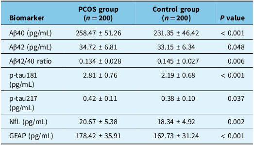
As shown in Table 2 and Figure S1, women with PCOS exhibited higher plasma Aβ40 (258.47 ± 51.26 pg/mL vs 231.35 ± 46.42 pg/mL, P < 0.001) and slightly elevated Aβ42 (34.72 ± 6.81 vs 33.15 ± 6.34 pg/mL, P = 0.048), resulting in a reduced Aβ42/40 ratio (0.134 ± 0.028 vs 0.145 ± 0.027, P = 0.006). Plasma p-tau181 levels were significantly higher in the PCOS group (2.81 ± 0.76 vs 2.19 ± 0.68 pg/mL, P < 0.001), while p-tau217 showed significant increase (0.42 ± 0.11 vs 0.38 ± 0.10 pg/mL, P = 0.037). Both NfL (20.67 ± 5.38 vs 18.34 ± 4.92 pg/mL, P = 0.002) and GFAP (178.42 ± 35.91 vs 162.73 ± 31.24 pg/mL, P < 0.001) were also elevated in PCOS participants.
Peripheral Alzheimer’s disease-related biomarkers in the PCOS and control groups

Note: Data are presented as mean ± SD. P values were derived from independent-samples t-tests. After adjusting for age, BMI, and ApoE ϵ4 genotype, all group differences remained statistically significant.
Aβ = β-amyloid; Aβ40 = amyloid-β 1–40; Aβ42 = amyloid-β 1–42; Aβ42/40 = Aβ42-to-Aβ40 ratio; p-tau181 = phosphorylated tau protein at threonine-181; p-tau217 = phosphorylated tau protein at threonine-217; NfL = neurofilament light chain; GFAP = glial fibrillary acidic protein.
Correlation analyses among biomarkers, insulin resistance and inflammatory factors
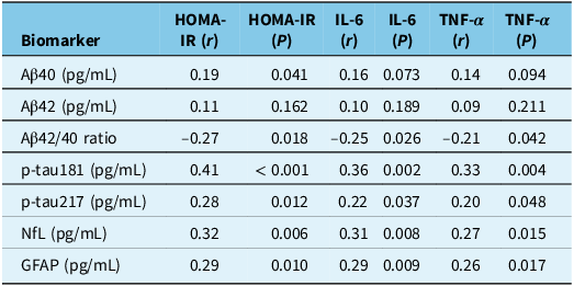
As summarized in Table 3 and Figure S2, after adjustment for age and BMI, plasma p-tau181 levels were positively correlated with HOMA-IR (r = 0.41, P < 0.001), IL-6 (r = 0.36, P = 0.002) and TNF-α (r = 0.33, P = 0.004). p-tau217 showed correlations with HOMA-IR (r = 0.28, P = 0.012). In contrast, the Aβ42/40 ratio was inversely correlated with HOMA-IR (r = –0.27, P = 0.018) and IL-6 (r = –0.25, P = 0.026). Both NfL and GFAP were positively associated with inflammatory cytokines, particularly IL-6 (r = 0.31 and 0.29, P < 0.01).
Correlations between insulin resistance, inflammatory markers and AD-related biomarkers

Note: Correlation coefficients (r) were computed using Pearson’s correlation for normally distributed variables and Spearman’s rank correlation otherwise. All analyses were adjusted for age and BMI.
Aβ = β-amyloid; Aβ40 = amyloid-β 1–40; Aβ42 = amyloid-β 1–42; Aβ42/40 = Aβ42-to-Aβ40 ratio; p-tau181 = phosphorylated tau protein at threonine-181; p-tau217 = phosphorylated tau protein at threonine-217; NfL = neurofilament light chain; GFAP = glial fibrillary acidic protein.
Multivariate regression analyses
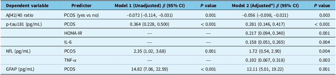
As presented in Table 4, unadjusted linear regression models (Model 1) showed that PCOS status was significantly associated with lower Aβ42/40 ratio (β = –0.072, 95% CI –0.114 to –0.031, P = 0.001) and higher p-tau181 (β = 0.364, 95% CI 0.228–0.500, P < 0.001), NfL (β = 2.35, 95% CI 1.02–3.68, P = 0.001) and GFAP (β = 14.82, 95% CI 7.06–22.59, P < 0.001). After further adjustment for HOMA-IR, IL-6 and TNF-α (Model 2), the magnitude of the PCOS effect was significant for all biomarkers. In the fully adjusted model, HOMA-IR (β = 0.217, 95% CI 0.094–0.340, P = 0.001) and IL-6 (β = 0.158, 95% CI 0.051–0.265, P = 0.004) emerged as independent predictors of p-tau181, while TNF-α was an independent predictor of NfL (β = 0.192, 95% CI 0.067–0.318, P = 0.003).
Multivariate linear regression models for AD-related biomarkers

Note: *Model 2 adjusted for age, BMI, ApoE ϵ4 status, HOMA-IR, IL-6, and TNF-α.β coefficients represent standardized effects. Significant associations remained after full adjustment, suggesting that insulin resistance and inflammation partially mediate but do not fully explain the observed elevations in AD-related biomarkers among PCOS participants.
Aβ = β-amyloid; Aβ40 = amyloid-β 1–40; Aβ42 = amyloid-β 1–42; Aβ42/40 = Aβ42-to-Aβ40 ratio; p-tau181 = phosphorylated tau protein at threonine-181; p-tau217 = phosphorylated tau protein at threonine-217; NfL = neurofilament light chain; GFAP = glial fibrillary acidic protein.
Logistic regression analyses for insulin resistance and p-tau181
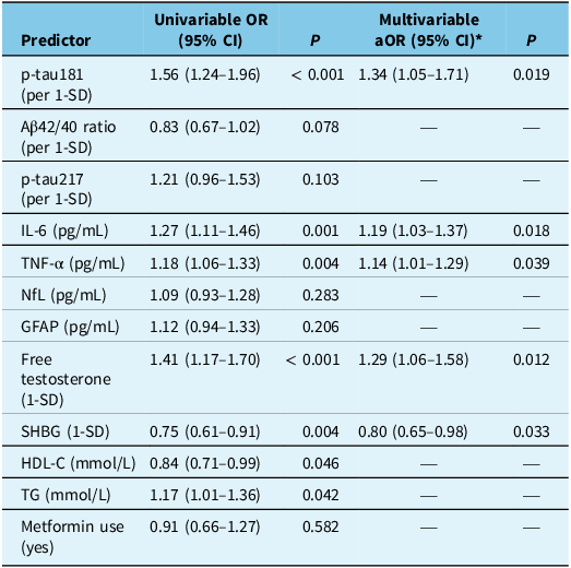
In the univariable logistic regression (Table 5), higher plasma p-tau181 levels were strongly associated with insulin-resistance (IR) status (OR = 1.56, 95% CI 1.24–1.96, P < 0.001). Inflammatory markers IL-6 (OR = 1.27, 95% CI 1.11–1.46, P = 0.001) and TNF-α (OR = 1.18, 95% CI 1.06–1.33, P = 0.004) were also positively correlated with IR. Endocrine variables, including Free T (OR = 1.41, 95% CI 1.17–1.70, P < 0.001) and SHBG (OR = 0.75, 95% CI 0.61–0.91, P = 0.004), demonstrated the expected opposite trends, indicating that androgen excess and low SHBG were linked to higher odds of IR. In contrast, Aβ42/40 ratio, p-tau217, NfL, GFAP, lipid indices and metformin use were not significantly associated (P > 0.05).
Univariable and multivariable logistic regression for insulin resistance (HOMA-IR ≥ 2.5)

Note: Univariable analyses were performed for all candidate variables; only those with POR = odds ratio; aOR = adjusted odds ratio; CI = confidence interval.
Aβ = β-amyloid; Aβ40 = amyloid-β 1–40; Aβ42 = amyloid-β 1–42; Aβ42/40 = Aβ42-to-Aβ40 ratio; p-tau181 = phosphorylated tau protein at threonine-181; p-tau217 = phosphorylated tau protein at threonine-217; NfL = neurofilament light chain; GFAP = glial fibrillary acidic protein.
In the multivariable model adjusted for age, BMI, education and ApoE ϵ4 status, variables that were significant in the univariable analysis remained independent predictors of IR. Specifically, p-tau181 (aOR = 1.34, 95% CI 1.05–1.71, P = 0.019), IL-6 (aOR = 1.19, 95% CI 1.03–1.37, P = 0.018), TNF-α (aOR = 1.14, 95% CI 1.01–1.29, P = 0.039), Free T (aOR = 1.29, 95% CI 1.06–1.58, P = 0.012) and SHBG (aOR = 0.80, 95% CI 0.65–0.98, P = 0.033) remained statistically significant.
Mediation analyses
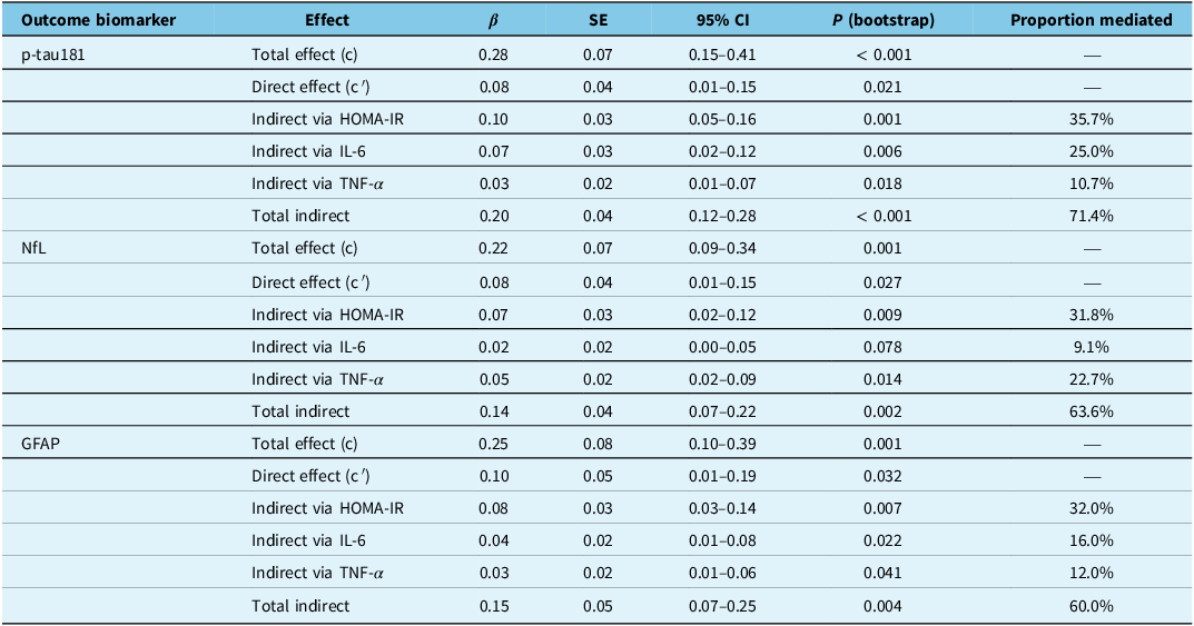
The mediation analyses examined whether insulin resistance and systemic inflammation mediated the associations between PCOS status and AD-related biomarkers (Table 6). The standardized indirect effect via HOMA-IR was β = 0.10 (95% CI 0.05–0.16, P = 0.001), accounting for approximately 36% of the total association. The indirect effects through IL-6 (β = 0.07, 95% CI 0.02–0.12, P = 0.006) and TNF-α (β = 0.03, 95% CI 0.01–0.07, P = 0.018) explained additional 25% and 11% of the total effect, respectively. Overall, about 71% of the PCOS–p-tau181 association was mediated jointly by HOMA-IR and inflammation, indicating a robust metabolic–inflammatory pathway underlying tau phosphorylation. For NfL, the total indirect effect (β = 0.14, 95% CI 0.07–0.22, P = 0.002) accounted for approximately 64% of the total association, with the largest contribution from HOMA-IR (β = 0.07, 95% CI 0.02–0.12, P = 0.009) and TNF-α (β = 0.05, 95% CI 0.02–0.09, P = 0.014). The IL-6 pathway showed only a marginal effect (P = 0.078). For GFAP, combined mediators explained about 60% of the total effect. The indirect effect through HOMA-IR (β = 0.08, 95% CI 0.03–0.14, P = 0.007) was dominant, followed by IL-6 (β = 0.04, P = 0.022) and TNF-α (β = 0.03, P = 0.041).
Mediation analysis of the association between PCOS and AD-related biomarkers via HOMA-IR and inflammatory pathways

Note: Standardized coefficients (β) are reported. P values and 95% confidence intervals were obtained using 5000-sample bias-corrected bootstrap estimation.Parallel mediation model included HOMA-IR, IL-6, and TNF-α as mediators, adjusted for age, BMI, education, and ApoE ϵ4 status.Model fit indices were satisfactory (CFI > 0.95, TLI > 0.95, RMSEA < 0.05).Proportion mediated = indirect / total effect × 100%.
Sensitivity and subgroup analyses
Sensitivity analyses showed that the association between p-tau181 and IR remained significant after excluding participants using metformin or oral contraceptives (aOR = 1.33, 95% CI 1.04–1.70, P = 0.024). As summarized in Supplementary Table S3, the positive association between p-tau181 and IR was stronger among obese women (BMI ≥ 25 kg/m2), ApoE ϵ4 carriers, and participants with higher Free T levels, whereas no significant associations were observed in their corresponding comparison groups.
Discussion
This study identified distinct AD-related biomarker alterations in women with PCOS. Compared with controls, PCOS participants showed a decreased Aβ42/40 ratio and elevated levels of p-tau181, NfL and GFAP, indicating impaired amyloid clearance, tau hyperphosphorylation and neuronal injury. Insulin resistance (HOMA-IR) and inflammatory cytokines (IL-6, TNF-α) were significantly increased and correlated with these biomarkers, suggesting metabolic–inflammatory involvement in AD-like pathology. Mediation analysis confirmed that insulin resistance and inflammation partially mediated the link between PCOS and AD biomarker abnormalities, while hyperandrogenism further amplified these effects. Collectively, the findings indicate that chronic metabolic and inflammatory dysregulation in PCOS may contribute to early neurodegenerative processes relevant to AD.
Despite growing recognition of the metabolic–neurological interface, few studies have directly examined AD-related biomarker profiles in women with PCOS. Reference Motafeghi, Akbarzadeh and Talebi22,Reference Jiang, Xu and Chen23 Most prior investigations have focused on metabolic or reproductive outcomes, leaving a critical gap in understanding how systemic insulin resistance, chronic inflammation and hormonal imbalance may translate into early neurodegenerative vulnerability. Reference Matuszewski, Tomaszek and Szklarz24,Reference Liu, Zhang, Fan, Bai, Zhang and Wang25 Recent epidemiological studies also suggest increased cognitive vulnerability in women with PCOS. Reference Showkath, Sinha, Ghate, Agrawal, Mandal and Sinha26–Reference Abdollahi, Mirghafourvand, Babapour and Mohammadi30 Our biomarker findings complement these data by providing molecular evidence of early AD-related alterations in metabolically affected PCOS populations.
Most previous studies on PCOS have primarily focused on its reproductive and metabolic abnormalities, whereas few have examined neurodegenerative mechanisms or AD-related biomarkers in this population. Reference Xu, Liu and Wang31–Reference Shi, Yu, Li, Wu and Luo35 To our knowledge, the present study is among the first to systematically evaluate peripheral AD-associated markers – including Aβ42/40 ratio, p-tau181, NfL and GFAP – in women with PCOS. Our findings are consistent with prior animal and clinical studies demonstrating that metabolic and inflammatory dysregulation can promote amyloid accumulation, tau hyperphosphorylation and neuronal injury. Reference Shao, Zhang, Xu, Pan and Chen36–Reference Ramasubbu and Devi Rajeswari40 Experimental evidence has shown that insulin resistance and chronic inflammation activate GSK3β signaling and oxidative stress pathways, leading to AD-like neuropathological changes. Reference Riachi, Khalife, Kedzia and Niechcial38,Reference Pszczolowska, Walczak and Miskow41 Similarly, population-based research has suggested that women with PCOS are at increased risk of cognitive impairment and dementia, supporting the existence of a shared metabolic–inflammatory–hormonal axis underlying both disorders. Reference Sabayan, Foroughinia, Borhani Haghighi and Mowla37,Reference Riachi, Khalife, Kedzia and Niechcial38,Reference Petrillo, Battipaglia, Virmani, Genazzani and Genazzani42,Reference Petrie43 By integrating biochemical evidence with clinical context, our study provides molecular-level support for the hypothesis that PCOS may represent a prodromal or at-risk state for neurodegenerative processes related to AD. These results extend the current understanding of PCOS beyond reproductive and metabolic dysfunction, highlighting its potential relevance to long-term brain health.
Early identification of PCOS subtypes with pronounced insulin resistance and inflammation may enable timely interventions to reduce neurocognitive risk. Our findings underscore the value of integrating metabolic and neurological assessment in PCOS management and suggest that improving insulin sensitivity and lowering systemic inflammation could offer downstream neuroprotective benefits. Longitudinal and mechanistic studies are still needed to clarify causal pathways and refine therapeutic targets.
The mediation findings clarify how metabolic–inflammatory disturbances may drive AD-related biomarker changes in PCOS. Insulin resistance can impair neuronal insulin signaling and activate GSK3β, promoting tau phosphorylation – consistent with the strong indirect effect of HOMA-IR on p-tau181. Reference Ghazeeri, Fakih, Abbas, Harajly and Awwad28-Reference Abdollahi, Mirghafourvand, Babapour and Mohammadi30 Elevated IL-6 and TNF-α may further intensify oxidative stress, microglial activation and impaired Aβ clearance, helping explain reductions in the Aβ42/40 ratio and increases in NfL and GFAP. Reference Showkath, Sinha, Ghate, Agrawal, Mandal and Sinha26,Reference Kumari, Manna, Marwah, Ahluwalia and Panwar27 That these mediators accounted for over 60–70% of the associations suggests that downstream metabolic stress and inflammation, rather than PCOS status itself, are major contributors to the early neurodegenerative signals observed. This provides a coherent mechanistic framework for interpreting the indirect pathways identified in our analyses.
Multiple interrelated mechanisms may explain the association between PCOS and Alzheimer’s-related pathology. First, insulin resistance – a core feature of PCOS – impairs neuronal insulin signaling, activates GSK3β, promotes tau hyperphosphorylation and reduces Aβ clearance, consistent with the “type 3 diabetes” hypothesis of AD. Reference Jiang, Xu and Chen23,Reference Kong, Yu, Wan, Gao and Zhang44,Reference Kiyama45 Second, chronic low-grade inflammation, characterized by elevated IL-6, TNF-α and hs-CRP, promotes oxidative stress and microglial activation, accelerating amyloid deposition and neuronal injury. Reference Yang, Duan, Luo and Tao P46–Reference Plantone, Pardini and Manco49 This supports the concept of a peripheral–central inflammatory continuum linking systemic inflammation to neurodegeneration. Reference Wang and Sun47,Reference Plantone, Pardini and Manco49–Reference Ng, Tam and Zhang51 Third, obesity and lipid dysregulation commonly seen in PCOS may further exacerbate oxidative stress, enhancing Aβ aggregation and tau pathology. Reference Raza, Saleem and Imran52–Reference Ak, Nilofar and Bahadirli56 Adipokine imbalance and endocrine inflammation create a self-reinforcing “obesity–inflammation–neurodegeneration” loop that amplifies cognitive vulnerability. Reference Zhao, Liu and Hua57 Finally, sleep disturbances, particularly obstructive sleep apnea (OSA), frequently co-occur with PCOS and contribute to intermittent hypoxia and glymphatic dysfunction, thereby impairing Aβ clearance and accelerating neurodegenerative processes. Reference Sam and Tasali58–Reference Cavailles, Andrews, Leng, Chatterjee, Daghlas and Yaffe62 Together, these interconnected pathways highlight a complex metabolic–inflammatory network through which PCOS may predispose to early AD-like changes.
Uur findings support a model in which metabolic dysfunction and systemic inflammation jointly shape neurobiological vulnerability in PCOS. Rather than acting through a single pathway, insulin resistance, inflammatory cytokines and androgen imbalance appear to contribute additively to AD-related biomarker alterations. These insights underscore the importance of early metabolic screening and targeted interventions in PCOS – not only for reproductive and cardiometabolic health, but also for long-term brain health.
Several limitations should be acknowledged when interpreting the present findings. First, the cross-sectional design precludes causal inference regarding the directionality between metabolic–inflammatory alterations and AD-related biomarker changes. Prospective longitudinal studies are needed to determine whether insulin resistance and inflammation precede or result from early neurodegenerative processes in PCOS. Second, the study cohort was relatively homogeneous in age and geographic background, which may limit the generalizability of the findings to broader or ethnically diverse populations. Multi-center validation across different age groups and ethnicities is warranted. Third, peripheral biomarkers provide indirect evidence of neurodegenerative risk; the absence of neuroimaging or cerebrospinal fluid (CSF) data limits interpretation of central nervous system pathology. Future research integrating PET or MRI-based measures of amyloid, tau and glymphatic function with plasma biomarkers could strengthen mechanistic insights. Finally, the current analyses focused on limited metabolic and inflammatory indicators. Incorporating multi-omics approaches – such as transcriptomics, metabolomics and microbiome profiling – may help elucidate the complex molecular networks linking reproductive–metabolic dysfunction to neurodegeneration. Sleep quality and obstructive sleep apnea (OSA) – both common in PCOS – were not evaluated in this study. Because sleep disturbances and OSA can affect inflammation, insulin resistance and Aβ clearance, their omission may have introduced residual confounding. Future studies incorporating objective sleep assessments are needed to clarify the contribution of sleep-related factors to AD-related biomarker changes in PCOS. Future work should aim to develop and validate an integrative “PCOS–AD risk prediction model” combining metabolic, hormonal and neurobiological parameters, ultimately supporting early identification and precision prevention strategies in at-risk women.
Conclusion
In conclusion, this study demonstrates that women with PCOS show peripheral alterations in AD-related biomarkers, including decreased Aβ42/40 ratio and elevated p-tau181, NfL and GFAP levels. Insulin resistance and inflammation partly mediated these associations, while hyperandrogenism further enhanced their effects. These results suggest that PCOS may represent a metabolic–inflammatory state linked to early neurodegenerative vulnerability. Early identification and management of insulin resistance and systemic inflammation may help reduce long-term cognitive risk in this population.
Supplementary material
The supplementary material for this article can be found at https://doi.org/10.1017/cjn.2025.10495.
Data availability
The datasets generated and/or analyzed during the current study are available from the corresponding author upon reasonable request.
Acknowledgements
The authors sincerely thank all study participants for their invaluable contributions.
Author contributions
Bao Xing conceived and designed the study, collected clinical data and performed statistical analyses. Pang Xiaoqing contributed to data interpretation, literature review and figure preparation. Zhang Yan supervised the study, critically revised the manuscript for important intellectual content and approved the final version for submission. All authors have read and approved the final manuscript and agree to be accountable for all aspects of the work.
Funding statement
Hebei Administration of Traditional Chinese Medicine Grant No.: 2025469.
Competing interests
The authors declare that they have no competing interests.
Ethics approval and consent to participate
This study was reviewed and approved by the Institutional Review Board of Shijiazhuang Maternal and Child Health Hospital. All participants provided written informed consent prior to enrollment. The research was conducted in accordance with the Declaration of Helsinki (2013 revision) and relevant national regulations on biomedical research involving human subjects.
Consent for publication
Not applicable.
Informed consent
Written informed consent was obtained from all participants prior to enrollment in the study.