Introduction
Microtrichia are cuticular microstructures occurring on the body surface of a variety of insects (Richards and Richards Reference Richards and Richards1979) and serve mainly mechanical functions such as the generation of frictional or adhesive forces (Gorb Reference Gorb1997, Reference Gorb1998, Reference Gorb1999; Perez Goodwyn and Gorb Reference Perez Goodwyn and Gorb2004). Microtrichial structures in the Heteroptera were described in the wing-locking mechanisms of aquatic bugs (Gorb and Perez Goodwyn Reference Gorb and Perez Goodwyn2003; Perez Goodwyn and Gorb Reference Perez Goodwyn and Gorb2003). The microsculpture of the frena surface in selected Heteroptera species was presented by Štys (Reference Štys1998) and Weirauch and Cassis (Reference Weirauch and Cassis2009).
Microtrichia of the wing coupling mechanism were described in a study on the morphology of mesothoracic wings in the Afrotropical Scutelleridae (Czaja Reference Czaja2012). Microtrichial structures are also visible on the dorsal and ventral surfaces of the corial areas of the forewings.
The aim of this study is to present the morphological variety of microtrichial patterns observed in different genera of the Afrotropical Scutelleridae (Hemiptera) and to determine whether they are of taxonomic value for this group of true bugs. New morphological data are important in testing and constructing the classification system within the Scutelleridae, where subfamiliar and tribal relationships remain controversial. A variety of suprageneric arrangements in the Scutelleridae have been proposed in the literature. An important recent study of Cassis and Vanags (Reference Cassis and Vanags2006) divided the Australian Scutellerids into five subfamilies: Elvisurinae, Odntotarsinae, Pachycorinae, Scutellerinae, and Tectocorinae. Göllner-Scheiding (Reference Göllner-Scheiding2006) recognised seven subfamilies in the catalogue of the Palaearctic Region: Elvisurinae, Eurygastrinae, Odontoscelinae, Odontotarsinae, Pachycorinae, Scutellerinae, and Tectocorinae. Carapezza (Reference Carapezza2009) distinguished the subfamily Hoteinae for two Afrotropical genera (Hotea Amyot and Serville, 1843 and Deroplax Mayr, 1864), which follows the classification of Tsai et al. (Reference Tsai, Redei, Yeh and Yang2011) in recognising eight subfamilies. Fischer (Reference Fischer2001) supported the monophyletic character of the Sphaerocorinae, Pachycorinae, and Elvisurinae. These proposed classifications should be tested by phylogenetic analyses of the entire family. Morphological data supported by descriptions of new features such as microtrichial wing patterns may provide additional evidence supporting suprageneric relationships within the Scutelleridae and formulate a homogenous classification system within this family.
Material and methods
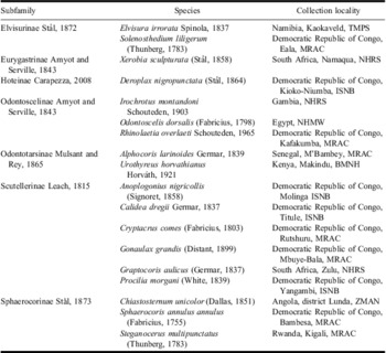
Representatives of 19 genera of Afrotropical Scutelleridae were examined. The classification within the family followed Fischer (Reference Fischer2001) and Göllner-Scheiding (Reference Göllner-Scheiding2006), with recent modifications by Carapezza (Reference Carapezza2009). Sphaerocorinae is treated in this study as a separate subfamily. The material examined is presented in Table 1.
Material examined.

The range of microtrichial areas was observed and drawn under the standard optical equipment (Olympus CX-41 light microscope; Olympus Corporation, Tokyo, Japan). The hemelytra of air-dried specimens were cleaned in 70% alcohol, coated with gold-palladium (20 mA) and examined with a scanning electron microscope (Hitachi S-4700; Hitachi, Tokyo, Japan, with Noran Vantage microanalysis system, Noran Instruments, Naarden, The Netherlands) at 20 kV.
The ground-plan and terminology for the forewing of the Heteroptera follows Betts (Reference Betts1986) and Wootton and Betts (Reference Wootton and Betts1986). The terminology of microtrichial structures is that of Gorb and Perez Goodwyn (Reference Gorb and Perez Goodwyn2003).
The following abbreviations were used in the text and figures: C – corium, MC – mesocorium, EC – exocorium, M – membrane, MA – microtrichial area, FM – flattened microtrichia.
The following institutional collections provided the study material (with acronyms used in the text): BMNH – The Natural History Museum, London, United Kingdom; ISNB – Institut Royal des Sciences Naturelles de Belgique, Brussels, Belgium; MNHN – Muséum National d'Histoire Naturelle, Paris, France; MRAC – Museé Royal de l'Afrique Centrale, Tervuren, Belgium; NHMW – Naturhistorisches Museum Wien, Austria; NHRS – Naturhistoriska Riksmuseet, Stockholm, Sweden; TMPS – Transvaal Museum, Pretoria, South Africa; ZMAN – Zoölogisch Museum, University of Amsterdam, Amsterdam, The Netherlands.
Results
The following features of the microtrichial sculpture were used to investigate morphological diversification of these structures:
1. Range of microtrichial fields present on the ventral corial surface.
2. Shape of microtrichia present on the ventral corial surface.
3. Relative sizes of microtrichia present on the ventral corial surface.
4. Density of microtrichia present on the ventral corial surface.
5. Range of microtrichial fields present on the dorsal corial surface.
6. Shape of microtrichia present on the dorsal corial surface.
7. Relative sizes of microtrichia present on the dorsal corial surface.
8. Density of microtrichia present on the dorsal corial surface.
Description
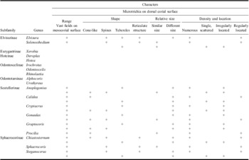
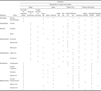
Microtrichial structures are different in shape (cone-like, tubercles, spines, tile-like, dome-like, reticulate structures with protruding margins with spines and tubercles) and size (similar and distinctly different in size). The location and density of microtrichia within the corial area are variable (vast fields on the dorsal and ventral corial surfaces, basal sclerotised parts of mesocorium, ventral convexity of corium, microtrichia regularly and irregularly located). Descriptions of the variability of these structures of 19 Scutelleridae species are given below. Summarised results are presented in Tables 2 and 3.
Summarised characters of microtrichial patterns on dorsal corial surface.

Summarised characters of microtrichial patterns on ventral corial surface

Subfamily Elvisurinae
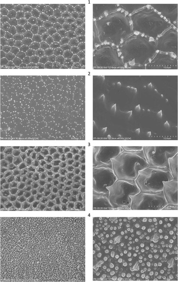
Elvisura irrorata (Figs. 1–2)
Microtrichia arranged in vast fields on the dorsal and ventral mesocorial surfaces; different in size and shape; dorsal mesocorial surface with spines and tubercles arranged in near regular reticulate structure with protruding margins; ventral fields with small tubercles, larger spines and cone-like structures forming irregular adjoining rings.
Microtrichia of dorsal and ventral corial surface. Elvisura irrorata Spinola: 1, dorsal surface of the mesocorium (left), the same, enlarged (right); 2, ventral surface of the mesocorium (left), the same, enlarged (right). Solenosthedium liligerum (Thunberg); 3, dorsal surface of the mesocorium (left), the same, enlarged (right); 4, ventral surface of the mesocorium (left), the same, enlarged (right).

Solenosthedium liligerum (Figs. 3–4)
Microtrichia arranged in vast fields on the dorsal and ventral mesocorial surfaces; dorsal surface with spines similar in size forming near regular reticulate structure with protruding margins and single small tubercles, similar in size, irregularly located; ventral mesocorial surface densely covered by numerous small tubercles different in size and scattered, larger cone-like structures crispated basally, almost regularly located within fields.
Subfamily Eurygastrinae
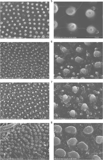
Xerobia sculpturata (Figs. 5, 29)
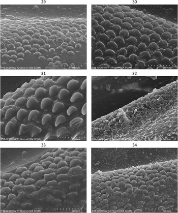
Microtrichia visible on ventral sclerotised parts of corium; sclerotised basal parts of mesocorium with very regularly located, cone-like structures similar in size and single, scattered small tubercles; microtrichia of ventral convexity of exocorium distinctive in medial part, numerous, flattened, tile-like with rounded apices, similar in size, nearly regularly located.
Subfamily Hoteinae
Deroplax nigropunctata (Figs. 6, 30)
Microtrichia visible on ventral sclerotised parts of corium; basal part of mesocorium with numerous cone-like structures, similar in size, almost regularly located, and small scattered tubercles, differing in size; microtrichia of ventral convexity of exocorium distinctive in medial part, numerous, flattened, tile-like with rounded apices, similar in size, nearly regularly located.
Hotea acuta (Fig. 7)
Microtrichia visible on ventral sclerotised parts of corium; basal part of mesocorium with numerous cone-like structures, similar in size, almost regularly located, and small scattered tubercles differing in size; microtrichia of ventral convexity of exocorium distinctive in medial part, numerous, flattened, tile-like with rounded apices, similar in size, nearly regularly located.
Microtrichia of ventral corial surface (left), the same enlarged (right). 5, Xerobia sculpturata (Stål); 6, Deroplax nigropunctata (Stål); 7, Hotea acuta Stål; 8, Odontoscelis dorsalis (Fabricius).

Subfamily Odontoscelinae
Irochrotus montandoni (Fig. 31)
Microtrichia visible on ventral sclerotised parts of corium; basal part of mesocorium with irregular, scattered larger dome-like structures different in size, small tubercles and scattered spines, differing in size; microtrichia of ventral convexity of exocorium distinctive in medial part, numerous, flattened, tile-like with rounded apices, similar in size, nearly regularly located.
Ventral surface of the mesocorium. 29, Xerobia sculpturata (Stål); 30, Deroplax nigropunctata (Stål); 31, Irochrotus montandoni Schouteden; 32, Rhinolaetia overlaeti Schouteden; 33, Alphocoris larinoides Germar; 34, Urothyreus horvathianus Horváth.

Odontoscelis dorsalis (Fig. 8)
Microtrichia visible on ventral sclerotised parts of corium; basal part of mesocorium with numerous larger dome-like structures, similar in size, almost regularly located, and single, scattered small tubercles; microtrichia of ventral convexity of exocorium distinctive in medial part, numerous, flattened, tile-like with rounded apices, similar in size, nearly regularly located.
Rhinolaetia overlaeti (Figs. 9, 32)
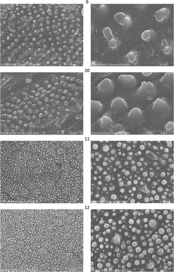
Microtrichia visible on ventral sclerotised parts of corium; basal part of mesocorium with numerous, cone-like structures, similar in size, almost regularly located, and single, scattered small tubercles; microtrichia of ventral convexity of exocorium distinctive in medial part, numerous, flattened, tile-like with rounded apices, similar in size, nearly regularly located.
Subfamily Odontotarsinae
Alphocoris larinoides (Fig. 33)
Microtrichia visible on ventral sclerotised parts of corium; basal part of mesocorium with numerous dome-like and tile-like structures, differing in size, almost regularly located, and single, scattered small tubercles; microtrichia of ventral convexity of exocorium distinctive in medial part, numerous, flattened, tile-like with rounded apices, similar in size, nearly regularly located.
Urothyreus horvathianus (Figs. 10, 34)
Microtrichia visible on ventral sclerotised parts of corium; basal part of mesocorium with microtrichia different in shape: numerous, larger dome-like and cone-like structures, different in size, almost regularly located and single, scattered small tubercles, differing in size; microtrichia of ventral convexity of exocorium distinctive in medial part, numerous, flattened, tile-like with rounded and dentated apices, similar in size, nearly regularly located.
Subfamily Scutellerinae
Anoplogonius nigricollis (Figs. 11–12)
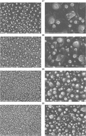
Microtrichia arranged in vast fields on dorsal and ventral mesocorial surfaces; both dorsal and ventral surface densely covered by numerous, small tubercles, differing in size and scattered larger spines and cone-like structures, similar in size, irregularly located within fields.
Microtrichia of dorsal and ventral corial surface. Rhinolaetia overlaeti Schouteden; 9, ventral surface of the mesocorium (left), the same, enlarged (right). Urothyreus horvathianus Horváth; 10, ventral surface of the mesocorium (left), the same, enlarged (right); Anoplogonius nigricollis (Signoret); 11, dorsal surface of the mesocorium (left), the same, enlarged (right); 12, ventral surface of the mesocorium (left), the same, enlarged (right).

Calidea dregii (Figs. 13–14)
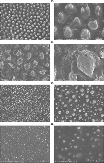
Microtrichia arranged in vast fields occurring on dorsal and ventral mesocorial surfaces; dorsal fields with regularly located, numerous cone-like structures, differing in size and single small tubercles; ventral mesocorial surface with larger, irregular dome-like structures, different in size; small tubercles absent.
Microtrichia of dorsal and ventral corial surface. Calidea dregii Germar: 13, dorsal surface of the mesocorium (left), the same, enlarged (right); 14, ventral surface of the mesocorium (left), the same, enlarged (right). Cryptacrus comes (Fabricius); 15, dorsal surface of the mesocorium (left), the same, enlarged (right); 16, ventral surface of the mesocorium (left), the same, enlarged (right).

Cryptacrus comes (Figs. 15–16)
Microtrichia arranged in vast fields on dorsal and ventral mesocorial surfaces; both dorsal and ventral mesocorial surfaces with numerous small tubercles, differing in size and scattered, larger spines and cone-like structures different in size, irregularly located within fields.
Gonaulax grandis (Figs. 17–18)
Microtrichia arranged in vast fields on dorsal and ventral mesocorial surfaces; both dorsal and ventral surfaces covered by numerous, small tubercles, differing in size, spines, and larger cone-like structures crispated basally, almost regularly located within fields, similar in size.
Microtrichia of dorsal and ventral corial surface. Gonaulax grandis (Distant): 17, dorsal surface of the mesocorium (left), the same, enlarged (right); 18, ventral surface of the mesocorium (left), the same, enlarged (right). Graptocoris aulicus (Germar); 19, dorsal surface of the mesocorium (left), the same, enlarged (right); 20, ventral surface of the mesocorium (left), the same, enlarged (right).

Graptocoris aulicus (Figs. 19–20)
Microtrichia arranged in vast fields on dorsal and ventral mesocorial surfaces; both dorsal and ventral surfaces densely covered by numerous small tubercles, differing in size and larger cone-like structures and spines irregularly located within fields, differing in size.
Procilia morgani (Figs 21–22)
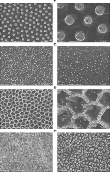
Microtrichia arranged in vast fields on dorsal and ventral mesocorial surfaces; dorsal fields with numerous, regularly located larger cone-like structures, similar in size, small tubercles absent; ventral surface densely covered by numerous small tubercles, differing in size.
Microtrichia of dorsal and ventral corial surface. Procilia morgani (White): 21, dorsal surface of the mesocorium (left), the same, enlarged (right); 22, ventral surface of the mesocorium (left), the same, enlarged (right). Chiastosternum unicolor (Dallas): 23, dorsal surface of the mesocorium (left), the same, enlarged (right); 24, ventral surface of the mesocorium (left), the same, enlarged (right).

Subfamily Sphaerocorinae
Chiastosternum unicolor (Figs. 23–24)
Microtrichia arranged in vast fields on dorsal and ventral mesocorial surfaces; dorsal surface with spines and cone-like structures, differing in size, forming near regular reticulate structure with protruding margins and small scattered tubercles; ventral mesocorial surface densely covered by numerous small tubercles, differing in size and scattered, irregular spines.
Sphaerocoris annulus annulus (Figs. 25–26)
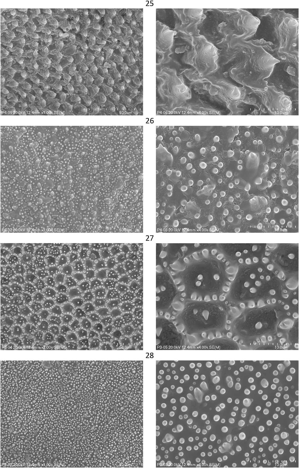
Microtrichia arranged in vast fields on dorsal and ventral mesocorial surfaces; dorsal surface with distinctive wide spines forming regular reticulate structure with protruding margins; ventral mesocorial surface densely covered by numerous small tubercles, differing in size and larger, numerous cone-like structures, crispated basally, almost regularly located within fields, similar in size.
Microtrichia of dorsal and ventral corial surface. Sphaerocoris annulus annulus (Fabricius): 25, dorsal surface of the mesocorium (left), the same, enlarged (right); 26, ventral surface of the mesocorium (left), the same, enlarged (right). Steganocerus multipunctatus (Thunberg): 27, dorsal surface of the mesocorium (left), the same, enlarged (right); 28, ventral surface of the mesocorium (left), the same, enlarged (right).
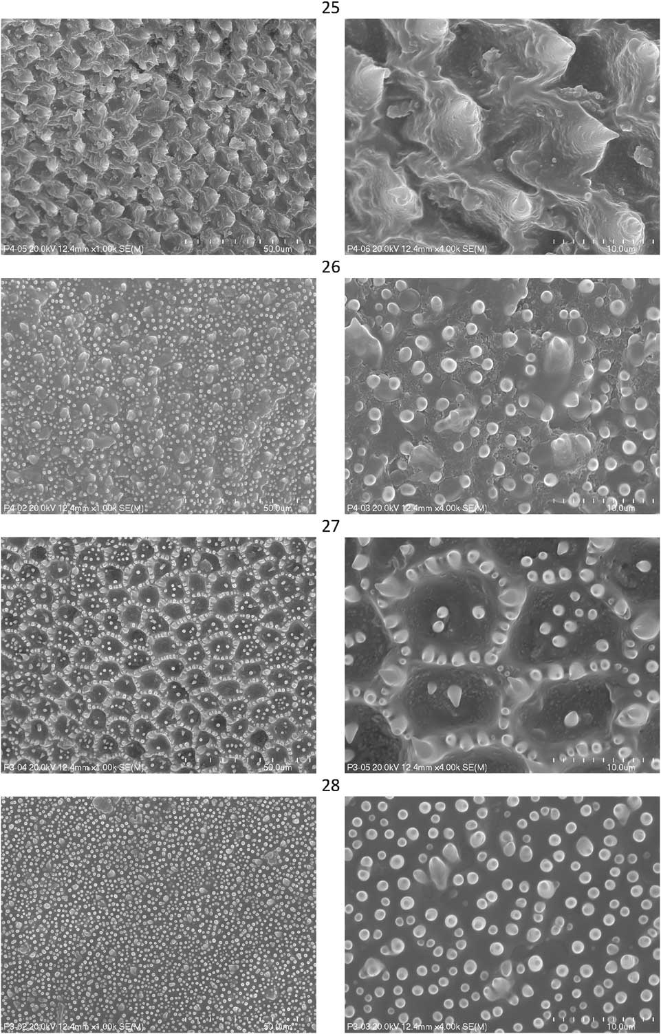
Steganocerus multipunctatus (Figs. 27–28)
Microtrichia arranged in vast fields on dorsal and ventral mesocorial surfaces; dorsal fields with spines differing in size, forming nearly regular reticulate structure with protruding margins and small scattered tubercles; ventral mesocorial surface densely covered by numerous small tubercles and larger scattered cone-like structures, differing in size.
Summary of the results
Four types of the microtrichial sculpture in Afrotropical Scutelleridae were identified in this study:
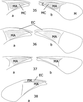
I type (Fig. 35)
Types of microtrichial patterns of mesothoracic wings in Scutelleridae: 35, I type, Elvisurinae: Elvisura, Solenosthedium; Sphaerocorinae: Chiastosternum, Sphaerocoris, Steganocerus; 36, II type, Scutellerinae: Anoplogonius, Cryptacrus, Gonaulax, Graptocoris; 37, III type, Scutellerinae: Calidea, Procilia; 38, IV type, Eurygastrinae: Xerobia; Hoteinae: Deroplax, Hotea; Odontoscelinae: Irochrotus, Odontoscelis, Rhinolaetia; Odontotarsinae: Alphocoris, Urothyreus. Ventral surface of corium (A); dorsal surface of corium (B).

Distinctive characters: Microtrichial fields well developed on dorsal and ventral corial surfaces; dorsal fields with microtrichia arranged in near regular reticulate structures with protruding margins, ventral surface with microtrichia, differing in shape and size (small tubercles, larger spines and cone-like structures).
Genera included: Elvisurinae: Elvisura, Solenosthedium, Sphaerocorinae: Chiastosternum, Sphaerocoris, Steganocerus.
II type (Fig. 36)
Distinctive characters: Microtrichial fields well developed on dorsal and ventral mesocorial surfaces; both dorsal and ventral fields with microtrichia differing in shape and size (numerous, small tubercles, larger, scattered spines and cone-like structures); dorsal surface without reticulate microtrichial structures.
Genera included: Scutellerinae: Anoplogonius, Cryptacrus, Gonaulax, Graptocoris.
III type (Fig. 37)
Distinctive characters: Microtrichial fields well developed on dorsal and ventral mesocorial surfaces; dorsal fields with regularly located cone-like microtrichia; ventral fields with microtrichia differing in shape and size (small tubercles, larger spines and cone-like and dome like structures).
Genera included: Scutellerinae: Calidea, Procilia
IV type (Fig. 38)
Distinctive characters: Microtrichial fields developed only on ventral sclerotised parts of mesocorial surface with microtrichia different in shape and size: larger dome-like and cone-like structures almost regularly located, small, scattered tubercles; microtrichia of ventral convexity of exocorium distinctive in medial part, tile-like with rounded apices.
Genera included: Eurygastrinae: Xerobia; Hoteinae: Deroplax, Hotea; Odontoscelinae: Irochrotus, Odontoscelis, Rhinolaetia; Odontotarsinae: Alphocoris, Urothyreus.
Discussion
The microtrichial sculpture on the corial surface is observed in all genera studied, but the characters of size, shape, density, and location varied.
Type IV is the most frequently occurring microtrichial sculpture observed in nine genera representing the subfamilies Eurygastrinae, Hoteinae, Odontoscelinae, and Odontotarsinae. It is characterised by a reduced microtrichial sculpture on the dorsal surface of the mesocorium and a reduced range of microtrichial fields on the ventral mesocorial surface. This is associated with morphological features of the mesothoracic wings in these genera. A considerable part of the mesocorium here is not heavily sclerotised and it is regarded as an apomorphic state (Fischer Reference Fischer2001). If we assume the degree of sclerotisation of the corium is correlated with the presence of the microtrichial sculpture in the Scutelleridae, then the reduced microtrichial sculpture can be regarded as an apomorphy.
The mesocorium of the remaining genera representing the subfamilies Elvisurinae, Scuttellerinae, Sphaerocorinae (types I, II, III) is heavily sclerotised. This character is regarded as a plesiomorphic state (Fischer Reference Fischer2001). Extended microtrichial fields are observed both on the dorsal and ventral mesocorial surfaces. Type I occurs in five genera. Distinctive characters of this type are microtrichial fields well developed on the dorsal and ventral corial surfaces and dorsal microtrichia arranged in regular reticulate structures with protruding margins. Type II is present in four genera of the subfamily Scutellerinae. Microtrichia of dorsal and ventral fields are different in size and shape: numerous, small tubercles, larger, scattered spines, and cone-like structures. Type III was found in species representing only two genera of the subfamily Scutellerinae, but they differ from type II, where the dorsal microtrichia are conical and regularly located.
There are several controversial points of scutellerid higher classification. The systematic position of subfamily Sphaerocorinae still remains unclear. Genera within this subfamily are either included as a tribe in the subfamily Scutellerinae (Schuh and Slater Reference Schuh and Slater1995; Göllner-Scheiding Reference Göllner-Scheiding2006; Tsai et al. Reference Tsai, Redei, Yeh and Yang2011) or are treated as a separate subfamily (Fischer Reference Fischer2001). Relationships between the subfamilies Eurygastrinae, Odontotarsinae, and Odontoscelinae are mostly undefined. McDonald (Reference McDonald1966), McDonald and Cassis (Reference McDonald and Cassis1984), Schuh and Slater (Reference Schuh and Slater1995) recognised the subfamilies Eurygastrinae and Odontotarsinae (with the tribes Eurygastrini, Odontotarsini, and Odontoscelini included within Odontotarsiane). Recently the rank of Odontoscelini has been raised to subfamily level (Göllner-Scheiding Reference Göllner-Scheiding2006; Tsai et al. Reference Tsai, Redei, Yeh and Yang2011) but this position needs to be reconsidered and verified. Hoteinae, with two genera formerly in the subfamily Pachycorinae, was reclassified by Carapezza (Reference Carapezza2009). Members of this subfamily are recognised by distinctive characters of the genital structures but their relationships to other Scutelleridae taxa should be further tested.
The results of this study indicate that Scutelleridae microtrichial sculpture of the mesocorium is diversified and can be used as a character of taxonomic importance. Based on the range, shape, size, and density of microtrichia on the ventral and dorsal corial surfaces representatives of Afrotropical genera of the Scutelleridae are divided into four groups. The subfamilies Eurygastrinane, Hoteinae, Odontoscelinae, and Odontotarsinae (included in type IV) are separated from the subfamilies Elvisurinae, Scutellerinae, and Sphaerocorinae, which are divided into three parts. This suprageneric separation is also visible in metathoracic wing venation patterns (Czaja Reference Czaja2007) and other characters associated with wing morphology (Czaja Reference Czaja2012). It may suggest a closer relationship among the subfamilies Eurygastrinae, Hoteinae, Odontoscelinae, and Odontotarsinae compared to the species of the subfamilies Elvisurinae, Sphaerocorinae, and Scutellerinae. However, these preliminary conclusions should be supported by an extended phylogenetic analysis for the entire family.
Acknowledgements
The author is grateful to Professor Jerzy Lis (Department of Biosystematics, University of Opole) for his comments on the manuscript and to Dr. Joanna Kazik (University of Lodz) and Jerome Constant (ISNB) for their linguistic corrections. Many thanks are due to Dr. Mick Web (BMNH), Dr. Dominique Pluot-Sigwalt (MNHN), Dr. Herbert Zettel (NHMW), Dr. Eliane De Coninck (MRAC), Gunvi Lindberg (NHRS), Dr. Petr Kment (NMPC), Robin Lyle (TMSA), and Dr. J.P. Duffels (ZMAN) for the loan of the material.