Introduction
In the course of a study by scanning electron microscopy with energy-dispersive spectrometry (SEM-EDS) and ore microscopy of the fragments of quartz-sulfide veins from the abandoned Boevskoe W-Be deposit, Southern Urals, Russia, we encountered a previously unknown Ag sulfotelluride. The grains of the new phase were numerous but their tiny size and completely polycrystalline nature prevented the investigation of its crystal structure. After long searching, one single 30 μm grain was found allowing the collection of powder micro-X-ray diffraction data which, along with the chemical composition, showed a perfect match between the new sulfotelluride and the synthetic phase Ag2TeS3 studied structurally by Pertlik (Reference Pertlik1997). The identity was also confirmed by means of electron back-scatter diffraction (EBSD). Due to the tiny grain size of the new sulfotelluride, some of its properties completing its description as a new mineral species were obtained on a synthetic analogue, Ag2TeS3, prepared by the authors.
The new mineral was named zavyalovite [pronounced: za vya lo vait; Russian cyrilic – завьяловит] to honour Russian mineralogist and crystallographer, Dr Evgeniy Nikolaevich Zavyalov (born 16.04.1944) in recognition of his outstanding contributions to the mineralogy, crystallography and crystal chemistry of ore minerals and, in particular, of tellurides and sulfotellurides. Dr Zavyalov is the author or co-author of 11 new minerals, including rucklidgeite PbBi2Te4 (Zavyalov and Begizov, Reference Zavyalov and Begizov1977), aleksite PbBi2Te2S2 (Lipovetskiy et al., Reference Lipovetskiy, Borodaev and Zavyalov1978), ingodite Bi2TeS (Zavyalov and Begizov, Reference Zavyalov and Begizov1981), sulphotsumoite Bi3Te2S (Zavyalov and Begizov, Reference Zavyalov and Begizov1982) and baksanite Bi6Te2S3 (Pekov et al., Reference Pekov, Zavyalov, Fedyushchenko, Shcherbachev, Borodayev and Dorokhova1996), as well as many papers and handbooks focused on ore minerals (Zavyalov and Begizov, Reference Zavyalov and Begizov1983; Zavyalov et al., Reference Zavyalov, Begizov and Fedchuk1984; Chvileva et al., Reference Chvileva, Bezsmertnaya, Spiridonov, Agroskin, Papayan, Vinogradova, Lebedeva, Zavyalov, Filimonova, Petrov, Rautian and Sveschnikova1988). Last but not least is Dr Zavyalov’s involvement as an author in the book Mineralogy of Urals: Elements, Carbides, Sulfides (Yushkin, Reference Yushkin1990). In view of this, our suggestion to name a new sulfotelluride from the Urals after him seemed particularly appropriate. The new mineral, its name and symbol (Zvv) have been approved by the Commission on New Minerals, Nomenclature and Classification of the International Mineralogical Association (IMA-CNMNC) (IMA2024-096, Kasatkin et al., Reference Kasatkin, Nestola, Vymazalová, Košek, Agakhanov and Kuznetsov2025a). The holotype specimen of zavyalovite, along with its synthetic analogue, were deposited in the systematic collection of the Fersman Mineralogical Museum of the Russian Academy of Sciences, Moscow, with the registration numbers 6214/1 and 6214/2, respectively.
Occurrence and general appearance
The new mineral occurs at the Boevskoe W-Be deposit, 35 km SW of the town of Kamensk-Uralskiy, Kaslinskiy District, Chelyabinsk Oblast, Southern Urals, Russia (56°14’44’’ N, 61°22’26’’ E). The Boevskoe deposit, known as the first W deposit in Russia, has been mined for tungsten since the end of 19th Century. At the end of the 1940s, due to the small tungsten content in the ores compared to other deposits discovered at that time, the Boevskoe deposit was classified as a small-scale mining site and abandoned. In 1957 prospecting workings resumed and led to the discovery of fluorite-muscovite greisens with beryllium mineralization but no industrial mining of Be was undertaken. Currently, the deposit consists of numerous forested ditches up to several metres deep and collapsed adits. Quartz veins with hübnerite and sulfides are confined to the subvolcanic body of metamorphosed gabbro as part of volcanic-sedimentary and carbonate-terrigenous sedimentary rocks of the Middle Devonian. The main minerals of the veins are quartz, muscovite, fluorite, beryl, hübnerite and pyrite. Subordinate minerals include phenakite, fluorapatite, microcline, albite, calcite, scheelite, stolzite, galena, sphalerite, pyrrhotite and rutile. The presence of sulfides in the ores of the deposit along with fluorite led to the formation of sulfuric and hydrofluoric acids in the oxidation zone and the active development of supergene processes to a depth of 100 m (Rundqvist and Chistyakov, Reference Rundqvist and Chistyakov1960; Zabolotnaya, Reference Zabolotnaya1978; Zoloev et al., Reference Zoloev, Levin, Mormil and Schardakova2004; Kupriyanova and Shpanov, Reference Kupriyanova and Shpanov2011; Ponomarev et al., Reference Ponomarev, Erokhin and Grigoriev2022). A more detailed description of the geology of the Boevskoe deposit was given recently by Kasatkin et al. (Reference Kasatkin, Zubkova, Škoda, Gurzhiy, Nestola, Biagioni, Agakhanov, Britvin, Plašil and Kuznetsov2025b).
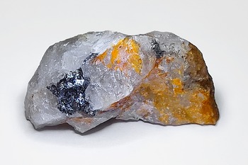
The samples containing the new mineral were collected in August 2024 by one of the authors (A.M. Kuznetsov) at the old dumps of an unnamed pit used for tungsten mining in the first half of 20th Century (Fig. 1). Macroscopically, the samples consist of fragments of a quartz vein with visible galena and sphalerite (Fig. 2). Zavyalovite occurs as numerous but usually very small anhedral grains (typically 5–15 μm with one grain found up to 30 μm across) or narrow veinlets (up to 100 × 5 μm), replacing galena or at the contact of galena and sphalerite. Both sulfides and zavyalovite are typically corroded by late anglesite (Fig. 3a–c).
Boevskoe W-Be deposit; old dumps where samples with zavyalovite were collected during August, 2024. (Photo courtesy of Pavel M. Andryushchenko).

Figure 1 Long description
The image shows a forested area with numerous trees and a rocky terrain. The trees are tall and slender, with a mix of foliage. The ground is uneven, featuring rocks and patches of moss. Sunlight filters through the canopy, creating a dappled light effect on the forest floor.
Sample Boev-14 showing sulfides (galena and sphalerite) on limonite-stained quartz. Polished sections where zavyalovite was discovered were made from samples of this and similar specimens. Size: 4 cm × 2 cm. (Photo courtesy of Maria D. Milshina).

Figure 2 Long description
The image shows a mineral sample featuring sulfides, including galena and sphalerite, on quartz. The quartz has orange limonite staining. The sample displays a mix of textures and colours, with distinct areas of sulfide deposits contrasting against the quartz matrix.
Back-scattered electron (a–c) and reflected-light (d) images showing zavyalovite (Zvv) and associated minerals: anglesite (Ang), galena (Gn) and sphalerite (Sp). Mineral symbols are after Warr (Reference Warr2021).

Figure 3 Long description
The image A shows a back-scattered electron image with zavyalovite labelled as Zvv, surrounded by anglesite (Ang) and galena (Gn). Zavyalovite is about 15 micrometres wide and 50 micrometres long. Zavyalovite is darker grey than anglesite, but not as dark as sphalerite. The image B shows zavyalovite (Zvv) of similar size between anglesite (Ang), sphalerite (Sp) and galena (Gn). The image C shows arrows pointing to small crystals of zavyalovite (Zvv) less than 10 micrometres long in anglesite (Ang) and at the contact with galena (Gn). The image D shows a reflected-light image with a roughly 20x20 micrometre light coloured zavyalovite (Zvv) grain in darker anglesite (Ang) in grey sphalerite (Sp) next to bright galena (Gn).
Other minerals also found in association include bismuth, bismutite, chalcopyrite, eskimoite, hessite, joséite-B, matildite, perite, pyrite, tellurite, zavaritskite and the new mineral boevskite (Kasatkin et al., Reference Kasatkin, Zubkova, Škoda, Gurzhiy, Nestola, Biagioni, Agakhanov, Britvin, Plašil and Kuznetsov2025b).
Synthetic analogue
The synthetic analogue of zavyalovite was prepared using an evacuated silica glass tube method in the Laboratory of Experimental Mineralogy of the Czech Geological Survey in Prague. Silver (99.99%), tellurium (99.99%) and sulfur (99.99%) were used as starting materials for the synthesis. The evacuated tube with its charge was sealed and then heated at 800°C for a week. After cooling by cold-water bath, the charge was ground into powder in acetone using an agate mortar, and mixed thoroughly to homogenize. The pulverized charge was sealed in an evacuated silica-glass tube again and reheated at 300°C for 3 months. The experimental product was quenched rapidly in cold water. The synthesized Ag2TeS3 is a fine-grained dark red powder.
Physical properties and optical data
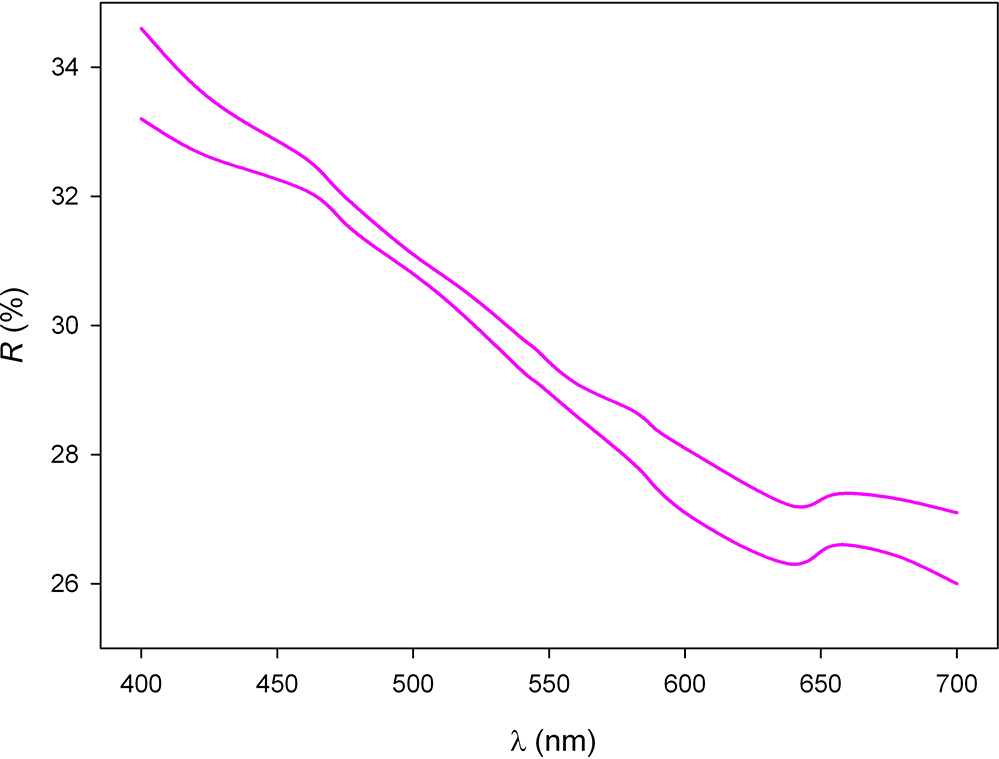
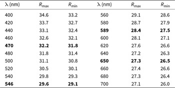
Zavyalovite is dark red in colour, is opaque with a sub-metallic lustre, and has brittle tenacity and uneven fracture. It has a red streak (determined on synthetic material). No cleavage or parting was observed. The Vickers’ micro-indentation hardness (VHN, 10 g load) for zavyalovite and its synthetic analogue are 95 kg/mm2 (range 93–98, n = 4) and 91 kg/mm2 (range 88–94, n = 4), respectively, corresponding to a Mohs hardness of 2.5. The density of the new mineral could not be measured due to the paucity of available material and the absence of the necessary heavy liquids. A density value calculated using the empirical formula and unit-cell volume obtained from powder X-ray diffraction (PXRD) data is 5.33 g/cm3. In reflected light of the ore microscope, zavyalovite is grey, darker than galena but lighter than sphalerite and anglesite (Fig. 3d). It is very weakly bireflectant and non-pleochroic. Under crossed polarizers, the new mineral exhibits strong anisotropy, in grey tones. Internal reflections are abundant and red in colour. The reflectance values were measured in air using an MSF-R (LOMO, Saint-Petersburg, Russia) microspectrophotometer. Silicon was used as a standard. The reflectance values (R max/R min) are given in Table 1 and plotted in Fig. 4.
Reflectance curves of zavyalovite.

Figure 4 Long description
A line graph showing two reflectance curves. The x-axis is labelled as wavelength in nanometres, ranging from 400 to 700. The y-axis is labelled as reflectance percentage, ranging from 26 to 34. The curves show a general downward trend with some fluctuations.
Reflectance values for zavyalovite (COM* standard wavelengths are given in bold)

Table 1 Long description
The table measures the reflectance values for zavyalovite across various wavelengths from 400 nm to 700 nm. The COM wavelengths in the order: wavelength, R max, R min are: 470, 32.2, 31.8, 546, 29.6, 29.1, 589, 28.4, 27.5, 650, 27.3, 26.0
* COM – The reference wavelengths required by the Commission on Ore Mineralogy.
Raman spectroscopy
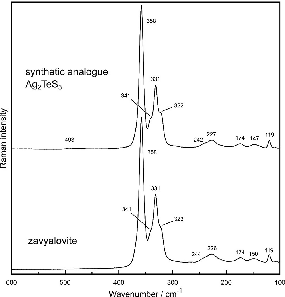
Raman spectra were obtained for both zavyalovite and its synthetic analogue (Fig. 5). The Raman spectroscopic investigation was carried out using a Renishaw inVia Reflex Raman system coupled with a Leica microscope. Individual grains were measured with a 100× objective lens with excitation provided by a 785-nm diode laser and the signal recorded by a thermoelectrically cooled CCD detector (100–650 cm–1 spectral range, spectral resolution of 2 cm–1). To achieve enhanced signal-to-noise ratios, 10–20 scans were accumulated, each having a 20 s exposure time, with laser power at source kept at maximum of 30 mW to avoid thermal degradation. A polystyrene standard was used to check the wavenumber calibration and the spectra were analysed using the GRAMS/AI 9.3 software package.
Raman spectra of zavyalovite and its synthetic analogue, Ag2TeS3.

Figure 5 Long description
The graph shows Raman spectra for zavyalovite and its synthetic analogue Ag2TeS3. The y-axis is labelled 'Raman Intensity' and the x-axis is labelled 'Wavenumber per cm superscript negative 1'. The spectrum for the synthetic analogue displays bands of various intensities at wavenumbers 493, 358, 341, 331 and 322, 242, 227, 174, 147, 119. The spectrum for zavyalovite shows bands of various intensities at wavenumbers 358, 341, 331, 323, 2244, 226, 174, 150 and 119. The strengths of the intensities for each band are described in the text.
For zavyalovite, the following Raman bands were observed: 119, 150, 174, 226, 244, 323, 331, 341, and 358 cm⁻1. The Raman spectrum of the synthetic analogue is almost identical, within analytical uncertainty, and displays Raman bands at 119, 147, 174, 227, 242, 322, 331, 341, 358, and 493 cm⁻1. By analogy with other phases containing the TeS3 group (Pfitzner, Reference Pfitzner1998; Kysliak and Beck, Reference Kysliak and Beck2012; Wolff et al., Reference Wolff, Pfitzner and Schleid2024), the prominent bands in the 300–400 cm⁻1 range are manifestations of the internal stretching vibrations in the TeS3 polyhedra, with the band at 358 cm⁻1 assigned to the symmetric stretch vibration. The three bands at 323, 331, and 341 cm⁻1 are attributed to antisymmetric stretch vibrations. Generally, weaker bands at 150, 174, 226 and 244 cm⁻1 correspond to deformation vibrations in the TeS3 polyhedra. A single band at 119 cm⁻1 can be assigned to Ag–S or lattice vibrations. A very weak band at 493 cm⁻1, observed for the synthetic analogue, could be a combination band or an overtone.
Chemical data
Chemical analyses (eight spots for zavyalovite and five spots for its synthetic analogue) were carried out using a JEOL JXA-8230 electron microprobe instrument (WDS mode, 15 kV, 10 nA, 3 μm beam diameter). The results (mean data) and list of standards are given in Table 2. Both the mineral and the synthetic Ag2TeS3 turned out to be very pure and stoichiometric. No elements other than Ag, Te and S were detected via SEM-EDS in either sample. The empirical formulae, based on six atoms per formula unit, are Ag2.01Te1.00S2.99 (zavyalovite) and Ag2.01Te1.00S2.99 (synthetic analogue). The ideal formula of zavyalovite is Ag2TeS3, which requires Ag 49.08, Te 29.04, S 21.88, total 100 wt.%.
Chemical data (wt.%) for zavyalovite and its synthetic analogue

Table 2 Long description
The table compares the chemical composition of zavyalovite and its synthetic analogue, focusing on silver (Ag), tellurium (Te), and sulfur (S) content. Silver content for zavyalovite is 49.31% and the synthetic analogue is 49.16%. Tellurium content is slightly higher in zavyalovite at 29.04% compared to 28.98% in the synthetic analogue. Sulfur content is nearly identical, with zavyalovite at 21.75% and the synthetic analogue at 21.79%. The total percentage of constituents is 100.10% for zavyalovite and 99.93% for the synthetic analogue, indicating a close match in overall composition. The standard deviations suggest that the synthetic analogue has slightly less variability in its composition.
n = number of analyses, S.D. = standard deviation
X-ray crystallography
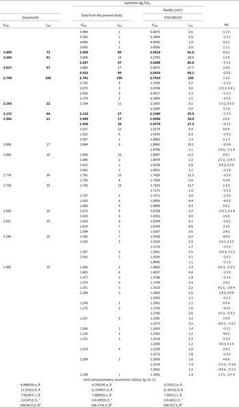
Single-crystal X-ray diffraction (SCXRD) studies of zavyalovite could not be carried out due to the completely polycrystalline nature of the grains and the absence of suitable single crystals. However, a grain which was 30 μm across in size (the largest found) was extracted from the polished section and powder micro-XRD data collected from it (Table 3) using a Rigaku Oxford Diffraction Supernova diffractometer working in micro-powder diffraction transmission mode. The diffractometer was equipped with a MoKα X-ray micro-source (conditions: 50 kV, 0.8 mA) and a Pilatus 200K Dectris detector with a detector-to-sample distance of 69 mm. 489 frames were collected over nine runs using an exposure time of 120 s per degree for a total time equal to 16 h.
Powder XRD (d in Å) and unit-cell parameters of zavyalovite and its synthetic analogue, Ag2TeS3

Table 3 Long description
The table compares powder XRD reflections and unit-cell parameters of zavyalovite and its synthetic analogue, Ag2TeS3 fom this work and the ICSD card fom the work of Pertlik (1997). The strongest lines for zavyalovite are also listed in the abstract and are, in the order d in Angstroms, intensity in percent and hkl: 3.495 (73) (0 0 2), 3.242 (61) (-1 3 1), 3.017 (57) (-2 0 2), 2.749 (100) (1 3 1), 2.163 (22) (1 3 2), 2.131 (64) (-1 3 3), 1.941 (21) (-3 3 1). Zavyalovite's unit-cell parameters are slightly larger than those of synthetic Ag2TeS3, with a volume of 548.96 ų compared to 548.37 ų. The monoclinic lattice parameters show minor variations, with zavyalovite having slightly larger dimensions in the a, b, and c axes. The β angle is nearly identical for both materials, indicating similar crystal symmetry.
Notes: Only reflections with intensities ≥1 are given. Strongest reflections are given in bold.
The powder XRD data of synthetic Ag2TeS3 (Table 3) were collected in Bragg-Brentano geometry on a Bruker D8 Advance powder diffractometer. The CuKα radiation, 10 mm automatic divergence slit and a Lynx Eye-XE detector were used. The data were collected in the angular range from 5 to 140°2θ with a 0.015°2θ step.
Unit-cell parameters for both zavyalovite and synthetic Ag2TeS3 were calculated from the observed d spacing data using UnitCell by Holland and Redfern (Reference Holland and Redfern1997) and are reported in Table 3. As shown, they are in very good agreement with each other as well as with the unit cell parameters obtained from SCXRD by Pertlik (Reference Pertlik1997).
Zavyalovite was also studied by means of EBSD using a TESCAN LYRA3 FEG SEM scanning electron microscope combined with EBSD system (Oxford Instruments AztecHKL with NordlysNano EDSD detector); the data were processed using the AZtec 6.0 software (Oxford Instruments). The sample surface was prepared for EBSD by polishing with colloidal silica (OP-U) for 20 min. The Kikuchi patterns obtained from the natural material (18 measurements on different spots on seven grains of natural zavyalovite) were found to match the patterns generated by the structural model described by Pertlik (Reference Pertlik1997) for synthetic Ag2TeS3 (Fig. 6). The Kikuchi bands (11 bands, containing 50 reflectors) were detected using the optimized accuracy indexing mode, using band edges, with a Hough resolution of 80. The values of the mean angular deviation (MAD; goodness-of-fit in the solution) between the calculated and measured Kikuchi bands range between 0.25 and 0.47°. These values reveal a very good match, therefore; mean angular deviations <1° are considered to be indicators of an acceptable fit (Fig. 6).
EBSD images of natural zavyalovite; in the parts on the right, the Kikuchi bands are indexed.

Figure 6 Long description
The composite image consists of four parts, each displaying Kikuchi band patterns. The top left and bottom left images show unindexed Kikuchi bands, characterized by intersecting lines forming a complex geometric pattern. The top right and bottom right images display similar Kikuchi band patterns, but with crystallographic labels such as 121, 112, 110, 211, 310, 531, 101, 112, 201 and 111. These labels are positioned at various intersections of the bands, indicating specific crystallographic orientations.
Crystal structure and discussion
The crystal structure of zavyalovite is the same as that of synthetic Ag2TeS3 (Pertlik, Reference Pertlik1997), as clearly demonstrated by PXRD, EBSD, chemical and Raman data. Some of the important details of the structure of the synthetic analogue of zavyalovite described by Pertlik (Reference Pertlik1997) are provided in the Supplementary Material file (Tables S1 and S2). The atomic arrangement is characterized by isolated trigonal pyramidal TeS3 and irregularly coordinated AgS4 polyhedra (Fig. 7). The TeS3 polyhedra can be described as trigonal pyramids with the Te at the apex and at a distance of ∼1 Å from the plane defined by the three S atoms. The AgS4 and TeS3 polyhedra are connected in a way that the S atoms are in layers parallel to (001) with Ag and Te located in between. The TeS3 polyhedron shows three similar Te–S distances with an average of 2.373 Å; on the other hand, the Ag1S4 and Ag2S4 polyhedra are significantly irregular with Ag–S distances varying between 2.5 and 2.8 Å and with an average of ∼2.64 Å for Ag1S4 and 2.62 Å for Ag2S4 (Table S1).
Crystal structure of zavyalovite (data after Pertlik, Reference Pertlik1997) as seen down a. The atom labels are given in the top left of the figure. The black line represents the unit cell. Drawn using Vesta (Momma and Izumi, Reference Momma and Izumi2011).

Figure 7 Long description
The image shows the crystal structure of zavyalovite, featuring a repeating pattern of trigonal pyramidal TeS3 and irregularly coordinated AgS4 polyhedra. The TeS3 polyhedra are depicted with Te at the apex and S atoms forming the base. The Ag1 and Ag2 atoms are labelled, along with S1, S2 and S3. The black line outlines the unit cell. The structure is arranged in layers parallel to the plane, with Ag and Te located between the S layers. A coordinate axis is shown at the bottom left, indicating orientation.
A very interesting point is the role of Te in the zavyalovite structure. It is well known that Te, being a chalcogen, occurs in minerals in three formal oxidation states 2-, 4+ and 6+ (see, e.g., Missen et al., Reference Missen, Ram, Mills, Etschmann, Reith, Shuster, Smith and Brugger2020). As of July 2025, the current IMA List of Minerals (available at https://cnmnc.units.it/files/editor/IMA_Master_List_(2025-07).pdf) includes more than 100 non-oxygen minerals with species-defining Te. In the vast majority of these, Te is an anion Te2–, e.g. in case in cervelleite Ag4TeS (Criddle et al., Reference Criddle, Chisholm and Stanley1989; Bindi et al., Reference Bindi, Stanley and Spry2015), the only other mineral with the same species-defining elements as zavyalovite. The structure of the former can be described as a body-centred array of tetrahedrally coordinated Te and S atoms with Ag2(Te,S)4 polyhedra in planes nearly parallel to (010) and the sheets linked by other silver position (Ag1) that exhibits a three-fold coordination (Bindi et al., Reference Bindi, Stanley and Spry2015). Zavyalovite, instead, is a very rare case of a primary non-oxygen mineral where Te is a Te4+ cation surrounded by S atoms. Other similar minerals are, e.g. Te-rich members of tetrahedrite group: arsenogoldfieldite Cu6Cu6(As2Te2)S13 (Sejkora et al., Reference Sejkora, Biagioni, Dolníček and Voudouris2023), arsenoústalečite Cu6Cu6(As2Te2)Se13 (Sejkora et al., Reference Sejkora, Biagioni, Škácha, Musetti and Mauro2024) goldfieldite (Cu4□2)Cu6Te4S13 (Trudu and Knittel, Reference Trudu and Knittel1998), stibiogoldfieldite Cu6Cu6(Sb2Te2)S13 (Biagioni et al., Reference Biagioni, Sejkora, Musetti, Makovicky, Pagano, Pasero and Dolníček2022) and stibioústalečite Cu6Cu6(Sb2Te2)Se13 (Sejkora et al., Reference Sejkora, Plášil and Makovicky2022).
Cui and Qi (Reference Cui and Qi1996) described a phase designated M1 from the Detiangau Au deposit in China as grains up to 100 μm across in pyrite-gold-polymetallic sulfide quartz veins in association with numerous tellurides and native Te. This phase is also named as UM1996-29:S-AgTe in the list of valid unnamed minerals (Smith and Nickel, Reference Smith and Nickel2007) updated in January 2023 (http://cnmnc.units.it/). The composition of this phase, given as Ag(S1.48Te0.52), can be recast to a zavyalovite formula of Ag2TeS3. The optical data for M1 are also quite similar to that of zavyalovite. However, the lack of XRD data does not allow us to conclude unambiguously that M1 is identical to zavyalovite.
Dincă et al. (Reference Dincă, Popescu, Ciobetea-Barbu and Bîrgăoanu2018) reported on a phase from the Săcărâmb Au-Ag-Te deposit, Romania, which has the composition Ag2.1TeS2.8, close to that of zavyalovite. No X-ray data were given by those authors and optically, the phase is described as isotropic, so it does not seem to correspond to zavyalovite.
The relationships between minerals in the samples studied allow several conclusions to be drawn about the stages of mineral-forming process at Boevskoe deposit. From a general and a somewhat simplistic perspective, the paragenetic sequence can be described as follows: quartz → simple sulfides (galena, sphalerite, chalcopyrite, pyrite) → tellurides and sulfotellurides (hessite, matildite, zavyalovite) → supergene minerals (anglesite, bismutite, boevskite, perite). Zavyalovite, thus, formed at a late stage of low-temperature hydrothermal activity.
Supplementary material
The supplementary material for this article can be found at https://doi.org/10.1180/mgm.2025.10142.
Acknowledgements
The authors thank Associate Editor, Owen Missen, and three anonymous reviewers for constructive comments that improved the manuscript. Maria D. Milshina is acknowledged for help with the figures.
Competing interests
The authors declare none.