Introduction
Nanostructured materials (i.e., forms of matter characterized by spatial patterns on length scales from a few to a few hundred nanometers) provide the basis for many nanotechnological applications and enable advances in fields as diverse as fuel cells, solar cells, membrane gas filters, bone scaffolds, and drug delivery agents. Nature itself boasts a range of multifunctional nanomaterials, including functional optical materials such as antireflection and antiwetting coatings, and polarization-sensitive visual organs.Reference Chiou, Kleinlogel, Cronin, Caldwell, Loeffler, Siddiqi, Goldizen and Marshall1,Reference Schroeder, Houghtaling, Wilts and Mayer2
The animal kingdom provides some of the most brilliant colored materials. In fact, the biophotonic nanostructures at the base of these colors are among the most impressive examples of nature’s use of nanostructural materials.Reference Srinivasarao3,Reference Vukusic and Sambles4 Diverse organisms from across nearly all animal phyla have evolved an arsenal of light manipulation strategies that rely on the ability to generate hierarchically structured, optical material designs, often involving soft components.Reference Kolle and Lee5 By tuning the dimensions and the complexity of such nanostructures, it is for example possible to achieve intense colors that span the entire visible wavelength spectrum, without employing pigments or colorants.
Bioinspired and biomimetic approaches that adopt nature’s designs to achieve material functions are commonly used (e.g., for wettable surfaces).Reference Telford, Hawkett, Such and Neto6 However, nature is less commonly used as a source of inspiration for the formation process of complex structures, that is, the question of how to design bioinspired protocols for self-assembly or growth of structures. This is particularly surprising since nature often demonstrates a far greater ability to control features of the nanostructural organization (such as symmetry or length scale) than synthetic self-assembly protocols, and since it shows enhanced efficiency, biocompatibility and scalability compared with state-of-the-art top-down nanofabrication strategies (notwithstanding recent progress in that field).Reference Gan, Turner and Gu7,Reference Dolan, Wilts, Vignolini, Baumberg, Steiner and Wilkinson8 In this article, we present different nanostructures in nature, describe what is currently known about their formation mechanisms, and what we can learn from them.
Bicontinuous nanostructures in nature
Bicontinuous geometries carve up space into two labyrinth-like domains.Reference Hyde, O’Keeffe and Proserpio9 These geometries are by now firmly established as a commonly observed structural motif of soft and hard nanostructured systems in equilibrium. In vitro, bicontinuous phases occur generically in synthetic self-assembled amphiphilic systems, such as lipidsReference Hyde, Andersson, Ericson and Larsson10–Reference Negrini and Mezzenga12 and block copolymers,Reference Dolan, Wilts, Vignolini, Baumberg, Steiner and Wilkinson8,Reference Khandpur, Foerster, Bates, Hamley, Ryan, Bras, Almdal and Mortensen13 but also in inorganic mesoporous solid materialsReference Attard, Glyde and Göltner14,Reference Alfredsson and Anderson15 and as “cubosomes” in pharmaceutical drug delivery systems.Reference Zabara, Negrini, Onaca-Fischer and Mezzenga16,Reference Drummond and Fong17 There is widespread consensus that these synthetic bicontinuous phases—and their derived structures—form in essence by equilibrium self-assembly that is likely due to specific material constituents and their (bio-)chemical interactions.
In living, biological systems, such bicontinuous phases have been observed in the convoluted membranes of cell organelles of both plant and animal cells.Reference Almsherqi, Landh, Kohlwein and Deng18 In that sense, structured biomaterials mimic the structure of bicontinuous cubic mesophases of synthetic systems in vitro, though at different length scales and for sizes often much larger than their synthetic counterparts.
Of particular relevance is the occurrence of bicontinuous structures in chitinous hardened solids in butterflies and beetles.Reference Dolan, Wilts, Vignolini, Baumberg, Steiner and Wilkinson8,Reference Saranathan, Seago, Sandy, Narayanan, Mochrie, Dufresne, Cao, Osuji and Prum19,Reference Wilts, Zubiri, Klatt, Butz, Fischer, Kelly, Spiecker, Steiner and Schröder-Turk20 This was also observed in fibrillar structures of keratin,Reference Evans and Roth21 in the bicontinuous lipid phases formed in the lung’s alveolar surface and in human digestion models.Reference Salentinig, Phan, Khan, Hawley and Boyd22 The formation process of these biological structures in vivo is far less understood than the equilibrium process underlying the formation of their synthetic counterparts in vitro. Next, we will present two extreme cases of biological optical structures, gyroid structured networks of butterfly scales and quasi-ordered networks of bird feathers, and describe the current knowledge about their formation processes.
Gyroid networks in butterflies
Triply periodic minimal surfaces are excellent examples of the importance of geometrical principles in understanding materials at nano-sized length scales. These can be most notably observed in the biophotonic structures of weevil and butterfly wing scales.
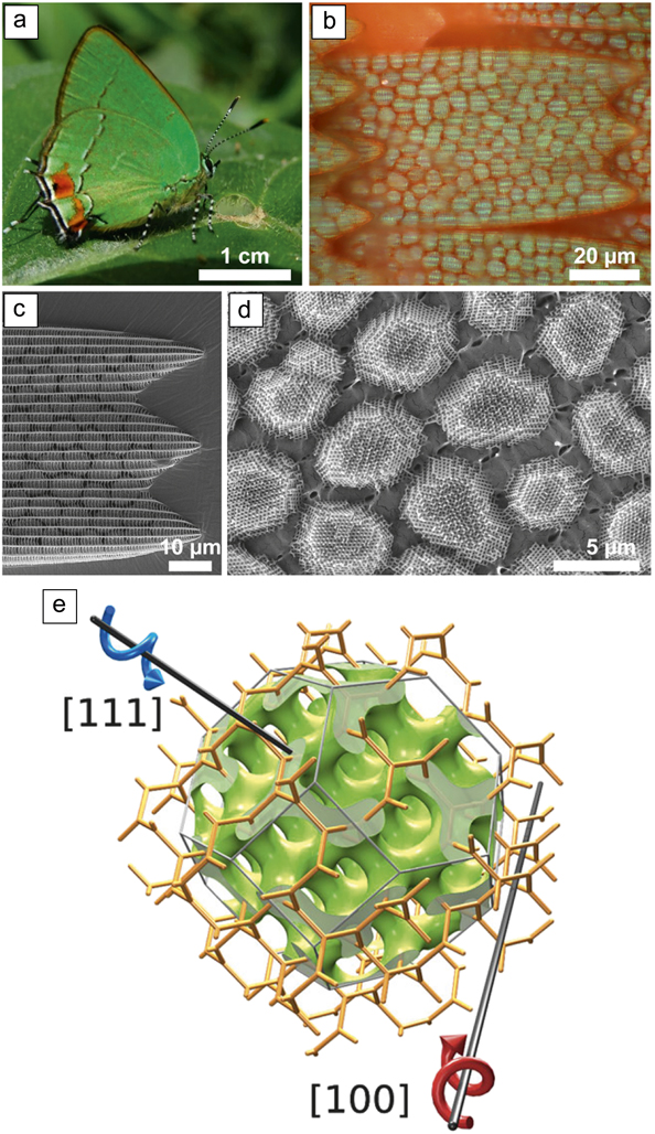
An uncommon occurrence of these complex structured solids is found in the hierarchical structure of the single gyroid chitin nanostructure in the butterfly species Thecla opisena (Figure 1),Reference Saba, Thiel, Turner, Hyde, Gu, Grosse-Brauckmann, Neshev, Mecke and Schröder-Turk23 where each “crystallite” of a wing scale is in fact a single crystal of a single network gyroid structure.Reference Wilts, Zubiri, Klatt, Butz, Fischer, Kelly, Spiecker, Steiner and Schröder-Turk20 In general, butterfly wing scales are hollow “casings” with dimensions of ∼200 × 60 × 5 µmReference Srinivasarao3 with the upper, outward-facing side forming a porous cross-hatched lattice of “ribs” and “cross-ribs.” Gyroid photonic structures, when present, are found inside the casing, which is called lumen, and are normally observed in a space-filling, multidomain nature.Reference Saranathan, Seago, Sandy, Narayanan, Mochrie, Dufresne, Cao, Osuji and Prum19,Reference Wilts, Zubiri, Klatt, Butz, Fischer, Kelly, Spiecker, Steiner and Schröder-Turk20,Reference Wilts, Michielsen, De Raedt and Stavenga24
(a–d) Hierarchical nanostructural organization of gyroid crystallites in the Neotropical Green Hairstreak butterfly, Thecla opisena.Reference Wilts, Zubiri, Klatt, Butz, Fischer, Kelly, Spiecker, Steiner and Schröder-Turk20 (e) Model of a single gyroid network with a demonstration of two of the high-symmetry orientations.Reference Saba, Thiel, Turner, Hyde, Gu, Grosse-Brauckmann, Neshev, Mecke and Schröder-Turk23

In contrast to other gyroid-forming butterfly species (e.g., the Emerald-patched Cattlehear, Parides sesostris; the Green Hairstreak, Callophrys rubi; or the Kaiser-i-Hind, Teinopalpus imperialis),Reference Wilts, Michielsen, De Raedt and Stavenga24–Reference Schröder-Turk, Wickham, Averdunk, Brink, Fitz Gerald, Poladian, Large and Hyde28 in T. opisena the gyroid nanostructure forms disconnected micron-sized crystallites of a single orientation and handedness—without internal defects. This structure is particularly exciting as it allows a glimpse into the development of these structures, as inferences from multidomain structures are strenuous and inconclusive.
Disordered networks in birds
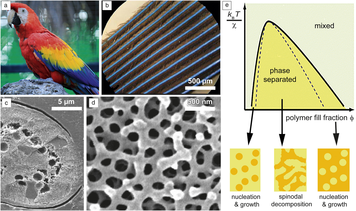
The amazing variety of structural color in nature is not restricted to ordered photonic structures such as the gyroid, but can also result from quasi-ordered materials.Reference John29,Reference Yablonovitch30 A prominent example is the feathers of the kingfisher and parrot, where bright structural color arises from the interference of light with disordered network-like morphologies created from keratin.Reference Saranathan, Forster, Noh, Liew, Mochrie, Cao, Dufresne and Prum31–Reference Stavenga, Tinbergen, Leertouwer and Wilts36 An isotropic bandgap can be exhibited from quasi-ordered nanostructures, resulting in angle-independent colors (Figure 2a–d).
Disordered photonics of a scarlet macaw feather. (a) Scarlet macaw, Ara macao. (b, c) The photonic structure is present in the outermost cells of the barbs, covered by a transparent envelope. (d) Scanning electron microscope image showing a disordered network. (e) The underlying growth process is assumed to be spinodal decomposition. Shown here is a schematic phase diagram of a polymer solution as a function of polymer volume fraction, ϕ, temperature, T, and interaction parameter, χ. The black line indicates the phase boundary that separates the single-phase system (above) from a solution that separates into two distinct phases either via spinodal decomposition or nucleation and growth. (b–d) Adapted with permission from Reference Reference Yin, Dong, Liu, Zhan, Shi, Zi and Yablonovitch38. © 2012 National Academy of Sciences. (e) Adapted with permission from References Reference Dufresne, Noh, Saranathan, Mochrie, Cao and Prum52 and Reference Prum, Dufresne, Quinn and Waters54. © 2009 Royal Society of Chemistry and The Royal Society, respectively. Note: k B, Boltzmann constant.

At first glance, these network morphologies look random, but at closer inspection, a dominant length scale of ∼180–250 nm can be found in the networks.Reference Saranathan, Forster, Noh, Liew, Mochrie, Cao, Dufresne and Prum31,Reference Prum, Torres, Williamson and Dyck34,Reference Stavenga, Tinbergen, Leertouwer and Wilts36 This length scale, together with the mean refractive index of keratin of ∼1.55,Reference Leertouwer, Wilts and Stavenga37 results in the blue and green colors observed in many bird barbs. Yin and colleaguesReference Yin, Dong, Liu, Zhan, Shi, Zi and Yablonovitch38 described these structures as “amorphous” diamond. Whether the growth and assembly of these disordered structures is identical to the growth of the ordered structures will be discussed next.
Biological bicontinuous structures’ unique properties
Biological optical nanostructures are remarkable for a number of reasons: (1) Large structural length scales—the length scale is often an order of magnitude or more above what can be achieved in synthetic self-assembled systems.Reference Dolan, Wilts, Vignolini, Baumberg, Steiner and Wilkinson8,Reference Angelov, Angelova, Drechsler, Garamus, Mutafchieva and Lesieur39 (2) Solid form—complex nanostructures in animals appear in a solid form, made from simple biomaterials, such as keratin or chitin, that have been synthesized at ambient conditions. (3) Single-network structures—bicontinuous nanostructures occur in their “single form” (i.e., consisting of a single solid component and a single void component). In synthetic systems,Reference Cao, Xu, Yao, Han, Terasaki and Che40 single systems are largely unknown. (4) Chirality—the chiral single gyroid structure in the Green Hairstreak butterfly reveals a bias for one of the two enantiomers.Reference Wilts, Zubiri, Klatt, Butz, Fischer, Kelly, Spiecker, Steiner and Schröder-Turk20,Reference Winter, Butz, Dieker, Schröder-Turk, Mecke and Spiecker26 All of these properties make them excellent candidates for bioinspired applications.
Deciphering nature’s strategies for the formation of nanostructures
An important question for understanding the function and evolution of structural coloration of organisms is how these nanostructures develop. In fact, the final nanostructural morphology must already be precisely controlled during development in order to produce the vibrant, observed color.
An understanding of the mechanisms employed by nature for achieving complex, nanostructured morphologies has so far been in its infancy, preventing science from systematically translating these mechanisms into synthetic systems. In detail, we do not know if or how nonequilibrium cell growth mechanisms might be involved or invoked in this process. In general, the key aspects of biological formation mechanisms within bird feather or insect wing scales are poorly understood.
Growth of ordered structures
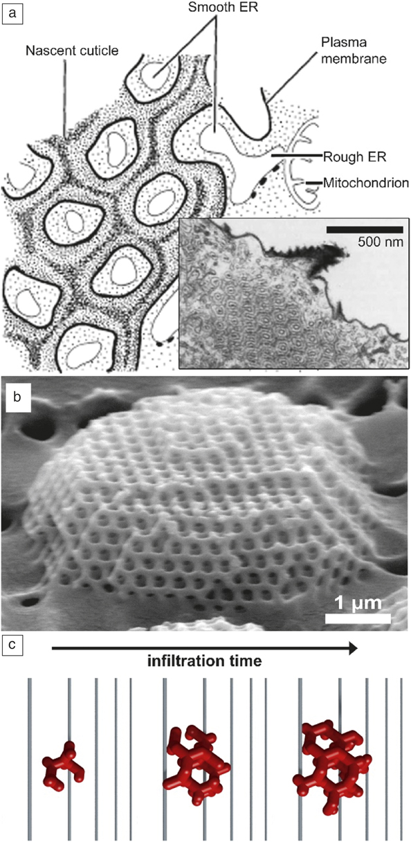
Conceptual models for the growth and development of butterfly cubic nanostructures have been proposed by GhiradellaReference Ghiradella41,Reference Ghiradella42 and others.Reference Saranathan, Osuji, Mochrie, Noh, Narayanan, Sandy, Dufresne and Prum43 Ghiradella associated this class of complex nanomorphologies with self-assembly of lipid membranes almost 30 years ago,Reference Ghiradella44,Reference Ghiradella and Locke45 based on detailed studies of the growth of wing scales within butterfly pupae (Figure 3). Figure 3a shows a cross-sectional scheme of a developing wing scale, derived from the transmission electron microscope (TEM) cross section shown in the inset. The resulting single gyroid structures (Figure 3b–c) can have a complex, single crystalline shape.
(a) Ghiradella’s model for the formation of wing scale nanostructures, as based on postmortem transmission electron micrographs of the developing wing scale. (b) Scanning electron microscope image of a faceted gyroid allows us to draw conclusions about the growth mechanism (c).Reference Wilts, Zubiri, Klatt, Butz, Fischer, Kelly, Spiecker, Steiner and Schröder-Turk20 (a) Adapted with permission from Reference Reference Ghiradella44. © 1989 Wiley.

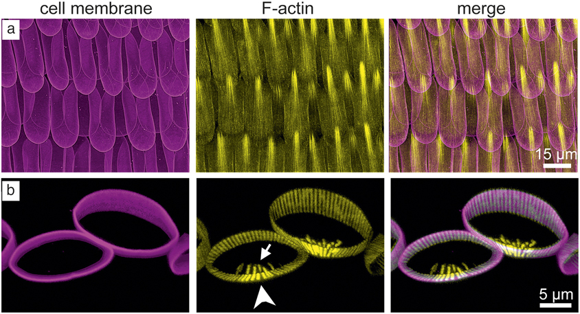
In fact, each wing scale of an adult butterfly corresponds to a single developing cell, which adopts (through F-actinReference Dinwiddie, Null, Pizzano, Chuong, Krup, Tan and Patel46) a highly anisotropic shape during development (Figure 4). During cell growth, the cell membrane (magenta) envelops the interior, while an internal network of F-actin (yellow) shapes the organization inside the cell resulting in three-dimensional forms. The internal nanostructure resulting in the complex structured networks has been suggested to form by a process where the intracellular endoplasmic reticulum membrane adopts an achiral double gyroid shape (similar to those reported in other intracellular membranes).Reference Almsherqi, Landh, Kohlwein and Deng18 Ghiradella’s model, a model that is now commonly used,Reference Saranathan, Seago, Sandy, Narayanan, Mochrie, Dufresne, Cao, Osuji and Prum19,Reference Saranathan, Osuji, Mochrie, Noh, Narayanan, Sandy, Dufresne and Prum47 states that the topologically complex chitin matrix seen in mature wing scales is formed by gradual polymerization of chitin oligomers within a water matrix defined by the bounding lipid membrane, where the lipid membrane spontaneously forms the gyroid structure. Chitin is then extruded into the network-like domain connected to the extracellular space, and solidifies as the cell dies off—leaving the observed single-network gyroid structures behind.
(a, b) Confocal laser scanning microscope allows 3D imaging of wing scale development of wing scale cells of Junonia coenia at 32% of pupal development, stained for the cell membrane (magenta) and F-actin (yellow). The wing scales are already elongated, anisotropic in shape and the lumen becomes discernible. A field of scales is viewed from the apical surface in (a), while a cross section of two scales is shown in (b). This level of detail can only be imaged postmortem with the appropriate fluorescent markers.Reference Dinwiddie, Null, Pizzano, Chuong, Krup, Tan and Patel46

This model is based on inferences from the postmortem study of butterfly pupae using two-dimensional TEM cross sections—and not on dynamic time-dependent studies. Performing such measurements is difficult, and little development has followed up on Ghiradella’s seminal studies. Recently, the study of the Neotropical Hairstreak butterfly T. opisena, with its isolated single-crystalline gyroid domains, has allowed the postmortem elucidation of details of this time-dependent process. By analyzing the pigment composition, the handedness of the single gyroids and the crystalline nature, Wilts et al. inferred that the gyroid is grown in a multistep process, by which an initially liquid water-lipid system is infiltrated by chitin and subsequently polymerized.Reference Wilts, Zubiri, Klatt, Butz, Fischer, Kelly, Spiecker, Steiner and Schröder-Turk20 Whether or not the membrane organelle takes the complex gyroid shapes upon membrane presence of the chitin or whether it is prefolded within the cell is still a debated topic.
The current model for the formation of ordered structures is inferred from scanning electron microscope images, and is ultimately somewhat speculative. To date, there has been no systematic experimental work confirming this mechanism due to difficulties in imaging folding tissue in vivo, due to the difficulty in imaging processes on this length scale deep within the optically thick tissue of a butterfly chrysalis over an extended period of time. Recent advances in the labeling of wing scale substructure during development (though primarily applied postmortem)Reference Dinwiddie, Null, Pizzano, Chuong, Krup, Tan and Patel46 and the isolated growth of individual wings within cell culturesReference Parker and Townley48 might lead to further insight into the nonequilibrium development processes in the near future.
There has been substantial recent progress in imaging techniques for biological tissues, with specific areas of application. Super-resolution microscopy allows imaging with a routine resolution of ∼40 nm, but relies on the availability of substructure-specific dyes and—usually—postmortem fixed samples.Reference Dinwiddie, Null, Pizzano, Chuong, Krup, Tan and Patel46,Reference Guo, Li, Zhang, Yang, Liu, Wang, Liu, Milkie, Moore, Tulu, Kiehart, Hu, Lippincott-Schwartz, Betzig and Li49 The imaging of developing tissue in vivo would require transgenic butterfly lines that express fluorescent proteins specific to the structure that one wishes to image in a nonlethal way. Environmental electron microscopy allows imaging of wet samples at reduced pressure, but is significantly limited to thin samples or surface observations.Reference Wagner, Cavalca, Damsgaard, Duchstein and Hansen50
For the butterfly system discussed here, it is now possible to grow butterfly species in a lab environment (with the pupae suitably dissected so that the developing wing scales can be observed using light microscopy from the outside. What is missing for this butterfly system and for many other membrane-structured systems with length scales in the range of 10–100 nm, including plant chloroplastsReference Pribil, Labs and Leister51) is an imaging system that allows imaging the cellular dynamics in vivo.
Growth or self-assembly of disordered structures?
While the previously discussed growth model holds true for periodic micromorphologies, quasi-ordered systems could also be produced in this fashion. This could be achieved either by incorporating elements (e.g., membrane proteins) that disturb the periodic assembly of the lipid membrane, or by “freezing-in” the (then disordered) structure far from its thermodynamic equilibrium.
However, the growth model most commonly assumed to lead to the quasi-ordered network morphologies within a feather barb is spinodal decompositionReference Dufresne, Noh, Saranathan, Mochrie, Cao and Prum52 (i.e., the process driving the spontaneous demixing of two immiscible components).Reference Saranathan, Forster, Noh, Liew, Mochrie, Cao, Dufresne and Prum31,Reference Dufresne, Noh, Saranathan, Mochrie, Cao and Prum52,Reference Dong, Zhan, Liu, Jiang, Liu, Hu and Zi53 Phase separation usually occurs through either spinodal decomposition or nucleation and growth, depending on the position in phase space and the kinetics of the system. Spinodally demixed structures are usually channel-like, can be described as self-similar, can be found from the cosmic scale down to the nanoscale and are most readily observed in phase-separating mixtures. Energetically, spinodal decomposition arises in mixtures with little (or no) energetic barrier for the nucleation of a phase, quite in contrast to nucleation and growth processes where the nucleation is subject to a large energetic penalty.
Prum and colleagues showed that the color-producing channel-type nanostructures develop intracellularly in the absence of any biological prepattern created by a cell membrane in a TEM study of developing feather barbs of the blue-and-yellow macaw (Ara ararauna) that were arrested in growth.Reference Prum, Dufresne, Quinn and Waters54 The authors argue that the color-producing, intracellular, quasi-ordered nanostructures develop their characteristic sizes and shapes by phase separation, via spinodal decomposition of keratin in the cellular watery matrix (Figure 2e). The shape and size of these self-assembled, intracellular nanostructures are determined by phase separation of keratin proteins from the cytoplasm of the cell rather than a membrane-supported growth process as in the ordered structures.
What can we learn from nature’s nanosolids?
Nanotechnology plays a large role in the development of future technologies, including energy materials, communication and computing, and pharmaceutical solutions. Each of these fields corresponds to large (and growing) industries.
The vast majority of developments in nanoscience and nanoengineering are driven by scientific advances in our understanding of how to control nanostructural features and nanoscale processes. Our ability to harness nanostructure functionality and design is, often, inferior to nature’s solutions. Deepening our understanding of the mechanisms used by nature will ultimately lead to advances of related nanostructural applications in a far broader scope, including pharmaceutical applications as drug delivery vehicles,Reference Negrini and Mezzenga12 or sustainable low-energy nanomaterials, as networked nanostructures have applications as functional energy materials.
Nature should provide inspiration and lend its formation processes—be it by growth or self-assembly, equilibrium or nonequilibrium—to provide an efficient and scalable process for their manufacture. The potential of nature’s nanostructures can only be unleashed with a comprehensive understanding of their properties in all detail. Developing this fundamental understanding of nanostructure formation in biology will lead to major significant benefits in advancing smart nanoengineering and nanobioengineering.
Summary
A comprehensive understanding of the design concepts, structure formation principles, material integration, and the control mechanisms employed in the in vivo development of biological photonic systems will allow us to challenge current paradigms in optical technology, as it will allow the creation of novel optical materials with widespread use (e.g., as toxin-free dyes in paint and arts).
“You can observe a lot by just watching.” While originally made in a different context, Yogi Berra’s famous quote rings true for understanding structure formation in biological growth and nonequilibrium processes. Significant recent progress has occurred for the in vivo imaging of several biological structures of single cells in bacteria,Reference Guo, Li, Zhang, Yang, Liu, Wang, Liu, Milkie, Moore, Tulu, Kiehart, Hu, Lippincott-Schwartz, Betzig and Li49 plants,Reference Pribil, Labs and Leister51 and butterflies.Reference Dinwiddie, Null, Pizzano, Chuong, Krup, Tan and Patel46 However, biology is complicated. The lack of easy methods to create transgenic butterflies or stain developing scales with nonlethal fluorescent dyes, and the degree and difference in the spatial organization of the living tissue, makes a method that is perfectly fine for one organism quickly unsuitable for another. For the many living systems where folded and spatially complicated membrane or membrane-derived structures exist on the scale of hundreds of nanometers, further suitable in vivo imaging methods are needed to answer the many questions about their development. It is interesting to speculate whether the optical effects caused by these nanostructures could be used in inverse methods to image the structures. Certainly, a method that allows us to watch the structure develop would be a substantial contribution toward understanding the dynamic nonequilibrium growth processes that generate these exquisite and beautiful natural ornaments, and ultimately help in taking inspiration from nature for replicating these in materials science.
Acknowledgments
This research was supported by the Swiss National Science Foundation through the National Centre of Competence in Research Bio-Inspired Materials and the Ambizione Program Grant No. 168223 (to B.D.W.).

Bodo D. Wilts is an Ambizione Fellow at the Swiss National Science Foundation and a group leader at the Adolphe Merkle Institute, Switzerland. He was a postdoctoral research associate in the Cavendish Laboratory at the University of Cambridge, UK. He received his PhD degree in 2013 from the Rijksuniversiteit Groningen, The Netherlands. His research focuses on the optical function, development, and design of biological and bioinspired photonic structures and polymer-templated plasmonic optical metamaterials. Wilts can be reached by email at bodo.wilts@unifr.ch.

Peta L. Clode is an associate professor in the Centre for Microscopy, Characterisation and Analysis at The University of Western Australia. She is currently head of Electron Microscopy (Biosciences), and prior to this, held the position of deputy director. Her research includes structure–function relationships over a range of length scales, and focuses on biomineralization, symbioses, and biomimetics. Clode can be reached by email at peta.clode@uwa.edu.au.

Nipam H. Patel is the director of the Marine Biological Laboratory (MBL) and a professor at The University of Chicago. Prior to his arrival at MBL, he was a professor in the Department of Molecular and Cell Biology and the Department of Integrative Biology at the University of California, Berkeley. His research interests include the development of novel, genetic model organisms for biological studies and the application of advanced imaging technologies to probe the fundamental dynamics of living systems. Patel can be reached by email at npatel@mbl.edu.

Gerd E. Schröder-Turk is an associate professor in the School of Engineering and Information Technology at Murdoch University, Australia. He received his PhD degree in 2005 from The Australian National University, Canberra, and his Habilitation degree in 2013 from the Friedrich-Alexander University Erlangen-Nuremberg, Germany. His research interests focus on the geometric structure, often on the nanometer scale, of both synthetic materials and biological materials. Schröder-Turk can be reached by email at G.Schroeder-Turk@murdoch.edu.au.