It is common in vertebrate palaeontology that the earliest collected specimens for many alpha-taxa, and thus the type materials, are often scrappy and poorly preserved. In the early days of the science, when the numbers of collected fossils in museums and other collections were minimal, the odds were significant that any newly collected material would not be referable to an existing taxon. There was also a tendency among the most prolific early vertebrate palaeontologists to be taxonomic ‘splitters’ (Benton Reference Benton2010), resulting in a plethora of novel taxonomic names. Moreover, some of these researchers were not conducting fieldwork, but instead working with material shipped in from various field collectors at a rapid rate, and competition between rival museums to generate results was high (see Brinkman Reference Brinkman2010). The hurried pace of that style of research, combined with the scrappy material and the general lack of good comparative specimens, resulted in many short and non-comprehensive published descriptions provided for these early specimens and the taxa erected for them. Generally, as better specimens were recovered subsequently, these early types were relegated simply to the role of “name bearers” (see discussion by Colbert Reference Colbert1989, p. 34), and more complete specimens came to be used as proxy “type” specimens. Current emphasis on detailed apomorphy-based comparisons for taxonomic work (e.g. Irmis et al. Reference Irmis, Parker, Nesbitt and Liu2007; Nesbitt et al. Reference Nesbitt, Irmis and Parker2007; Nesbitt & Stocker Reference Nesbitt and Stocker2008; Bell et al. Reference Bell, Gauthier and Bever2010) has demonstrated that many of these early descriptions and subsequent referrals are problematic and in dire need of revision (also see Padian Reference Padian and Padian1986).
Aetosaurs are a clade of suchian archosaurs with a global distribution during the Late Triassic; however, like many other extinct vertebrate groups, their early taxonomic history is confusing. The first described aetosaur specimen, Stagonolepis robertsoni, represented by the impression of a series of ventral osteoderms, was thought to represent a ganoid fish (Agassiz Reference Agassiz1844) until subsequent finds allowed for its determination as a “reptile” (Huxley Reference Huxley1859, Reference Huxley1875). The first North American aetosaur specimens were thought to represent a subgroup of “rhynchocephalians” (Cope Reference Cope1887a; McGregor Reference McGregor1906), or often as representing pseudosuchian (Cope Reference Cope1896) or parasuchian (phytosaurian) archosaurs (e.g. Marsh Reference Marsh1896; Case Reference Case1920, Reference Case1922, Reference Case1932; Camp Reference Camp1930). The concept of what exactly was an aetosaur (i.e. a monophyletic assemblage (clade) of armoured suchian archosaurs) was not recognised clearly until the work of Sawin (Reference Sawin1947) on Longosuchus meadei, or solidified until Walker's (Reference Walker1961) groundbreaking osteological description of Stagonolepis robertsoni and his thorough review of all known aetosaur taxa.
Although well preserved skeletons of Aetosaurus had been described from Germany as early as 1877 (Fraas Reference Fraas1877, Reference Fraas1907), the close phylogenetic relationships between Aetosaurus and morphologically similar forms such as S. robertsoni (e.g. Huxley Reference Huxley1875), were not recognised originally (e.g. Huene Reference Huene1902, Reference Huene1936; McGregor Reference McGregor1906; Camp Reference Camp1930; but see Cope Reference Cope1887a, Reference Cope1896). In part, this was because of the lack of good skull reconstructions of Stagonolepis in the earliest descriptions (e.g. Huxley Reference Huxley1875, reproduced by Huene Reference Huene1902) and the misinterpretation of the dentition of Aetosaurus (see discussion by Walker Reference Walker1961; Parker Reference Parker2007; Schoch Reference Schoch2007). Distinctions were blurred further by the common recovery of aetosaur osteoderms taphonomically mixed with the skulls and postcrania of phytosaurs (Gregory Reference Gregory1962; Murry Reference Murry1982), as well as the overall rarity of aetosaur cranial material. Indeed, the type materials of Typothorax coccinarum and Episcoposaurus horridus, the first aetosaurs described from North America, are mixed assemblages of phytosaur and aetosaur material (Gregory Reference Gregory1953; Parker Reference Parker2008a), with the material referable to aetosaurs consisting chiefly of osteoderms.
Despite the subsequent recognition that the short-snouted “stagonolepids” were actually distinct from the long snouted parasuchians (first attributed to Romer Reference Romer1945 by Gregory Reference Gregory1953), the idea that some phytosaurs (i.e. Nicrosaurus) shared a similar osteoderm morphology with aetosaurs distorted the systematics of both of these groups, as well as their relationships within Archosauria, for decades (e.g. Huene Reference Huene1902, Reference Huene1913, Reference Huene1915, Reference Huene1936, Reference Huene1940; McGregor Reference McGregor1906; Case Reference Case1920, Reference Case1922, Reference Case1932; Camp Reference Camp1930; Gregory Reference Gregory1962; Gregory & Westphal Reference Gregory and Westphal1969; Murry Reference Murry1982). That idea was not discarded fully until the work of Long & Ballew (Reference Long and Ballew1985), who assigned the rectangular German ‘phytosaur’ osteoderms to a new taxon Paratypothorax andressorum, which they considered unambiguously to be an aetosaur. Interestingly, this reassignment was made utilising no further evidence (e.g. associated skulls or postcranial material) than was available to past researchers. Fortunately, studies of other specimens (e.g. SMNS 19003; DMNH 9942) subsequently have verified Long & Ballew's (Reference Long and Ballew1985) hypothesis.
Another key argument by Long & Ballew (Reference Long and Ballew1985) was the recognition that the surface ornamentation pattern of osteoderms was taxonomically informative. Originally it was suggested that patterns were diagnostic at the species- (Long & Ballew Reference Long and Ballew1985; Long & Murry Reference Long and Murry1995) or genus-group (Heckert & Lucas Reference Heckert and Lucas2000) levels, such that single osteoderms or osteoderm fragments were taxonomically informative and diagnosable. However, current thinking is that in many cases ornamentation is homoplastic (i.e. shared between taxa in different clades) and that aetosaur taxonomy should also rely on non-osteoderm elements, or on complete rather than fragmentary osteoderms (Desojo Reference Desojo2005; Martz & Small Reference Martz and Small2006; Parker Reference Parker2008b).
The purpose of this paper is to redescribe the original type and referred materials of three historic aetosaur taxa from North America, Typothorax coccinarum Cope, Reference Cope1875a, Episcoposaurus horridus Cope, Reference Cope1877, and Episcoposaurus haplocerus Cope, Reference Cope1892, in a modern context, given our recent advances in aetosaur osteology and systematics, and to review the alpha taxonomy of these specimens. Since the original descriptions by Cope (Reference Cope1875a, Reference Cope1877, Reference Cope1892), these specimens were variously redescribed by Huene (Reference Huene1915), Gregory (Reference Gregory1953) and, most recently, by Heckert & Lucas (Reference Heckert, Lucas, Heckert and Lucas2002) and Lucas et al. (Reference Lucas, Spielmann, Heckert, Hunt, Lucas and Spielmann2007). However, some of those descriptions are vague, contain inaccuracies and, most importantly, have not separated out the aetosaurian material from other taxa mixed with these type materials. Both Huene (Reference Huene1915) and Gregory (Reference Gregory1953) recognised that the specimens represented a mix of phytosaur and aetosaur material, but considered the task of separating them impossible, a reasonable conclusion given the morphological confusion that existed between the two groups until the 1980s. Heckert & Lucas (Reference Heckert, Lucas, Heckert and Lucas2002) recognised some of the phytosaur material but not all of it, whereas Lucas et al. (Reference Lucas, Spielmann, Heckert, Hunt, Lucas and Spielmann2007) did not differentiate between much of the phytosaur and aetosaur material, considering it all to belong to aetosaurs. The goal of the present contribution is to provide the necessary morphological and thus taxonomic distinctions for this material, as well as to reexamine the taxonomic assignments of these specimens.
Institutional abbreviations. AMNH, American Museum of Natural History, New York, New York, USA; ANSP, The Academy of Natural Sciences of Drexel University, Philadelphia, Pennsylvania, USA; DMNH, Dallas Museum of Natural History, Dallas, Texas, USA; MCZ, Museum of Comparative Zoology, Harvard University, Cambridge, Massachusetts, USA; MNA, Museum of Northern Arizona, Flagstaff, USA; NMMNH, New Mexico Museum of Natural History and Science, Albuquerque, Mexico, USA; PEFO, Petrified Forest National Park, Petrified Forest, Arizona, USA; SMNS, Staatliches Museum für Naturkunde, Stuttgart, Germany; TMM, Texas Memorial Museum, Austin, Texas, USA; TTU, Texas Tech University, Lubbock, Texas, USA; UCMP, University of California Museum of Paleontology, Berkeley, California, USA; UMMP, University of Michigan Museum of Paleontology, Ann Arbor, Michigan, USA; USNM, United States National Museum, Washington, DC, USA.
1. Previous work
1.1. Early collections
Early field collecting trips to Upper Triassic rocks in New Mexico and Texas in the 1870s and 1880s by Edward Cope and his field collector David Baldwin resulted in the collection of numerous fossils, including two phytosaurs (e.g. AMNH FR 2318, type specimen of Machaeroprosopus buceros; unnumbered type specimen of Belodon scolopax, possibly lost) and much early dinosauriform material, including the original type materials of Coelophysis bauri (Cope Reference Cope1887b), C. longicollis (Cope Reference Cope1887b) and C. willistoni (Cope Reference Cope1887a), as well as material recently recognised as belonging to the dinosauromorph Dromomeron romeri (Nesbitt et al. Reference Nesbitt, Irmis, Parker, Smith, Turner and Rowe2009). In addition, materials were collected that formed the basis for two new archosaurian taxa. The first of these was Typothorax coccinarum, named by Cope (Reference Cope1875a) for osteoderms fragments, vertebral fragments and coprolites (USNM 2585, USNM 2586) from the Gallina area in the Chama Basin of north-central New Mexico. Cope (Reference Cope1887a) subsequently assigned better preserved material collected by Baldwin from the same locality in 1881 to T. coccinarum, including two femora and more complete osteoderms (AMNH FR 2709, AMNH FR 2710). Baldwin's collection contained other osteoderms, vertebrae and limb bones (AMNH FR 2713) that Cope utilised to erect a second new taxon, Episcoposaurus horridus. At some point, presumably in the late 1800s, a small collection of material of putative syntype material of Typothorax coccinarum was made and reposited at the Academy of Natural Sciences in Philadelphia (ANSP 14695; Spamer & Daeschler Reference Spamer and Daeschler1995; Heckert & Lucas Reference Heckert, Lucas, Heckert and Lucas2002).
Cope (Reference Cope1892) erected a third taxon, Episcoposaurus haplocerus, for another collection made by Baldwin, this time from the panhandle of Texas, which included osteoderms and vertebrae (ANSP 14688). Apparently, Cope personally returned to the Texas site after Baldwin's initial visit and collected additional materials from the site, including a complete ilium (currently part of ANSP 14688) and more vertebrae and osteoderms (TMM 40041-3, TMM 40041-5); however, these materials were not included in the original description (Wilson Reference Wilson1950; Gregory Reference Gregory1953; Parker Reference Parker2008a).
Case (Reference Case1920) erected the name Desmatosuchus spurensis for a skeleton (UMMP 7476) of another purported phytosaur from the panhandle of Texas, not far from where Baldwin had made his collection, and subsequently provided more thorough descriptions of that material (Case Reference Case1921, Reference Case1922). Desmatosuchus was diagnosed by the presence of enlarged recurved horns on the armour of the cervical region, a character also found in Episcoposaurus haplocerus, which led some researchers to suggest that the two were synonymous (e.g. Sawin Reference Sawin1947). That specimen (UMMP 7476) was also the first North American form to preserve the skull, which demonstrated that the cranium in these specimens was short-snouted and very different from typical phytosaurs (Case Reference Case1920, Reference Case1922).
1.2. Subsequent reviews
Huene (Reference Huene1915) redescribed much of Cope's New Mexican material, including the referred material of T. coccinarum (mistakenly referred to by Huene as the type specimen), and the syntype material of E. horridus. Huene (Reference Huene1915) correctly recognised that the E. horridus material belonged to more than one individual, and removed elements from the syntype material. In particular, he removed the forelimb materials, which he assigned to a small ‘mystriosuchid’ phytosaur; however, this was mostly done based on element size (in comparison to the massive hind limb elements in the material), rather than by utilising specific morphological characters.
The first comprehensive review of these materials was completed by Gregory (Reference Gregory1953) in his study of North American phytosaurs. Gregory (Reference Gregory1953) argued that Huene's (Reference Huene1915) removal of the forelimb material from the type of E. horridus (AMNH FR 2713) was premature and that, in comparison to the skeleton of the recently described “Typothorax” (=Longosuchus) meadei (Sawin Reference Sawin1947), the hindlimb and forelimb material could belong to the same animal. Gregory (Reference Gregory1953) also provided the first detailed description and figures of the type material of Episcoposaurus haplocerus (ANSP 14688). That author argued convincingly for the synonymy of Desmatosuchus spurensis and Episcoposaurus haplocerus, as well as that of Episcoposaurus horridus and Typothorax coccinarum. Because Episcoposaurus was now a junior synonym of Typothorax, the E. haplocerus material was assigned to Desmatosuchus as D. haplocerus. As noted above, Gregory recognised that the aetosaur material was mixed with some phytosaur material and removed some elements (e.g. ilium from ANSP 14688), but considered an attempt to completely separate the specimens “hopeless”.
Heckert & Lucas (Reference Heckert, Lucas, Heckert and Lucas2002) figured and provided brief redescriptions of some of this material (USNM 2585, USNM 2586, ANSP 14688, ANSP 14865) and provided lectotype designations. Lucas et al. (Reference Lucas, Spielmann, Heckert, Hunt, Lucas and Spielmann2007) briefly discussed and figured the referred specimens of Typothorax coccinarum (AMNH FR 2709, AMNH FR 2710) and the syntypes of Episcoposaurus horridus (AMNH FR 2713). They also proposed a lectotype for E. horridus, but unfortunately they mistakenly considered much of AMNH FR 2709 and AMNH FR 2710 to be part of the syntype series (Lucas et al. Reference Lucas, Spielmann, Heckert, Hunt, Lucas and Spielmann2007, p. 242).
2. Methods
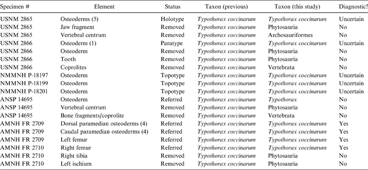
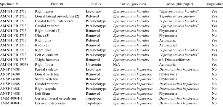
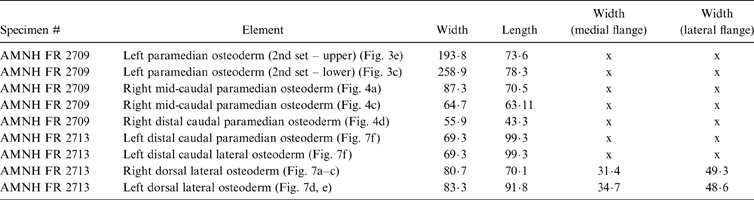
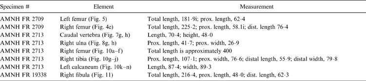
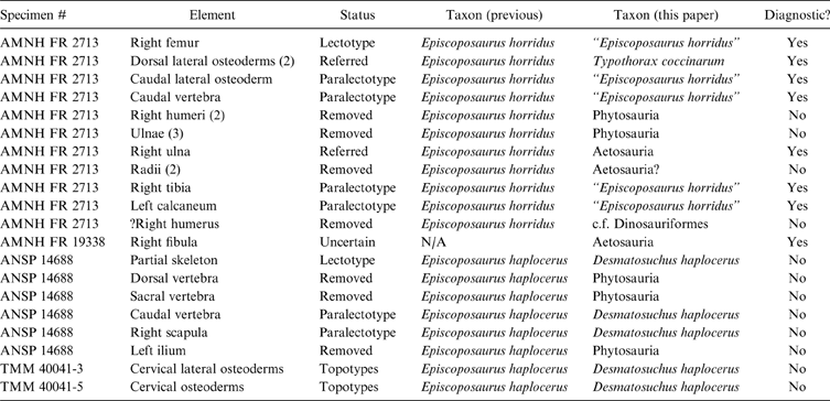
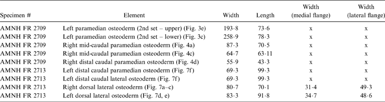
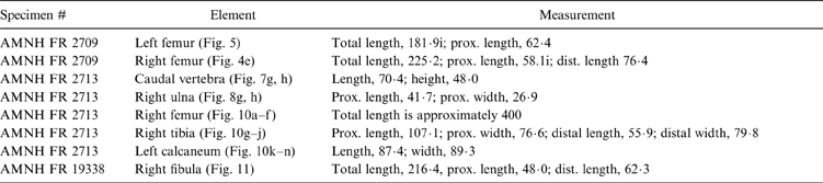
For this present study, the holotype specimen of Episcoposaurus horridus (AMNH FR 2713) and referred materials of Typothorax (AMNH FR 2709, AMNH FR 2710) were borrowed to allow detailed first hand observation. The holotype material of Episcoposaurus haplocerus (ANSP 14688) was also studied firsthand. Because of the historic nature of these specimens, further preparation was not conducted. Previously published literature (Huene Reference Huene1915; Gregory Reference Gregory1953; Heckert & Lucas Reference Heckert, Lucas, Heckert and Lucas2002; Lucas et al. Reference Lucas, Spielmann, Heckert, Hunt, Lucas and Spielmann2007) contains many misconceptions about what material has been included and excluded in the various syntypes, when and by whom. However, a careful reading of the primary literature elucidates much of this and will be discussed throughout the following text. These assignments are also provided in tabular form (Tables 1, 2). Measurements for specimens are provided in Tables 3–5.
Type status of specimens described in text. Diagnostic status refers to the species level taxa.
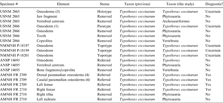
Type status of specimens described in text. Diagnostic status refers to the species level taxa.
Measurements (in mm) of paramedian and lateral osteoderms.
Measurements (in mm) of limb and vertebral elements discussed in text. “i” indicates that element is incomplete.
Measurements (in mm) of osteoderms discussed in text. ‘i’ indicates that element is incomplete.

Taxonomic assignments of these materials are accomplished using an apomorphy-based approach as advocated by Bell et al. (Reference Bell, Head, Mead and Barnosky2004, Reference Bell, Gauthier and Bever2010), Bever (Reference Bever2005), Nesbitt et al. (Reference Nesbitt, Irmis and Parker2007) and Nesbitt & Stocker (Reference Nesbitt and Stocker2008). This is an explicit, conservative and, more importantly, a testable method that uses the presence of discrete synapomorphies in a phylogenetic framework to determine the taxonomic placement of individual specimens. This approach allows broader comparisons to be made, reduces the influence of geographical or stratigraphic factors in taxonomic assignments, and removes the inherent circularity involved with those influences (Bever Reference Bever2005; Bell et al. Reference Bell, Gauthier and Bever2010).
3. Systematic palaeontology
Archosauria Cope Reference Cope1869sensu Gauthier & Padian Reference Gauthier, Padian, Hecht, Ostrom, Viohl and Wellnhofer1985
Pseudosuchia Zittel Reference Zittel1887–90sensu Gauthier & Padian Reference Gauthier, Padian, Hecht, Ostrom, Viohl and Wellnhofer1985
Suchia Krebs Reference Krebs1974sensu Sereno Reference Sereno1991
Aetosauria Marsh Reference Marsh1884sensu Parker Reference Parker2007
Typothorax Cope, Reference Cope1875a
Typothorax coccinarum Cope, Reference Cope1875a
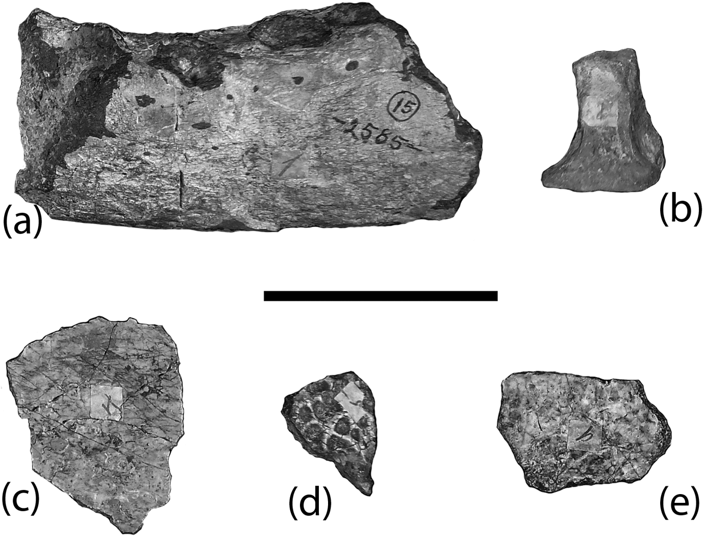
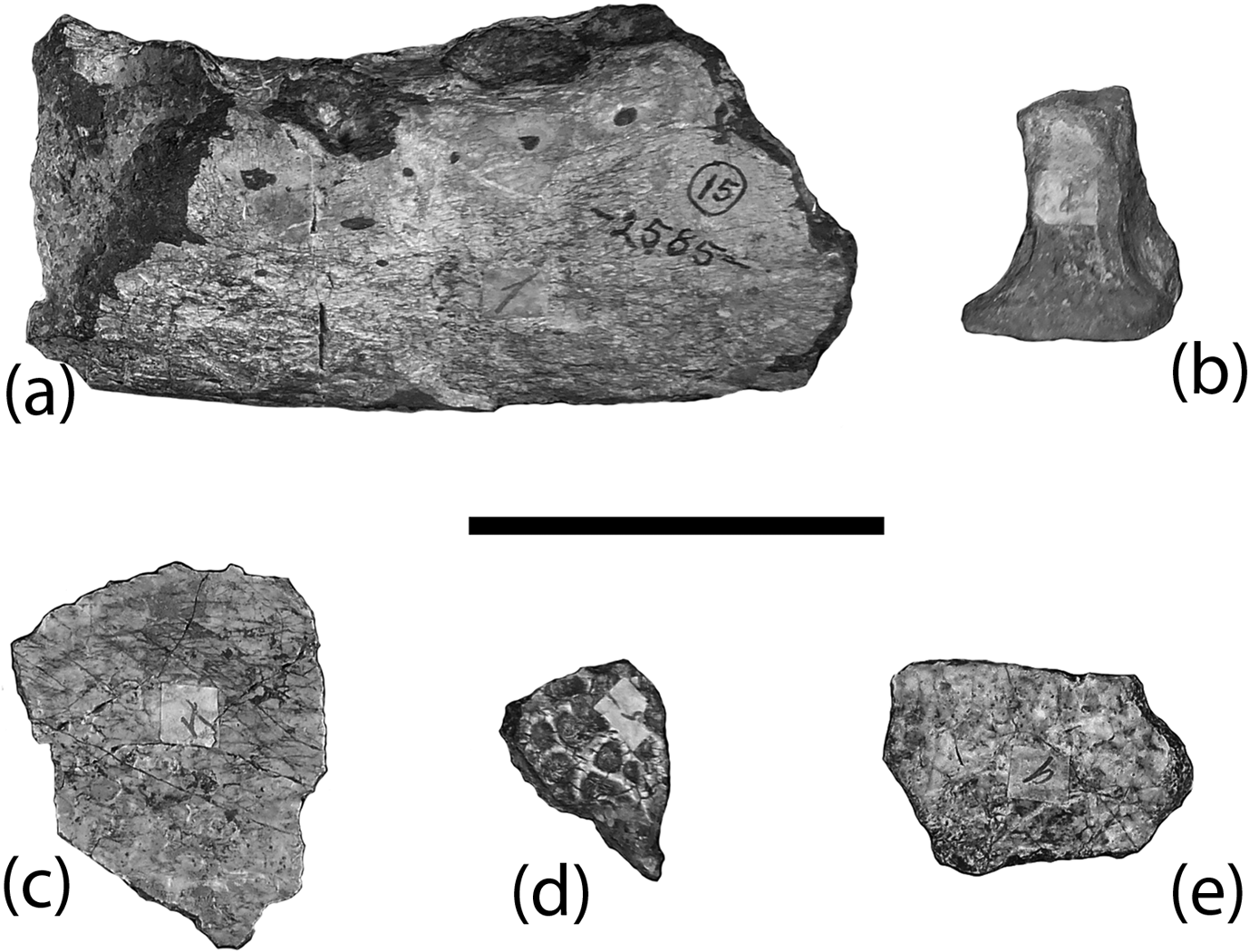
USNM 2585: (a) phytosaur dentary fragment in lateral view; (b) archosaur caudal vertebra in ventral view; (c–e) Typothorax coccinarum holotype and paratype paramedian osteoderm fragments in dorsal view. Scale bar=5 cm.
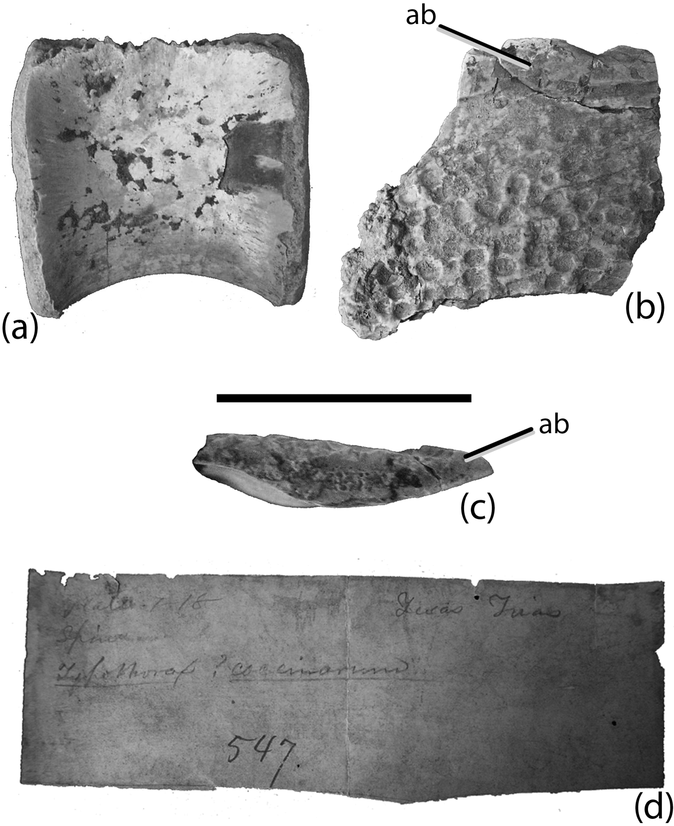
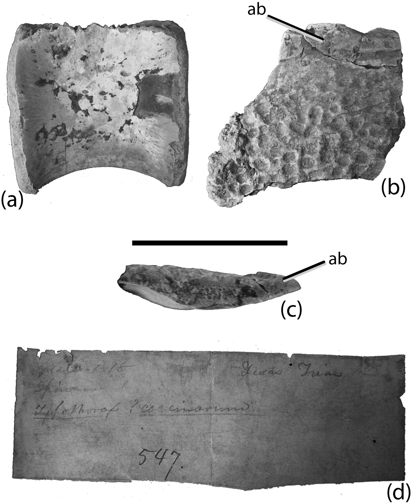
ANSP 14695: (a) phytosaur dorsal centrum in lateral view; (b–c) Typothorax coccinarum paramedian plate fragment in dorsal (b) and lateral (c) views; (d) note written by Cope that reads “Typothorax coccinarum, Texas (?), Trias.” The printed number 547 is Cope's original specimen number. Scale bar=5 cm. Abbreviations: ab=anterior bar.
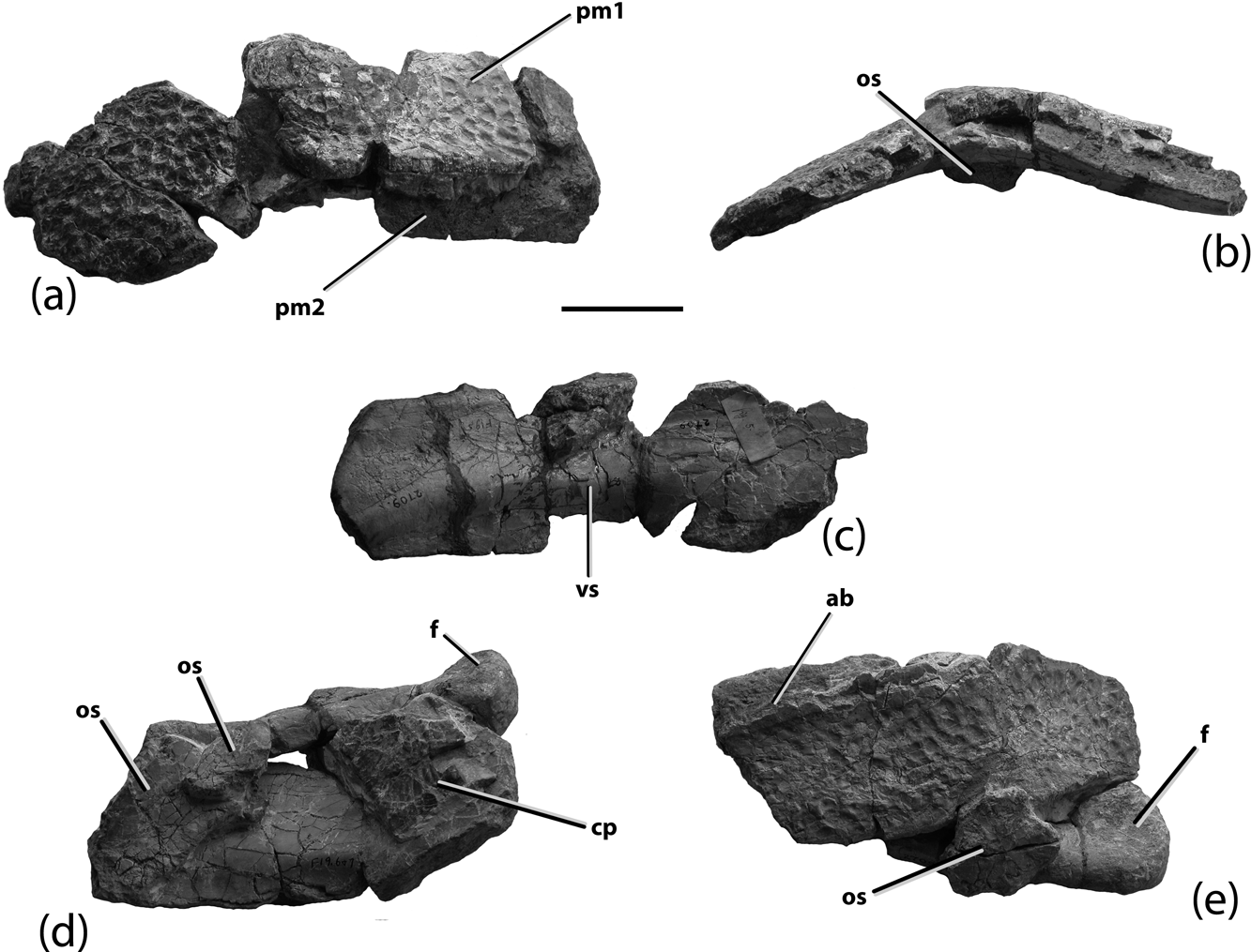
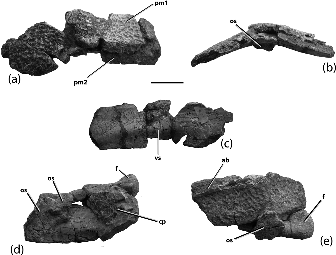
Typothorax coccinarum referred specimens (AMNH FR 2709): (a) first stacked set of two dorsal paramedian osteoderms in posterodorsal view; (b) same set in posterior view; (c) same set in ventral view; (d) stacked set of two dorsal paramedian osteoderms with attached femur and other osteoderms with the paramedian osteoderms oriented in ventral view; (e) same stacked set with the osteoderms oriented to dorsal view. Scale bar=5 cm. Abbreviations: ab=anterior bar; cp=caudal paramedian osteoderm; f=femur; os=osteoderm; pm=paramedian osteoderm; vs=ventral strut.
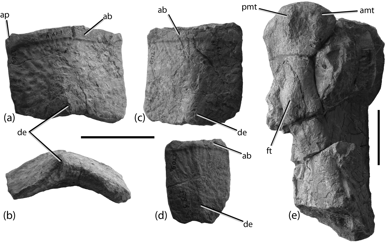
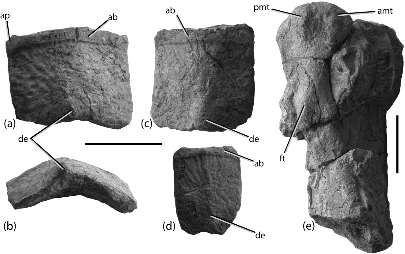
Typothorax coccinarum referred specimens (AMNH FR 2709): (a–b) right anterior mid-caudal dorsal paramedian osteoderm in dorsolateral (a) and posterior (b) views; (c) left mid-caudal dorsal paramedian osteoderm in dorsomedial view; (d) right posterior caudal dorsal paramedian in dorsolateral view; (e) proximal end of left femur in posterior view. Scale bar=5 cm. Abbreviations: ab=anterior bar; amt=anteromedial tuber; ap=anterior process; de=dorsal eminence; ft=fourth trochanter; pmt=posteromedial tuber.
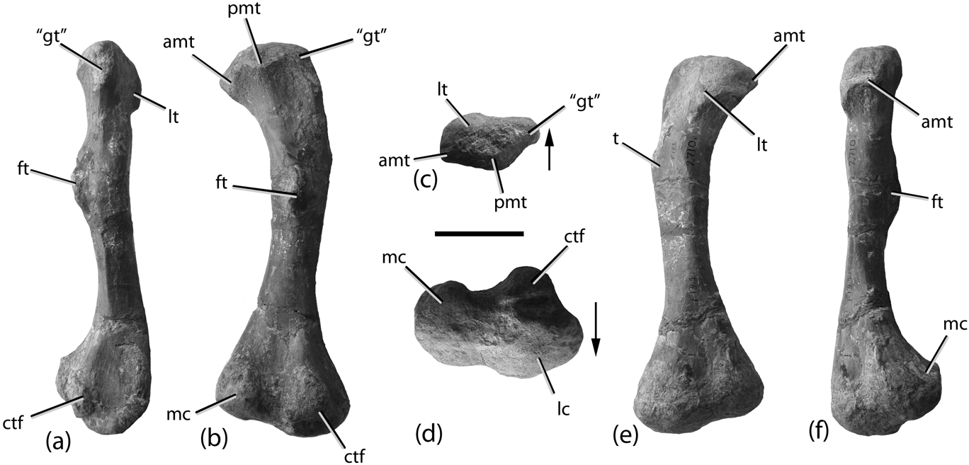
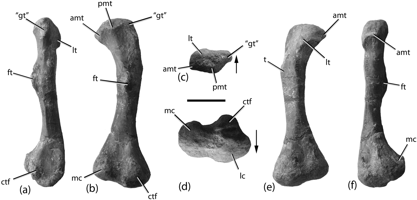
Typothorax coccinarum referred right femur (AMNH FR 2710): (a) lateral view; (b) posterior view; (c) proximal view; (d) distal view; (e) anterior view; (f) medial view. Scale bar=5 cm. Abbreviations: amt=anteromedial tuber; ctf=crista tibiofibularis; ft=fourth trochanter; “gt”=greater trochanter; lc=lateral condyle; lt=lateral tuber; mc=medial condyle; pmt=posteromedial tuber; t=tuber.
Holotype. USNM 2585, five osteoderm fragments. A single osteoderm fragment (Cope Reference Cope1877, pl. 22, fig. 4) from this specimen was designated as the lectotype by Heckert & Lucas (Reference Heckert, Lucas, Heckert and Lucas2002) following Long & Ballew (Reference Long and Ballew1985). Those authors stated that Cope did not explicitly list a holotype for Typothorax coccinarum; however, Cope (Reference Cope1875b, p. 266) clearly referred to this material as the “type specimen”. Thus USNM 2585 is fixed as the type specimen by original designation (ICZN 1999, Article 73.1.1) and any subsequent lectotype designations are invalid.
Paratype. USNM 2586, osteoderm fragment.
Original material. Cope's original material consists of a dentary fragment with four alveoli, a broken vertebra, five fragmentary osteoderms and other bone fragments. According to Cope (Reference Cope1875a, Reference Copeb) another locality produced osteoderms and skull fragments, a tooth and coprolites that may have an affinity with the previous specimens. Cope (Reference Cope1877) figured that material, whereas Cope (Reference Cope1887a) explicitly excluded the non-osteoderm material from the type specimen.
Original diagnosis. Distinguished from “Belodon” by the flat and regularly pitted dermal bones (Cope Reference Cope1875a, Reference Copeb).
Revised diagnosis. Heckert et al. (Reference Heckert, Lucas, Rinehart, Celeskey, Spielmann and Hunt2010) provided the most recent revised diagnosis for Typothorax coccinarum; however, this is based mainly on referred material (NMMNH P-12964, NMMNH P-56299). A diagnosis based on only the type materials (USNM 2585, USNM 2586) is problematic because it can only recognise the presence of a single character: dorsal surface osteoderm ornamentation consisting of randomly arranged shallow circular to subcircular pits.
Original referred material. AMNH FR 2709, AMNH FR 2710 (Cope Reference Cope1887a; Huene Reference Huene1915). Lucas et al. (Reference Lucas, Spielmann, Heckert, Hunt, Lucas and Spielmann2007) mistakenly considered this material to be part of the syntype series of Episcoposaurus horridus. Lucas & Hunt (Reference Lucas and Hunt1992, fig. 14 l–q) referred osteoderm fragments (NMMNH P-18197, NMMNH P-18199, NMMNH P-18201) from the Chama Basin of New Mexico to Typothorax coccinarum and considered these to be topotypes.
4. Description
4.1. Holotype specimens
USNM 2585 and USNM 2586 consist of a total of eleven bone fragments and three coprolites. These specimens were described originally by Cope (Reference Cope1875 a, Reference Copeb) and figured by Cope (Reference Cope1877). They were later redescribed by Heckert & Lucas (Reference Heckert, Lucas, Heckert and Lucas2002). The figure numbering from Cope's Reference Cope1877 publication is still apparent on each specimen, making correlation of elements with Cope's description simple. Moreover, although all of the elements are mentioned here, Cope (Reference Cope1887a) emended the type specimens to only include the osteoderm fragments. Heckert & Lucas (Reference Heckert, Lucas, Heckert and Lucas2002, fig. 4g–h, k–l) also figured three fragments as part of USNM 2585, but these are actually NMMNH specimens collected in the 1980s (NMMNH P-18197, NMMNH P-18199, NMMNH P-18201) purportedly from the type locality, and are therefore nothing more than possible topotypes (Lucas & Hunt Reference Lucas and Hunt1992).
The jaw fragment from USNM 2585 (Fig. 1a; Cope Reference Cope1877, pl. 12, fig. 1a–c) is a robust element about 10 cm in length, with a flat medial symphyseal surface and at least four alveoli on the dorsal surface. The morphology of this element is consistent with that of a partial fragment of the left dentary of a phytosaur, a conclusion reached by Cope (Reference Cope1887a), who explicitly removed this element from the syntypes of Typothorax coccinarum.
The partial vertebral centrum from USNM 2585 (Fig. 1b; Cope Reference Cope1877, pl. 12, fig. 2) is fragmentary and poorly preserved. It is mediolaterally compressed, with flared rims consistent with an archosauriform caudal centrum; however, no chevron facets are preserved on the ventral surface, suggesting that if this identification is correct it is the anterior portion of the centrum that is preserved. Given the presence of a partial phytosaur dentary with the material, its referral to Typothorax cannot be confirmed. Cope (Reference Cope1887a) also removed this specimen from the syntype materials.
The largest osteoderm fragment in USNM 2585 (Fig. 1c; Cope Reference Cope1877, pl. 12, fig. 4; Heckert & Lucas Reference Heckert, Lucas, Heckert and Lucas2002, fig. 4c–d) is a weathered portion of a paramedian osteoderm. The dorsal surface is ornamented with densely packed small oval pits typical on portions of the paramedian osteoderms of several aetosaur taxa, including various paratypothoracisines (e.g. PEFO 26690, PEFO 26725) and T. coccinarum. The ventral surface is flat and smooth.
A small (>3 cm length) osteoderm fragment in USNM 2585 (Fig. 1d; Cope Reference Cope1877, pl. 12, fig. 5; Heckert & Lucas Reference Heckert, Lucas, Heckert and Lucas2002, fig. 4e–f) probably also originates from a paramedian osteoderm. Heckert & Lucas (Reference Heckert, Lucas, Heckert and Lucas2002, p. 198) described this as a left caudal lateral osteoderm based on the presence of a “pronounced spike” and presumably deeper pitting. However, the ornamentation is no deeper than osteoderms from the dorsal series and the ‘spike’ is actually the osteoderm broken into a jagged fragment.
USNM 2586 contains a larger (∼4 cm long) paramedian osteoderm fragment (Fig. 1e; Cope Reference Cope1877, pl. 12, fig. 9; Heckert & Lucas Reference Heckert, Lucas, Heckert and Lucas2002, fig. 4a–b). This fragment possesses an dorsal surface ornamentation of ovate pits separated by a reticulate pattern of ridges, consistent with that found in Typothorax, but which also occurs in portions of paratypothoracisin osteoderms (e.g. PEFO 26690, PEFO 26725). Heckert & Lucas (Reference Heckert, Lucas, Heckert and Lucas2002) stated that this fragment is referable to Typothorax by the presence of a transverse ventral strut, but this feature is also found in some paratypothoracisins (e.g. Paratypothorax, PEFO 3004).
USNM 2586 also contains two unidentifiable bone fragments (Cope Reference Cope1877, pl. 12, figs 7–8), a phytosaur osteoderm fragment (Cope Reference Cope1877, pl. 12, fig. 5), a fragmentary tooth crown (Cope Reference Cope1877, pl. 12, fig. 13), and three coprolites (Cope Reference Cope1877, pl. 12, figs 10–12). The three bone fragments are morphologically and taxonomically indeterminate and were removed from the type materials by Cope (Reference Cope1887a). Cope (Reference Cope1875a, Reference Copeb) originally referred the tooth crown to Typothorax, but was less certain of this referral in his 1887 paper and removed it accordingly. The tooth is not aetosaurian and instead belongs to a phytosaur, because it is elongate, D-shaped in apical view and possesses distinct mesial and distal carinae with fine serrations (Hungerbühler Reference Hungerbühler2000). Cope (Reference Cope1875a, Reference Copeb) also described the coprolites as full of fish scales, but argued that they were too small to belong to the associated Typothorax specimen.
In summary, Cope (Reference Cope1887a) removed from his type specimens all of the materials that were not osteoderm fragments with the characteristic pitted sculpturing. That emendation of the type materials is supported here because none of the removed specimens possess apomorphies of Typothorax. However, assignment of the remainder of the material to Typothorax is also problematic because the fragments are very small, and the characteristics possessed by the fragments that are used to assign them to Typothorax coccinarum (e.g. ovate pitting, reticular sculpture, ventral strut) are found also in portions of paratypothoracisin osteoderms. The possible taxonomic implications of this will be discussed in more detail below.
4.2. ANSP specimens
ANSP 14695 consists of a paramedian osteoderm fragment, a dorsal vertebral centrum, four bone fragments, and a coprolite which are purported to represent syntypes of Typothorax coccinarum (Spamer & Daeschler Reference Spamer and Daeschler1995, fig. 15a–g; Heckert & Lucas Reference Heckert, Lucas, Heckert and Lucas2002, fig. 5a–g). Of this material, only the osteoderm fragment is referable to Typothorax; the centrum (Fig. 2a) most likely belongs to a phytosaur (Heckert & Lucas Reference Heckert, Lucas, Heckert and Lucas2002), and the other fragments are taxonomically indeterminate. The osteoderm fragment (Fig. 2b) possesses a smooth raised anterior bar as well as the dorsal surface pitting that is characteristic of the genus. The element is too incomplete to determine if a dorsal eminence was present; however, the ventral surface possesses a thickened ventral strut (Fig. 2c).
The provenance of these specimens is problematic. There is a field note (Fig. 2d) with the material in Cope's hand which reads “Plate 7–18, spine, Typothorax? coccinarum, 547”. There are two more words written on the tag which Spamer & Daeshler (Reference Spamer and Daeschler1995) interpreted as “Texas Trias”; the word Trias is written clearly, however, the word “Texas” is not. If this specimen is indeed from Texas, it cannot be part of the syntype series of Typothorax coccinarum, which was collected from New Mexico. Because they are reposited in the ANSP with the specimens of Episcoposaurus haplocerus, which are from Texas, and because Cope's field note shows uncertainty in the taxonomic assignment by adding the question mark it would appear that these specimens are also probably from Texas and not part of the type series.
4.3. Original referred specimens (AMNH FR 2709, AMNH FR 2710)
4.3.1. Dorsal paramedian osteoderms
The first set of osteoderms in AMNH FR 2709 (Fig. 3a–c; Cope Reference Cope1887a, pl. 1, fig. 1; Huene Reference Huene1915, fig. 5; Lucas et al. Reference Lucas, Spielmann, Heckert, Hunt, Lucas and Spielmann2007, fig. 2f–g) consists of two dorsal paramedian osteoderms fused together by matrix. Cope (Reference Cope1887a) and Huene (Reference Huene1915) mistakenly considered these to be a broad flattened rib covered by a single osteoderm. Gregory (Reference Gregory1953) recognised that the specimen consisted of two osteoderms rather than a rib and osteoderm, whereas Lucas et al. (Reference Lucas, Spielmann, Heckert, Hunt, Lucas and Spielmann2007) described the specimen as consisting of only a single osteoderm.
The lower osteoderm (the ‘rib’ of Cope and Huene) is nearly complete, missing only portions of the mid portions of the posterior and anterior edges, the anteromedial corner and the anterolateral projection (Fig. 3c). The ventral surface is smooth, with a longitudinally-oriented significantly thickened area (‘rib’), termed a ventral strut by Long & Ballew (Reference Long and Ballew1985). The dorsal surface is completely covered by matrix and the other osteoderm, making determination of orientation difficult; however, the ventral strut is closer to the anterior edge in Typothorax and there are distinct longitudinal grooves along the posterior portion of the ventral surface, as in all aetosaurs, where the osteoderm articulated ventrally with the next posteriorly situated osteoderm in life (see Cope Reference Cope1887a, pl. 1, fig. 1). The lateral edge is sigmoidal, missing only the anterolateral projection, thus this is an osteoderm from the left side of the carapace. In anterior view (Cope Reference Cope1887a, pl. 1, fig. 1a; Huene Reference Huene1915, fig. 5b) the osteoderm is strongly flexed (not arched; see discussion in Spielmann et al. Reference Spielmann, Hunt, Lucas, Heckert, Harris, Lucas, Spielmann, Lockley, Milner and Kirkland2006) ventrally, lateral to where the dorsal eminence would be located. Thus, it is from the posterior dorsal or anterior caudal region (Martz Reference Martz2002). The lateral portion of the osteoderm is longer than the median portion, as in all aetosaurs (Parker Reference Parker2007). An incomplete, badly preserved portion of a lateral osteoderm is fused to the underside of this osteoderm.
The upper paramedian osteoderm is preserved as two large, and one very small, fragments, adhered by matrix to the dorsal surface of the first osteoderm (Fig. 3a). Only slight remnants of the medial, lateral and posterior edges, and nothing of the anterior edge, are preserved; thus the presence of an anterior bar cannot be verified. The dorsal surface possesses the distinct circular, reticulate pitting characteristic of Typothorax (Long & Ballew Reference Long and Ballew1985). This osteoderm is also strongly flexed ventrally and appears to be slightly smaller than the underlying osteoderm, suggesting that the osteoderms were fused together with matrix before the centre portion of the upper osteoderm was lost (Fig. 3b). The size difference and overlying position suggests that the upper osteoderm is from a life position anterior (if not directly anterior) to the lower osteoderm.
The second osteoderm set is AMNH FR 2709 (Fig. 3d–e; Cope Reference Cope1887a, pl. 1, fig. 2; Huene Reference Huene1915, fig. 6; Lucas et al. Reference Lucas, Spielmann, Heckert, Hunt, Lucas and Spielmann2007, fig. 2d–e). As with the previous specimen, this set also is comprised mainly of two fused dorsal paramedian osteoderms rather than an articulated ‘rib’ and osteoderm, or a single osteoderm (Gregory Reference Gregory1953; contra Cope Reference Cope1887a; Huene Reference Huene1915; Lucas et al. Reference Lucas, Spielmann, Heckert, Hunt, Lucas and Spielmann2007). The overall preservation is worse for this set, making orientation determination difficult; however, the upper osteoderm preserves a portion of the raised anterior bar. A portion of the anterior bar on the lower osteoderm appears to also be present because of a thinning margin, but is covered by matrix. The dorsal surface sculpturing is the typical pattern for Typothorax and the dorsal eminence is just slightly raised along the posterior osteoderm margin on the upper osteoderm (Fig. 3e). These osteoderms are also flexed in line with this dorsal eminence, as is typical for Typothorax osteoderms (Martz Reference Martz2002) and, based on the preserved lengths of the lateral and medial flanges, is probably also from the left side, although this is not completely certain because the medial and lateral margins are incomplete.
No details of the dorsal surface of the lower osteoderm can be seen. The ventral surface preserves a portion of the thickened ventral strut. Portions of four lateral and appendicular osteoderms and the proximal end of a left femur are attached by matrix to the ventral surface of the lower paramedian osteoderm and posterior edges of both paramedian osteoderms (Fig. 3d–e). These elements will be described in more detail below.
Cope (Reference Cope1887a) suggested a posterior placement for these osteoderms based on the association with this femur. This placement is consistent with the overall morphology of the paramedian osteoderms (e.g. flexion, slight dorsal eminence) as described by Martz (Reference Martz2002). Personal examination of NMMNH C-4638, which is an exhibited cast of NMMNH P-12964 (Heckert et al. Reference Heckert, Lucas, Rinehart, Celeskey, Spielmann and Hunt2010), also demonstrates that the anterior paramedians in the carapace of Typothorax are arched, whereas the more posterior dorsal paramedians possess are flexed. This also appears to be the case in the posterior dorsal paramedians in NMMNH P-56299, although Heckert et al. (Reference Heckert, Lucas, Rinehart, Celeskey, Spielmann and Hunt2010) described them as ‘flat’; however, Heckert et al.'s (Reference Heckert, Lucas, Rinehart, Celeskey, Spielmann and Hunt2010) figure 2 clearly shows that the posterior dorsal paramedians (P15?, P17) are not flat, but rather strongly flexed. Furthermore, the posterior paramedians of the right side are telescoped beneath the associated lateral osteoderms, as the dorsal eminences of the paramedian osteoderms are nearly touching the laterals, thus their degree of flexion is difficult to determine.
4.3.2. Caudal paramedian osteoderms
Anterior mid-caudal paramedian (AMNH FR 2709; Huene Reference Huene1915, fig. 9; Lucas et al. Reference Lucas, Spielmann, Heckert, Hunt, Lucas and Spielmann2007, fig. 3e). This rectangular, wider than long, osteoderm is complete, although the ventral surface is still obscured by matrix (Fig. 4a). The anterior margin is arched in anterior view, whereas the posterior osteoderm margin is flexed (Fig. 4b) so that is there is a distinct sharp angle in the curvature consistent with osteoderms from the caudal region of Typothorax (Martz Reference Martz2002). The anterior bar is raised and strongly developed although the anteromedial projection is broken off (Fig. 4a). There is no distinct anterolateral projection of the anterior bar and the lateral margin of the osteoderm is straight, not sinuous. The dorsal eminence is a distinct, but low, slightly elongate ridge with a posterior apex that projects slightly past the posterior osteoderm margin. The low width/length ratio and the presence of a distinct dorsal eminence suggest that this is a right caudal paramedian osteoderm (Lucas et al. Reference Lucas, Heckert, Hunt, Heckert and Lucas2002; Martz Reference Martz2002).
The dorsal surface ornamentation is mostly obscured by matrix although, where visible, consists mainly of rounded pits, and a distinct grooved pit is apparent in the anterior portion of the medial half of the osteoderm. Distinct small anteroposteriorly trending grooves are present on the dorsal surface just posterior to the anterior bar. This last feature is typical for aetosaur osteoderm ornamentation and can help provide an orientation when the anterior bar is broken away (Parker Reference Parker2007). Huene (Reference Huene1915) interpreted this osteoderm reversed with the anterior bar along the posterior edge, whereas Lucas et al. (Reference Lucas, Spielmann, Heckert, Hunt, Lucas and Spielmann2007) considered this osteoderm to be from the left side. However, in aetosaurs, including Typothorax, the dorsal eminence of the paramedian osteoderm is always situated closest to the medial edge (Parker Reference Parker2007), thus this osteoderm is from the right side. The presence of a partially complete anteromedial projection of the anterior bar supports this interpretation.
Mid-caudal paramedian osteoderm (AMNH FR 2709; Huene Reference Huene1915, fig. 10; Lucas et al. Reference Lucas, Spielmann, Heckert, Hunt, Lucas and Spielmann2007, fig. 3f–g). This square (equant) osteoderm is also complete, but covered with matrix on its ventral surface. The near equal width–length ratio of this element and the pronounced dorsal eminence suggests that it is a paramedian osteoderm from the mid-caudal region. It possesses an anteroposteriorly narrow raised anterior bar and a pronounced knob-like dorsal eminence on the posterior margin of the dorsal surface (Fig. 4c). The eminence is situated slightly medially, so the osteoderm is from the left side. The medial margin is straight, whereas the lateral margin is slightly sinuous. An anteromedial projection of the anterior bar is present, but the tip is broken away. The anterolateral edge of the anterior bar is rounded. The dorsal surface ornamentation is weathered; however, there are a few anterolateral trending grooves and ridges in the anterolateral corner. As with the more anteriorly position plate described previously in anterior view the osteoderm is gently arched, but in posterior view it is flexed at the position of the dorsal eminence.
Posterior caudal osteoderm (Figure 3d; AMNH FR 2709; Huene Reference Huene1915, fig. 7; Lucas et al. Reference Lucas, Spielmann, Heckert, Hunt, Lucas and Spielmann2007, fig. 2e). This osteoderm is incomplete, missing its medial portion, although the ventral surface and thus the overall shape of the osteoderm has been preserved as a natural mould in the matrix (Fig. 3d). The dorsal surface is badly preserved; however, a distinct anterior bar is present, and the overall ornamentation consists of distinct grooves radiating from where the dorsal eminence would have been. The dorsal surface possesses a distinct anteroposteriorly oriented keel, dividing the surface into medial and lateral portions (see Huene Reference Huene1915, fig. 7). The preserved lateral osteoderm margin is complete and straight, and is considered here to be the lateral edge because the osteoderm surface it bounds is wider than its counterpart (considered here to be the medial portion of the osteoderm). Huene (Reference Huene1915) interpreted this osteoderm as part of a lateral series, but it is very similar to posterior dorsal caudal paramedians of Typothorax from the Canjilon Quarry (UCMP V2816 158670 70/Fa52; Martz Reference Martz2002).
A second osteoderm of this form (AMNH FR 2709; Huene Reference Huene1915, fig. 8; Lucas et al. Reference Lucas, Spielmann, Heckert, Hunt, Lucas and Spielmann2007, fig. 3h) is much longer than wide (Table 3) and also from the right side (Fig. 4d). The ornamentation is very faint and still consists of elongate grooves consistent with posterior caudal paramedians of Typothorax from the Canjilon Quarry (Martz Reference Martz2002). The posterior margin, including the dorsal eminence, is broken away. The ventral surface of this osteoderm is completely obscured by matrix. This osteoderm is very similar in form to one in the holotype specimen of Typothorax antiquum that Lucas et al. (Reference Lucas, Heckert, Hunt, Heckert and Lucas2002, fig. 3a) considered to represent a cervical ventral (?) paramedian. However, comparison with known Typothorax specimens (e.g. NMMNH P-12964) shows that the cervical paramedians are wider than long, cervical ventral paramedians are unknown, and that the osteoderms that are longer than wide are distal caudal paramedians.
4.3.3. Other osteoderms
Several partial osteoderms are adhered to the paramedian osteoderm blocks; however, they are worn, broken and often strongly adhered by matrix to the other elements, and thus details of these specimens cannot be clearly distinguished. The ventral surface of one osteoderm on the second paramedian osteoderm block is exposed; the dorsal surface adhered to the block (Fig. 3d). This osteoderm is transversely arched, providing an anteroposterior orientation that demonstrates that the element was much wider than long. Therefore it represents a caudal plate. The others are irregularly shaped and could be appendicular osteoderms (Fig. 3d–e), and one on the first paramedian block (Fig. 3b) appears to be acutely flexed and thus it is probably a dorsal lateral osteoderm.
4.3.4. Femur
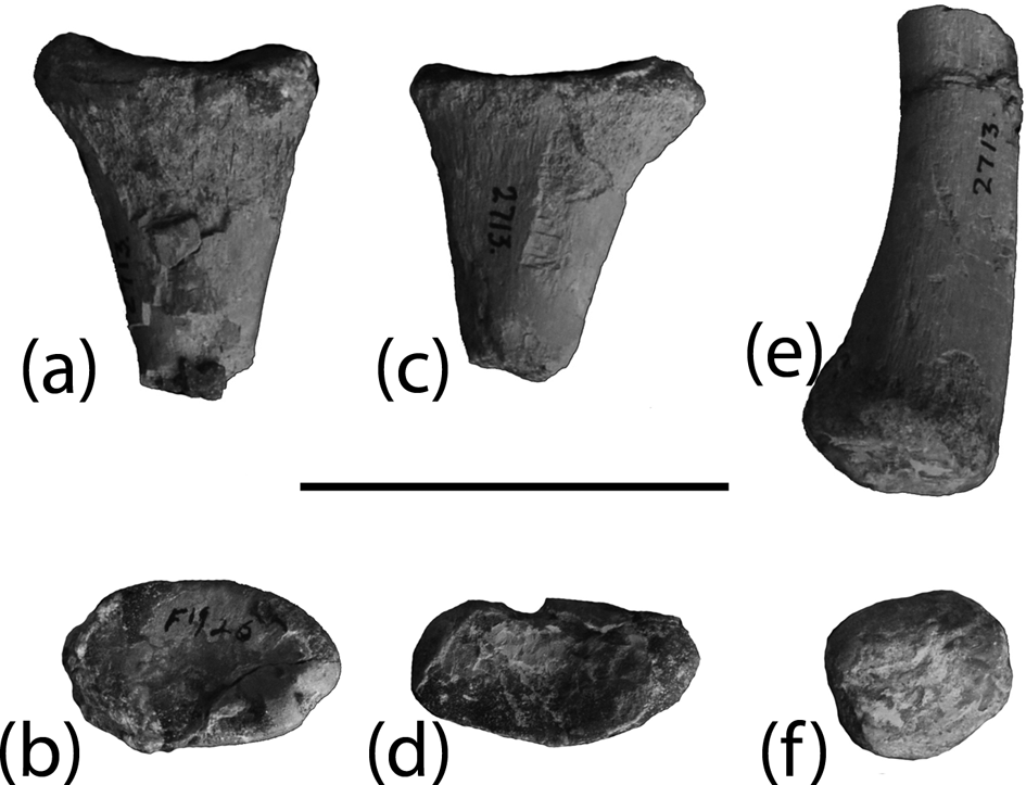
The proximal end of a left femur (AMNH FR 2709; Fig. 4e; Lucas et al. Reference Lucas, Spielmann, Heckert, Hunt, Lucas and Spielmann2007, fig. 2e) and a complete right femur (AMNH FR 2710; Fig. 5a–f; Cope Reference Cope1887a, pl. 1, figs 3, 3a–b; Huene Reference Huene1915, fig. 1; Lucas et al. Reference Lucas, Spielmann, Heckert, Hunt, Lucas and Spielmann2007, fig. 5a) are preserved in the referred material. Despite the differences in catalogue numbers, the femora are roughly the same size (Table 4), very similar morphologically, and from opposite sides, therefore they are probably from the same individual. The right femur is very well preserved. The shaft is straight in lateral view (Fig. 5a; Huene Reference Huene1915, fig. 1) and in posterior view the head is greatly expanded medially and on a distinct neck (Fig. 5b). In proximal view (Fig. 5c), the femoral head possesses three distinct anteromedial, posteromedial and anterolateral tubers (following the terminology of Nesbitt Reference Nesbitt2011). In Typothorax and aetosaurs, the posteromedial tuber is larger than the anteromedial tuber (Nesbitt Reference Nesbitt2011), but the anterolateral tuber is the largest of the three. The presence of three distinct tubers in the femoral head distinguishes aetosaur femora from those of phytosaurs, which lack a well developed anteromedial tuber. The anterolateral tuber extends distally to the base of the femoral neck. Martz (Reference Martz2002) considered this extension to be homologous with the proximal condylar fold found in Alligator (Brochu Reference Brochu1992); however, according to Nesbitt (Reference Nesbitt2011), this structure is absent in aetosaurs. The proximal surface of the femoral head is rounded and smooth, with no presence of a pronounced groove seen in some archosaur specimens.
The shaft is mediolaterally ovate in cross-section and at a point on the posterior surface just proximal to the midpoint of the shaft is a well-developed fourth trochanter (Fig. 5b). This trochanter is in the form of a crescentic ridge that opens medially (Fig. 5f). On the anterolateral face of the femoral shaft, between the position of the fourth trochanter and the femoral head, is a rugose area that may be homologous to the lateral insertion point for the pubo–ischio–femoralis internus pars dorsalis in Alligator (Rowe Reference Rowe1986; Brochu Reference Brochu1992). Just ventrolateral to this rugose surface is a distinct raised tuber (Fig. 5e) that provides this portion of the shaft with a ‘kinked’ shape in anterior view. Long & Murry (Reference Long and Murry1995) and Martz (Reference Martz2002) suggested that this distinct raised area could be the lesser (or anterior) trochanter; however, Martz (Reference Martz2002) cautioned that this structure may not be homologous with the anterior trochanter of ornithodirans. The anterior trochanter is an osteological correlate of the insertion area of the M. iliotrochantericus caudalis and M. iliofemoralis externus, both of which are derivatives of the ancestral reptilian M. iliofemoralis and insert farther down the shaft in pseudosuchians (Brochu Reference Brochu1992; Hutchinson Reference Hutchinson2001; Nesbitt et al. Reference Nesbitt, Irmis, Parker, Smith, Turner and Rowe2009). The “obturator flange” of Martz (Reference Martz2002) is the “greater trochanter” and the posterior surface of this structure has a broad, shallow depression that serves as the insertion area for the M. pubo-ischio-femoralis externus (Brochu Reference Brochu1992; Hutchinson Reference Hutchinson2001).
The distal end of the femur is expanded from the shaft and consists of the two distal condyles, separated by a shallow, posteriorly opening intercondylar groove. The distal surface between the condyles is nearly flat, with no distinct dividing groove. The lateral condyle is larger than the medial and possesses a distinct posteriorly directed tubercle, the crista tibiofibularis (Fig. 5d). In phytosaurs (e.g. AMNH FR 19337) the distal end of the femur is less expanded, the two distal condyles are more equant in size, and a distinct crista tibiofibularis is lacking. In distal view, the lateral edge of the crista tibiofibularis in AMNH FR 2710 is separated from the lateral margin of the main body of the lateral condyle by a shallow obtuse groove. On the ventral surface, the condyles are separated by a marked ovate depression. The outer margins of both condyles are rounded and the lateral edge of the lateral condyle tapers slightly. The femur of the specimen AMNH FR 2710 appears to lack the strongly tapering medial condyle seen in UCMP V2816/122674 (Long & Murry Reference Long and Murry1995, fig. 110a–c), but this could be because of breakage. Lucas et al. (Reference Lucas, Heckert, Hunt, Heckert and Lucas2002, p. 226) stated that the holotype femur of Typothorax antiquum (NMMNH P-36075) lacks the “well-developed medial condyles” of T. coccinarum, but this cannot be verified from the figures provided.
4.3.5. Other elements from AMNH FR 2710
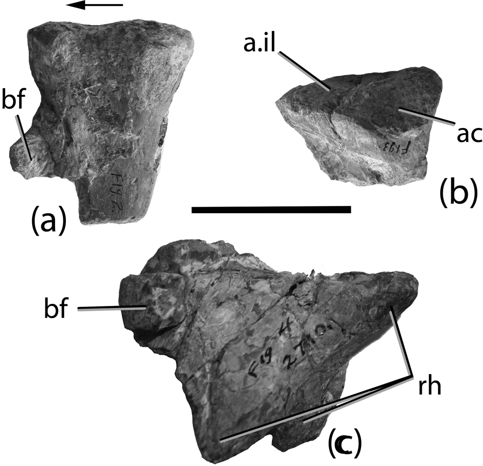
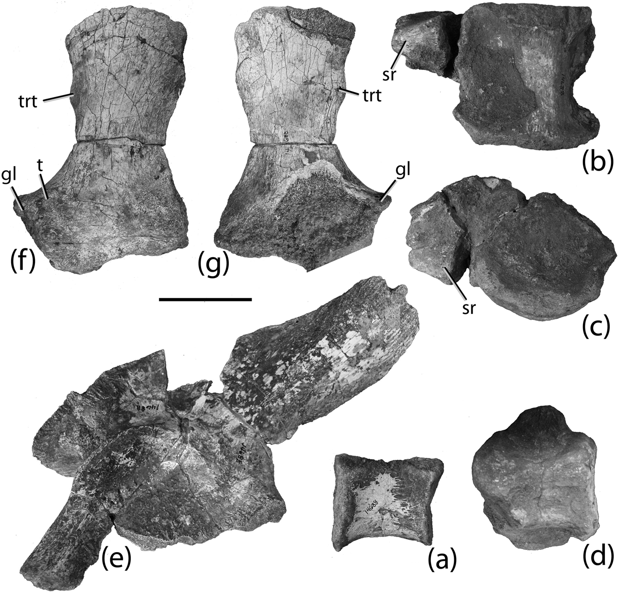
Other material from AMNH FR 2710 and considered to be part of the referred materials by Huene (Reference Huene1915) include a proximal end of a right tibia (Fig. 6a; Huene Reference Huene1915, fig. 2; Lucas et al. Reference Lucas, Spielmann, Heckert, Hunt, Lucas and Spielmann2007, fig. 5b) and the proximal end of a left ischium (Fig. 6b; distal scapula of Huene Reference Huene1915, fig. 3; Lucas et al. Reference Lucas, Spielmann, Heckert, Hunt, Lucas and Spielmann2007, fig. 5f–g). These specimens were also considered to be topotypes of T. coccinarum by Lucas et al. (Reference Lucas, Spielmann, Heckert, Hunt, Lucas and Spielmann2007); however, they both belong to a phytosaur and not to any aetosaur.
AMNH FR 2710: (a) proximal end of right phytosaur tibia in medial view; (b) proximal end of left phytosaur ischium in lateral view; (c) proximal end of phytosaur cervical rib in oblique view. Scale bar=5 cm. Arrow shows anterior direction. Abbreviations: a.il=articular surface with the ilium; ac=acetabulum; bf=bone fragment; rh=rib head.
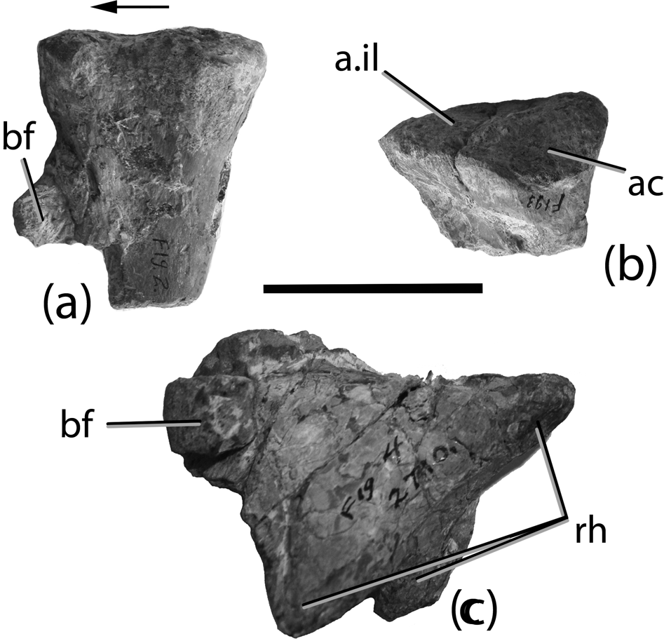
Lucas et al. (Reference Lucas, Spielmann, Heckert, Hunt, Lucas and Spielmann2007, fig. 2j) identified an element from AMNH FR 2710 (Huene Reference Huene1915, fig. 4) as a right (?) lateral osteoderm; however, the morphology of this element is not consistent with any osteoderm, and the presence of a three-pronged proximal surface (Fig. 6c) is strongly suggestive of cervical ribs from a phytosaur and consistent with Huene's (Reference Huene1915) original identification.
4.4. Summary
Reexamination of the original type and referred materials (USNM 2585, USNM 2586, ANSP 14695, AMNH FR 2709, AMNH FR 2710) demonstrates that only the following elements are most likely referable to Typothorax coccinarum. In USNM 2865, only three osteoderm fragments possess characters considered to be diagnostic of Typothorax, specifically the random pattern of circular round pits (Long & Ballew Reference Long and Ballew1985). This is also the case for the single osteoderm fragment from the purported syntype specimen ANSP 14695. Furthermore, this latter element possesses the ventral thickening also considered typical for the genus. The referred material (AMNH FR 2709, AMNH FR 2710) mostly is assignable to T. coccinarum based on the ornamentation and morphology of the osteoderms, as well as on the association of the partial left femur and morphological correspondence of the right femur. Moreover, these femora possess characteristics found only in aetosaurs and not in phytosaurs supporting their referral. The other material in these samples is either non-diagnostic or referable to a phytosaur.
4.5. Discussion
In the last century, much material from North America has been referred to Typothorax; however, the material used for the basis of these referrals has not been the original syntype material, but rather the referred material that David Baldwin collected in 1881. Thus, this specimen has served as a proxy “holotype”.
This is because the type materials of Typothorax coccinarum (USNM 2585) are extremely fragmentary and, although the dorsal surface possesses a random patterning of shallow rounded pits that is found in osteoderms referred to this taxon (e.g. AMNH FR 2709, UCMP V2816/34227), this ornamentation pattern is also found in portions of paratypothoracisin osteoderms. Furthermore, Lucas et al. (Reference Lucas, Heckert, Hunt, Heckert and Lucas2002) erected a second species of Typothorax, T. antiquum, which possesses very similar osteoderm ornamentation (Parker & Martz Reference Parker and Martz2011). Although Lucas et al. (Reference Lucas, Heckert, Hunt, Heckert and Lucas2002) listed slight differences in this patterning, the fragments from USNM 2585 are not complete enough to allow differentiation at the species level. I have argued elsewhere (Parker Reference Parker, Parker, Ash and Irmis2006; Parker & Martz Reference Parker and Martz2011) that T. antiquum simply represents a smaller individual of T. coccinarum, rendering this lack of differentiation extraneous; however, this does not resolve the similarities found in paratypothoracisin plates and suggests that T. coccinarum, might possibly be a nomen dubium. Certainly, the majority of aetosaur workers would like to see this name preserved for taxonomic stability and AMNH FR 2709 would serve superbly as a neotype; however, the original type specimens have not been lost, and therefore a case would need to be made to the International Committee on Zoological Nomenclature to create a neotype. Unfortunately, another name is available, Episcoposaurus horridus (see description below), which could severely weaken any proposal to designate a neotype. However, the lectotype of E. horridus is a large femur (AMNH FR 2713) that lacks key diagnostic features. The only other element in AMNH FR 2713 that possesses discrete characters found in referred specimens of Typothorax coccinarum (e.g. NMMNH P-56299) is a caudal paramedian osteoderm. Thus, AMNH FR 2713 is lacking the dorsal paramedian osteoderms of the genus that have come to characterise the taxon over the last three decades (see Long & Ballew Reference Long and Ballew1985). Furthermore, distinct differences between more distal caudal paramedians of T. coccinarum and T. antiquum have not been demonstrated. As mentioned above, the presence of dorsal paramedian osteoderms, the fact that this specimen is purportedly from the type locality and most likely a topotype, and its historic use as a “proxy holotype” makes AMNH FR 2709 a good candidate for a neotype designation.
Episcoposaurus horridus Cope, Reference Cope1887a
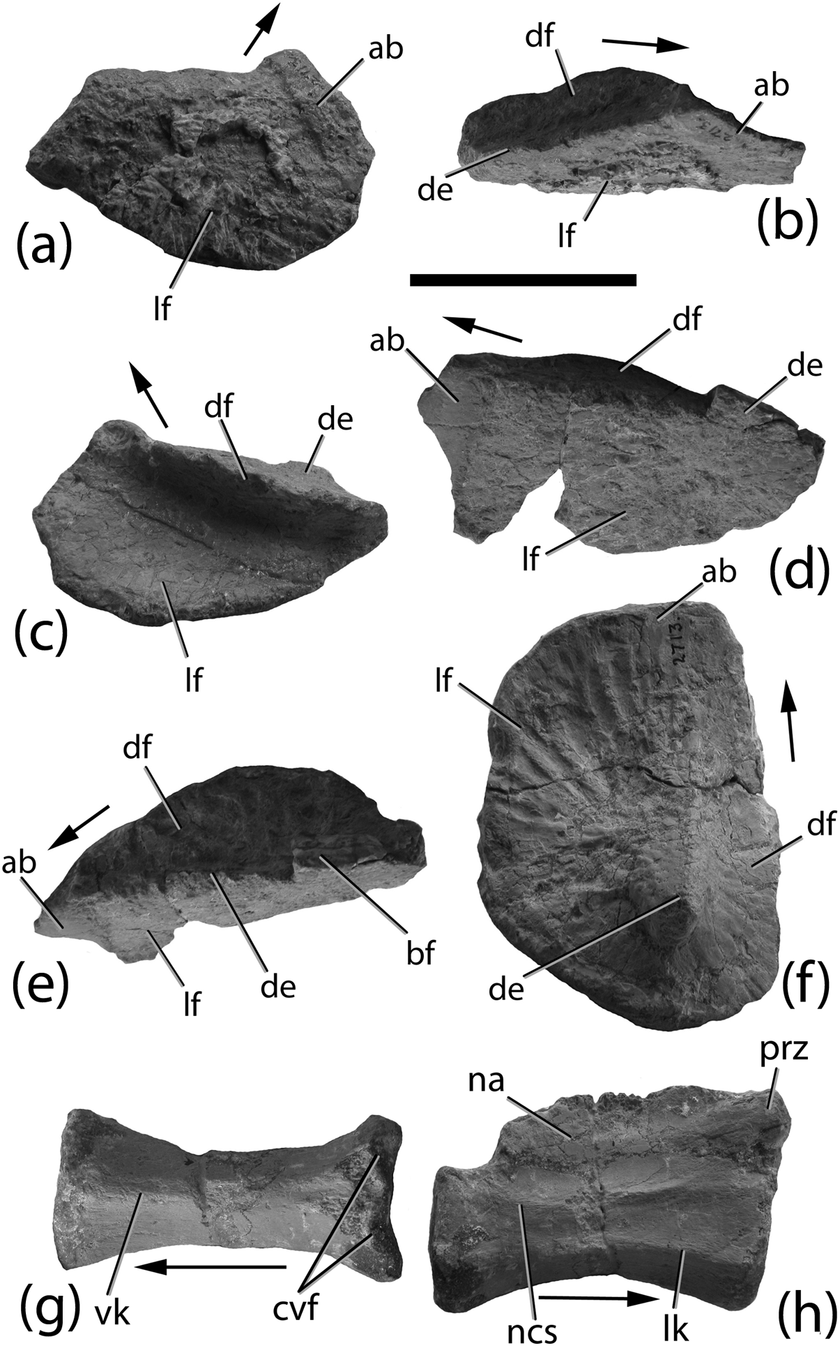
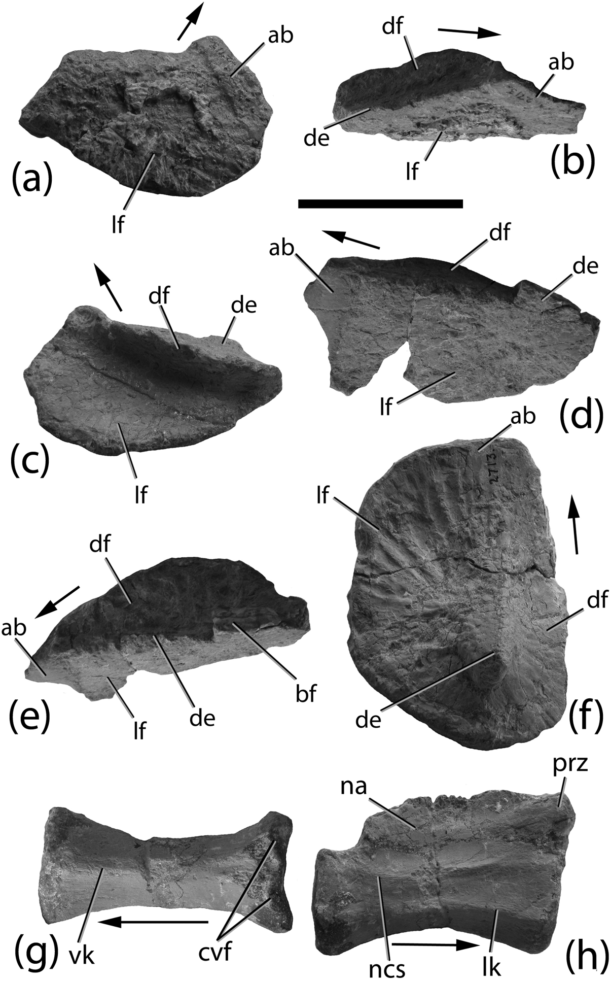
Episcoposaurus horridus referred osteoderms and vertebra (AMNH FR 2713): (a–c) right mid-dorsal lateral osteoderm in (a) lateral, (b) dorsolateral and (c) medial views; (d–e) left posterior mid-dorsal lateral osteoderm in (d) lateral and (e) dorsolateral views; (f) left dorsal posterior mid-caudal osteoderm in dorsolateral view; (g–h) distal caudal vertebra in ventral (g) and right lateral (h) views. Scale bar=5 cm. Arrows indicate anterior direction. Abbreviations: ab=anterior bar; bf=bone fragment; cvf=chevron facets; de=dorsal eminence; df=dorsal flange; lf=lateral flange; lk=lateral keel; na=neural arch; ncs=neurocentral suture; prz= prezygapophyses; vk=ventral keel.
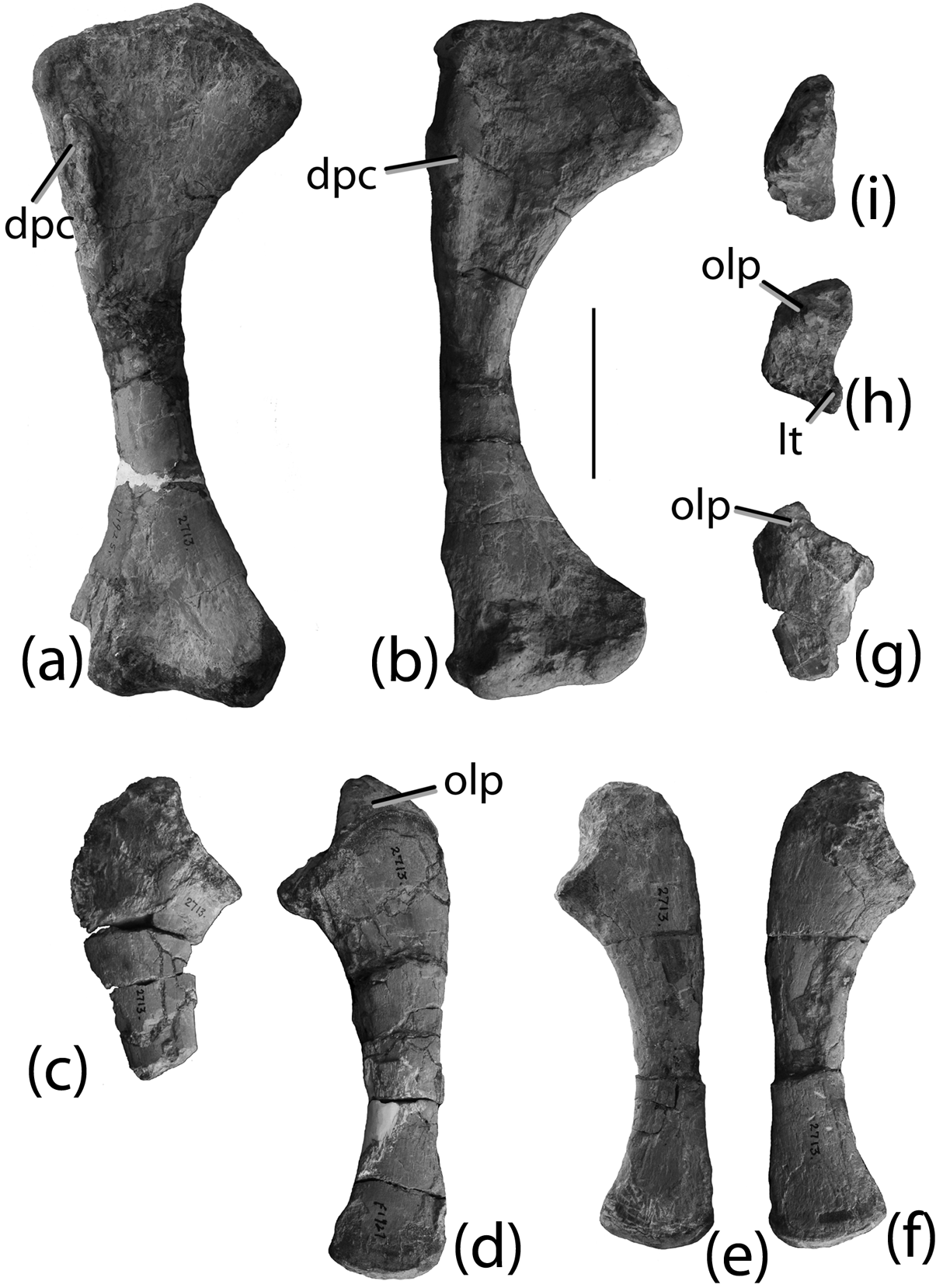
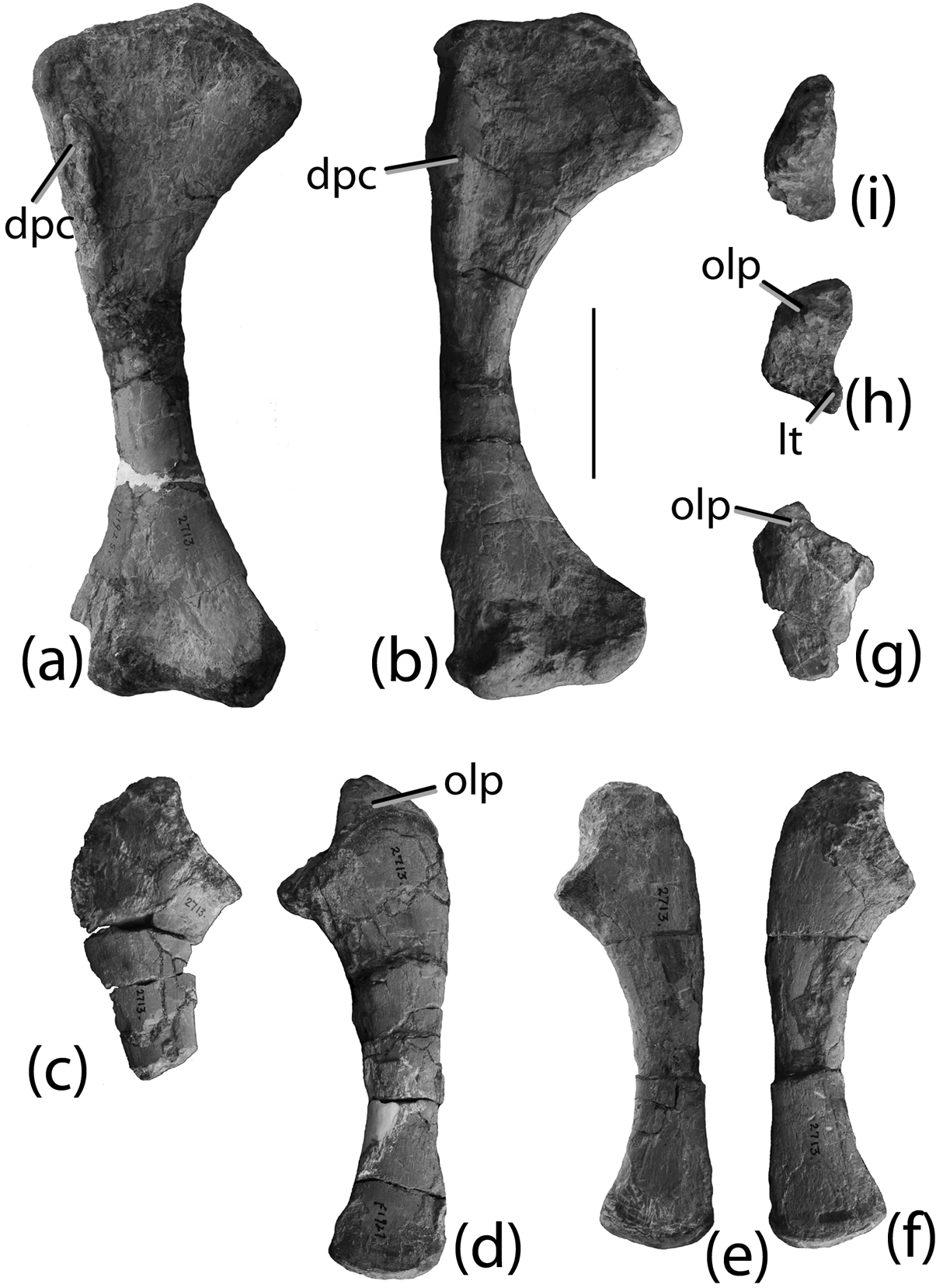
Forelimb elements (AMNH FR 2713): (a) phytosaur right humerus in anterior view; (b) phytosaur right humerus in anterior view; (c) proximal end of phytosaur right ulna in lateral view; (d) phytosaur left ulna in lateral view; (e–f) phytosaur right ulna in medial (e) and lateral (f) views; (g–h) proximal end of aetosaur right ulna in lateral (g) and proximal (h) views; (i) right phytosaur ulna in proximal view (same specimen as e and f). Scale bar=5 cm. Abbreviations: dpc=deltopectoral crest; lt=lateral tuber; olp=olecranon process.
Archosauriform radii (AMNH FR 2713): (a–b) proximal end in either anterior or posterior (a) and proximal views (b); (c–d) proximal end in either anterior or posterior (c) and proximal views (d); (e–f) distal end of an aetosaur radius in either anterior or posterior (e) and proximal views (f). Scale bar=5 cm.
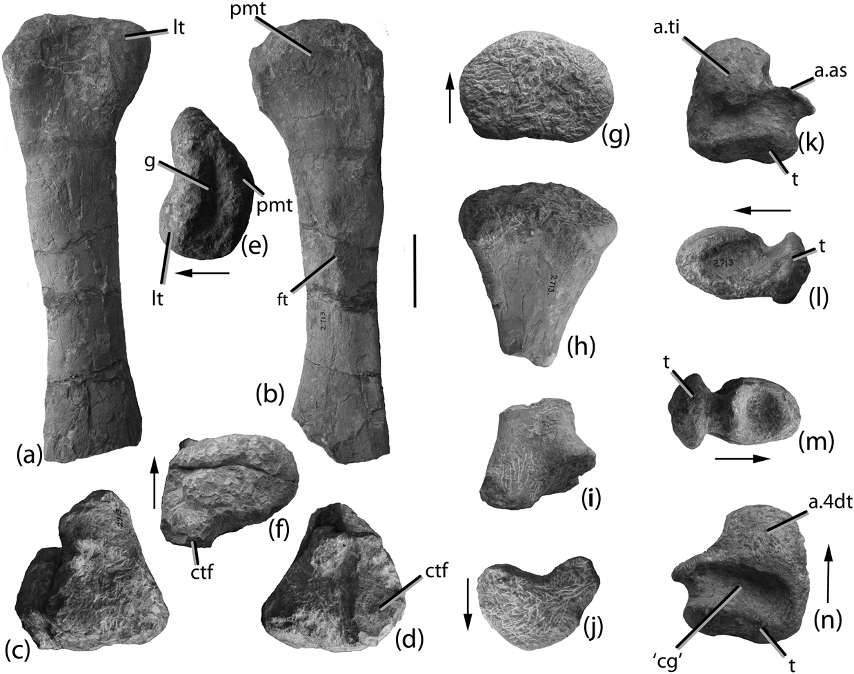
Episcoposaurus horridus lectotype and referred specimens (AMNH FR 2713): (a–b) proximal end of right femur in anterior (a) and posterior (b) views; (c–d) distal end of right femur in anterior (c) and posterior (d) views; (e) right femur in proximal view; (f) right femur in distal view; (g–h) proximal end of right tibia in proximal (g) and posterior (h) views; (i–j) distal end of right tibia in posterior (i) and distal (j) views; (k–n) left calcaneum in proximal (k), lateral (l), medial (m) and distal (n) views. Scale bar=5 cm. Arrows indicate anterior direction. Abbreviations: a.as=articular area for the astragalus; a.ti=articular area for the tibia; a.4dt=articular area for distal tarsal 4; ‘cg’=’clear gap’; ctf=crista tibiofibularis; ft=fourth trochanter; g=groove; lt=lateral tuber; pmt=posteromedial tuber; t=tuber.

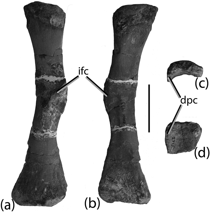
Other material: (a–b) aetosaur right fibula (AMNH FR 19338) in lateral (a) and medial (b) views; (c–d) silesaurid proximal humerus (AMNH FR 2713) in proximal (c) and anterior (d) views. Scale bar=5 cm. Abbreviations: dpc=deltopectoral crest; ifc=iliofibularis crest.

Lectotype. AMNH FR 2713 (formally AMNH FR 2307), right femur. Lucas et al. (Reference Lucas, Spielmann, Heckert, Hunt, Lucas and Spielmann2007) designated a complete left dorsal caudal paramedian osteoderm as the lectotype of E. horridus; however, in the same paper they noted that Huene (Reference Huene1915) had previously designated the femur as the lectotype. This was also noted by Gregory (Reference Gregory1953).
Paralectotypes. AMNH FR 2713, tibia, calcaneum, caudal centrum, caudal osteoderm. These specimens are apparently from same locality as the type of T. coccinarum (Huene Reference Huene1915); however, Huene (Reference Huene1915) erroneously considered the material referred by Cope (Reference Cope1887a) (AMNH FR 2709, AMNH FR 2710) to be the type specimen of T. coccinarum so this statement is ambiguous. Huene (Reference Huene1915) restricted the type materials of E. horridus to a large femur and referred forelimb bones from AMNH FR 2713 to a small “mystriosuchid” phytosaur.
Note. The material catalogued under AMNH FR 2713 clearly belongs to several different animals, a very large aetosaur, a smaller aetosaur, at least one phytosaur, and possibly a silesaurid. Mainly the aetosaur material is described in detail below and the lectotype and paralectotype specimens are restricted to the larger aetosaur material (i.e. femur, tibia, calcaneum, caudal osteoderm, distal caudal vertebra). The material matches well in size and preservation without an overlap of elements and therefore probably belongs to a single individual. The smaller aetosaur elements (i.e. dorsal lateral osteoderms, proximal end of ulna) match well with and could actually belong to the referred Typothorax specimen (ANMH FR 2709, AMNH FR 2710).
5. Description
5.1. Dorsal region osteoderms
AMNH FR 2713 contains two dorsal lateral osteoderms, one from the left side and the other from the right. The right side osteoderm (Fig. 7a–c; Huene Reference Huene1915, fig. 23a–b; Lucas et al. Reference Lucas, Spielmann, Heckert, Hunt, Lucas and Spielmann2007, fig. 3c–d) is the smaller of the two, with a length of 84 mm, and consists of distinct dorsal and lateral flanges at about 90° to each other and separated by a distinct sharp anteroposteriorly aligned ridge (dorsal eminence). In medial view, the ridge rises gently to an apex situated near the posterior osteoderm margin, and posterior to this apex plunges sharply posteroventrally terminating at the osteoderm margin (Fig. 7a). The dorsal flange is reduced greatly compared to the lateral flange, as is typical for typothoracisin aetosaurs, and it has a gently rounded medial edge that articulates with a corresponding paramedian osteoderm (Fig. 7b–c). The lateral flange is dorsoventrally deep, and possesses a gently rounded ventral margin (Fig. 7c). The dorsal portion of the anterolateral margin bears a raised anterior bar that extends slightly onto the dorsal flange. The posterior margin of the anterior bar is straight and slopes dorsolaterally and anteriorly. In lateral view, the posteroventral margin of the lateral flange slopes posteroventrally and medially, forming a 90° angle with the anterior bar. The dorsal surface ornamentation appears to have been obliterated during removal of the adhering matrix and large patches of matrix remain. Comparison with known articulated Typothorax specimens (e.g. NMMNH P-56299; Heckert et al. Reference Heckert, Lucas, Rinehart, Celeskey, Spielmann and Hunt2010) suggests that this is an anterior dorsal lateral osteoderm and was originally situated anterior to the tenth dorsal position.
The left side osteoderm (Fig. 7d–e; Huene Reference Huene1915, fig. 22a–b; Lucas et al. Reference Lucas, Spielmann, Heckert, Hunt, Lucas and Spielmann2007, fig. 2i) is larger (length of 94 mm) and therefore from further back in the carapace. This osteoderm is very similar to the one previously described, except that the angle between the flanges is more acute and the dorsal flange is larger in relation to the lateral flange, nearly two-thirds the size (Table 3). As with the right side, osteoderm the dorsal ornamentation is also obliterated or covered with matrix on this specimen; however, the raised smooth anterior bar is visible, despite the anteroventral portion being broken away. The inner surface of the osteoderm is still buried in matrix, giving it the triangular cross section depicted by Huene (Reference Huene1915, fig. 22b) and a small bone fragment of another element is adhered to the posterodorsal surface.
5.2. Caudal region osteoderms
Caudal paramedian osteoderm (Fig. 7f; Huene Reference Huene1915, fig. 24a, b; Gregory Reference Gregory1953, fig. 17a, b; Lucas et al. Reference Lucas, Spielmann, Heckert, Hunt, Lucas and Spielmann2007, fig. 3a). The dorsal surface of this osteoderm is well preserved, but the ventral surface is still covered in matrix. Comparison with MCZ 1488 shows that this is a left dorsal caudal osteoderm from the distal portion of the middle of the tail (Lucas et al. Reference Lucas, Spielmann, Heckert, Hunt, Lucas and Spielmann2007). In dorsal view, the osteoderm is suboval in shape, with a straight medial edge and rounded anterior, lateral and posterior edges. The anterior portion possesses an anterior bar, which is flush with, rather than raised above, the rest of the dorsal surface sculpturing. There is a large pyramidal dorsal eminence in the posterior portion of the osteoderm, which does not contact the posterior plate margin. This eminence is situated slightly medial of centre and from its apex a thin ridge extends obliquely (anteromedially) to the anteromedial corner of the osteoderm, effectively dividing it into dorsal and lateral flanges. The area of the dorsal flange is approximately half that of the lateral flange. An ornamentation of elongate grooves and flat ridges radiates from the dorsal eminence.
5.3. Caudal vertebra
A single elongate amphicoelous vertebra is present in the material (Fig. 7g–h; Huene Reference Huene1915, fig. 19; Lucas et al. Reference Lucas, Spielmann, Heckert, Hunt, Lucas and Spielmann2007, fig. 2a–c). The specimen is much longer than wide and hour-glass shaped in ventral view (Fig. 7g). Ventrally there are two chevron facets and a broad, yet weak ventral keel. Laterally, near the base of the centrum, there are horizontal keels that extend between the mid-points of the circular articular faces (Fig. 7h). A portion of the neural arch is present. The neural spine is broken, but must have been elongate and low. The postzygapophyses are broken away, whereas the prezygopophyses are mostly present but encased in matrix. The morphology of this element is consistent with that of an aetosaur and the size of the element is predicted by the size of the lectotype femur and the paralectotype caudal osteoderm. Thus, this element is retained in the lectotype material.
The anterior caudal vertebra (Huene Reference Huene1915, fig. 18) could not be located in the material.
5.4. Humeri
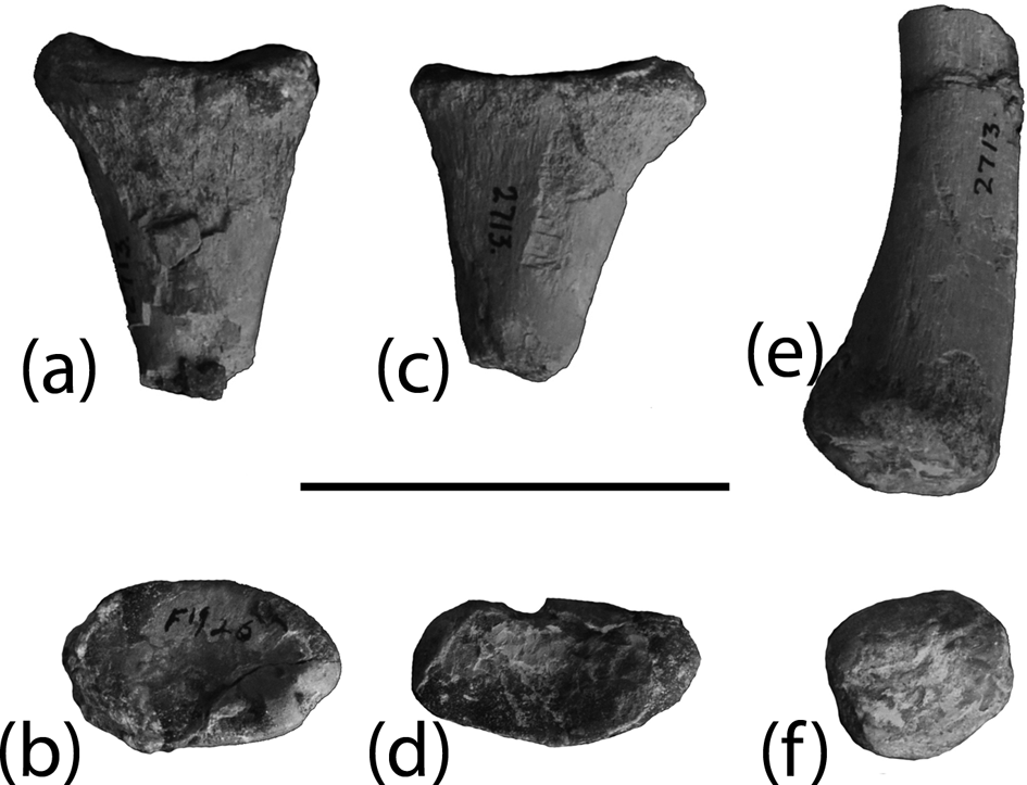
There are two right humeri under the catalog number AMNH FR 2713 (Fig. 8a–b). Because only a single humerus (Fig. 8a) was mentioned by Cope (Reference Cope1887a) and Huene (Reference Huene1915), the provenance of the second specimen (Fig. 8b) is not certain; however, both possess straight lateral margins and belong to phytosaurs (Nesbitt Reference Nesbitt2011). Furthermore, the area between the distal articular surfaces is flat, without the distinct deep sulcus found in aetosaurs (e.g. Martz Reference Martz2002, fig. 4.10). The larger of the two measures 220 mm in length and was described as part of the syntype material by Cope (Reference Cope1887a) and Huene (Reference Huene1915, fig. 25). Huene (Reference Huene1915) argued that the forelimb bones were too reduced in comparison to the hindlimb bones to belong to the same animal, and removed them from the syntype series and subsequently assigned them to a ‘mystriosuchid’-type phytosaur. In contrast, Gregory (Reference Gregory1953) argued that on the basis of comparison with the skeleton of Longosuchus meadei (Sawin, Reference Sawin1947), the limb proportions were acceptable and thus reinstated the forelimb elements as part of the syntype series. Unfortunately, he was not able to recognise that these elements were phytosaurian and therefore not referable to Typothorax or E. horridus. Likewise, Lucas et al. (Reference Lucas, Spielmann, Heckert, Hunt, Lucas and Spielmann2007, fig. 4a) also did not recognise the phytosaurian identity of these elements. Furthermore, they (p. 242) noted that the humerus was similar in morphology to a humerus (UCMP V2816/34240) figured by Long & Murry (Reference Long and Murry1995, fig. 105d–e) as Typothorax coccinarum. Unfortunately, this humerus was also misidentified and is phytosaurian as well (Martz Reference Martz2002). Therefore, the humerus of E. horridus is unknown.
5.5. Ulnae
AMNH FR 2713 contains portions of four ulnae, two complete (left and right; Fig. 8d–f) and two proximal ends (both rights; Fig. 8c, g). The left ulna (Fig. 8d) and a proximal end of a right (Fig. 8c) are about the same size and may represent a matching pair. Huene (Reference Huene1915, fig. 27) figured the left element and assigned it to a small phytosaur, whereas Gregory (Reference Gregory1953) and Lucas et al. (Reference Lucas, Spielmann, Heckert, Hunt, Lucas and Spielmann2007) considered it to be aetosaurian. Lucas et al. (Reference Lucas, Spielmann, Heckert, Hunt, Lucas and Spielmann2007, fig. 4d–e) also figured a 5th specimen, which the present author was unable to locate in the materials.
Phytosaurian and aetosaurian ulnae are superficially similar in their overall morphology except for one key detail; in aetosaurs the glenoid surface is much expanded and possesses a well-defined rim and lateral tuber (Nesbitt Reference Nesbitt2011; Fig. 8h). Thus the left ulna figured by Huene (Reference Huene1915) and Lucas et al. (Reference Lucas, Spielmann, Heckert, Hunt, Lucas and Spielmann2007) (Fig. 8d) belongs to a phytosaur, as does its possible counterpart (Fig. 8c) and the complete right ulna (Fig. 8e, f, i). However, one of the right proximal ends (Fig. 8g–h) possesses aetosaurian characteristics. In proximal view, the concave glenoid fossa is subrectangular, unlike the semilunate surface in phytosaurs (compare Figs 8h and 8i). The olecranon process is a raised and thickened knob on the posterior portion of the glenoid fossa. It does not taper posteriorly as described for T. antiquum (Lucas et al. Reference Lucas, Heckert, Hunt, Heckert and Lucas2002). The lateral portion of the glenoid fossa is a thickened rim that is expanded in relation to the lateral surface of the bone shaft, and ends in a distinct lateral (radial) tuber. The medial surface of the proximal head is flattened. The anteromedial corner of the glenoid fossa has a distinct anteromedially projecting lip that slightly overhangs the flattened medial surface of the head and is the medial extension of the radial tuber. The midshaft and distal end of this element is not preserved.
5.6. Radii
The proximal ends of two radii are present in the material (Fig. 9a–d). Huene (Reference Huene1915, fig. 26) figured one of them (Fig. 9a–b), although the more distal portion now appears to be missing (see Lucas et al. Reference Lucas, Spielmann, Heckert, Hunt, Lucas and Spielmann2007, fig. 4F). The proximal surfaces are convex, ovate in proximal view and possess a flaring rim. The shaft is slightly ovate in shape. Among phytosaurs and aetosaurs these specimens most closely resemble the proximal radii of Typothorax antiquum (Lucas et al. Reference Lucas, Heckert, Hunt, Heckert and Lucas2002, fig. 7c), although assignment to a phytosaur cannot be completely ruled out because they are also similar. A distal end is also present with a rounded rather than compressed cross-section that corresponds nicely to an aetosaur rather than to a phytosaur (Fig. 9e–f; Nesbitt Reference Nesbitt2011); however, it is not possible to determine to which proximal head it belongs.
5.7. Femur
A very large and mostly complete right femur is the most striking bone of the collection, predominantly because of its size (Fig. 10a–f). Cope (Reference Cope1887a) considered this element to be characteristic for Episcoposaurus in that it was straighter and more robust than the femur of Typothorax (AMNH FR 2710), which prompted Huene (Reference Huene1915, fig. 12) to designate it as the lectotype. The specimen is separated into two pieces: (1) the proximal end and much of the shaft, which is 29·5·cm in length (Fig. 10a–b); and (2) the distal condyles, which are 9·8 cm in length (Fig. 10c–d). The complete element would have measured over 40 cm in length (Huene Reference Huene1915). The shaft is robust and straight, different from the more sigmoidal shaft in phytosaurs (e.g. PEFO 31219). The fourth trochanter is situated approximately 14 cm distal from the proximal end of the element. The trochanter is damaged, but is moundlike and rounded (sensu Nesbitt Reference Nesbitt2011, char. 316:0), not a sharp crescentic ridge as in AMNH FR 2710, and is located on the posterior face of the shaft (Fig. 10b). The shaft is oval in cross section and mediolaterally narrows slightly distal to the position of the 4th trochanter before expanding again more distally.
The femoral head is also badly abraded and much of the anteromedial tuber has been broken or worn away (Fig. 10e). Nonetheless, the three tubers are still apparent distinguishing this femur from that of phytosaurs (Nesbitt Reference Nesbitt2011). The proximal surface is marked by a deep, but narrow mediolaterally oriented sulcus, similar to what is present in other archosauriforms such as Effigia (Nesbitt Reference Nesbitt2007). Similar sulci are randomly present in other aetosaur and phytosaur specimens and appear to represent incomplete ossification of the proximal articular surface. The section with the distal condyles is poorly preserved, not completely prepared and relatively unremarkable, except for the presence of a broken, but prominent crista tibiofibularis (Fig. 10c–d, f).
5.8. Tibia
Distal and proximal portions of a large right tibia are present in AMNH FR 2713 (Huene Reference Huene1915, figs 13–14; Lucas et al. Reference Lucas, Spielmann, Heckert, Hunt, Lucas and Spielmann2007, fig. 5c–e). Overall it is much more similar to TTU P-9214, than to the Canjilon Quarry material (UCMP V2816/122678; Martz Reference Martz2002), as it lacks the profound proximal expansion found in the UCMP specimens (Long & Murry Reference Long and Murry1995, fig. 111). The proximal end is poorly preserved and still marked with numerous tool marks from mechanical preparation (Fig. 10g). The posterior face possesses a distinct concave surface (Fig. 10h), whereas the anterior surface is convex. The lateral edge is thinner anteroposteriorly than the medial edge, broadening proximally into a distinct but broken posterolaterally directed tuber. Although the majority of the shaft is missing, the proximal portion that is preserved is smooth and slightly anteroposteriorly compressed. The distal end of the tibia (Fig. 10i–j) is crescent-shaped in ventral view, with a dorsoventrally oriented groove that provides the specimen with the typical “screw-joint” morphology (sensu Gauthier Reference Gauthier1986) found in suchians and not in phytosaurs (Nesbitt Reference Nesbitt2011).
5.9. Calcaneum
AMNH FR 2713 includes a very large and well preserved left calcaneum (Fig. 10k–n; Huene Reference Huene1915, fig. 15; Long & Murry Reference Long and Murry1995, fig. 112a–d; Lucas et al. Reference Lucas, Spielmann, Heckert, Hunt, Lucas and Spielmann2007, fig. 5k–m). This element is dorsoventrally compressed and very broad. The facet for the tibia on the dorsal surface is well-developed, convex and hemi-cylindrical in shape, typical for suchians (Fig. 10k; Sereno Reference Sereno1991; Nesbitt Reference Nesbitt2011). Posterior to this is a deep transverse trough which is confluent with the dorsal surface of the medial projection or lip that outlines the concave facet that is the articulation point for the astragalus (Brochu Reference Brochu1992). This trough (the dorsal portion of the calcaneal shaft sensu Nesbitt Reference Nesbitt2011) effectively separates the tibial facet from the posterior portion of the posteriorly directed calcaneal tuber, which is delimitated by a dorsoventrally expanded lip. Lateral to the tibial facet and dorsal trough is an ovate strongly concave depression that opens dorsolaterally, and probably articulated in part with the 5th metatarsal (Fig. 10l; Walker Reference Walker1961). The concave articulation point for the astragalus is shallow. The lip or spout for this articulation extends medially just past the medial expansion of the posterior tuber. The medial surface of the posterior tuber between the tuber distal expansion and the lip or spout is strongly concave (Fig. 10m). Ventrally, the articular surface for the 4th distal tarsal is flat. Posterior to this surface, the main body of the posterior tuber is deeply excavated into a mediolaterally broad rectangular fossa (Fig. 10n; the “clear gap” of Nesbitt Reference Nesbitt2011). A similarly deep depression is found in all aetosaurs and varies in form among taxa, but appears to be much less developed in the holotype specimen of Typothorax “antiquum” (NMMNH P-36075; Lucas et al. Reference Lucas, Heckert, Hunt, Heckert and Lucas2002, fig. 9a). The posterior surface of the calcaneal tuber in AMNH FR 2713 is only slightly concave, lacking the deep groove found in Alligator (Brochu Reference Brochu1992) and other paracrocodylomorphs (Nesbitt Reference Nesbitt2011).
5.10. Other material
5.10.1. Fibula
An isolated right fibula numbered AMNH FR 19338 was also collected by David Baldwin near Gallina (Fig. 11a–b). Its association with the other specimens is unclear and it was never described by Cope or Huene; however, the presence of a hypertrophied iliofibularis trochanter, which gives the element a distinctive kinked appearance, is diagnostic of aetosaurian fibulae. Both ends are abraded and much of the iliofibularis trochanter is broken away. Overall, the specimen compares nicely with UCMP V2816/34238 (Martz Reference Martz2002); however, it cannot be directly referred to Typothorax coccinarum without knowing the association with the other material.
5.10.2. Fragmentary elements
AMNH FR 2713 includes fragments of other bones collected with the main body of material. Several of these appear to be rounded long bone ends and fragments of metatarsals/carpals. There is a flat fragment that could represent the proximal end of a phytosaur interclavicle.
One very interesting fragment (Fig. 11c–d) appears to possibly represent the proximal end of a right humerus of a dinosauriform (e.g. Silesaurus; Dzik Reference Dzik2003, fig. 9b). The bone is relatively anteroposteriorly thin and featureless. The dorsal surface is horizontal, unlike the concave surfaces of phytosaurs and pseudosuchians. In proximal view, it is crescentic in shape, with the lateral edge curving and tapering anteriorly forming a slight deltopectoral ridge, similar to that of Silesaurus. Unfortunately, the humerus is not known for Eucoelophysis baldwini, the holotype of which was also found in Rio Arriba County (Nesbitt et al. Reference Nesbitt, Irmis and Parker2007), so a direct comparison with that taxon cannot be made.
5.11. Summary
Material from AMNH FR 2713 referable to Typothorax coccinarum includes the three osteoderms, a proximal end of a right ulna, the distal radius, the right femur, the left calcaneum, the tibia fragments and, probably, the proximal ends of the two radii. These elements can be grouped by size, with the caudal paramedian osteoderm, femur, tibia and calcaneum belonging to a larger specimen of Typothorax. A right fibula (AMNH FR 19338) from the same locality is also aetosaurian and most likely belongs to the smaller Typothorax specimen. The two humeri and the one complete and two partial ulnae belong to phytosaurs. Finally, a fragment of the proximal end of a humerus is consistent in its morphology to a silesaurid dinosauriform.
5.12. Discussion
Cope (Reference Cope1887a) separated the osteoderm types, assigning those with rectangular plates to T. coccinarum, and those with sharp ‘spines’ to E. horridus. This is probably the rationale to assigning the species haplocerus (see description below) to Episcoposaurus, which would be plausible given the understanding of aetosaur morphology in 1892. Because he had access to better material (e.g. MCZ 1488), Gregory (Reference Gregory1953) correctly recognised that the osteoderms with the sharp spines simply represent elements from the tail of Typothorax coccinarum. The latter author also noted that despite the differences in size, the femora of AMNH FR 2710 and AMNH FR 2713 corresponded morphologically. Gregory (Reference Gregory1953) used these characteristics to support his proposed synonymy of T. coccinarum and E. horridus. Indeed, there are no characteristics in the aetosaur material of AMNH FR 2713 that do not support referral of the material to T. coccinarum.
Episcoposaurus haplocerus Cope, Reference Cope1892
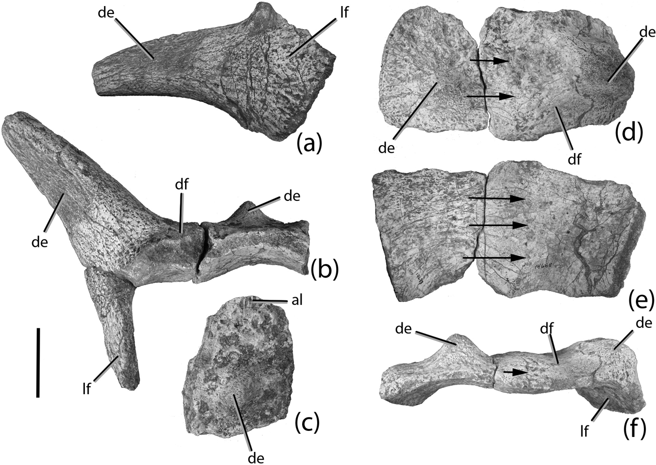
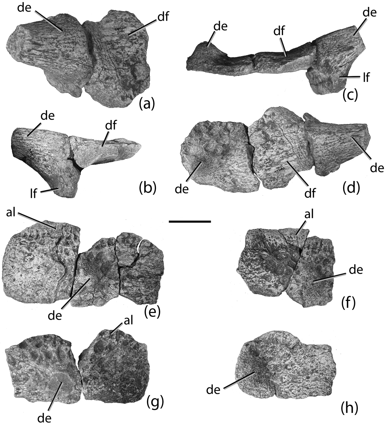
“Episcoposaurus” haplocerus cervical and anterior dorsal osteoderms (ANSP 14688): (a) right posterior cervical lateral osteoderm in ventral view; (b) articulated right posterior cervical lateral and paramedian osteoderms in anterior view; (c) left posterior cervical paramedian osteoderm in dorsal view; (d–f) fused right anterior dorsal lateral and paramedian osteoderms in dorsal (d), ventral (e) and posterior (f) views. Scale bar=5 cm. Arrows indicate suture between paramedian and lateral osteoderms. Abbreviations: al=anterior lamina; de=dorsal eminence; df=dorsal flange; lf=lateral flange.

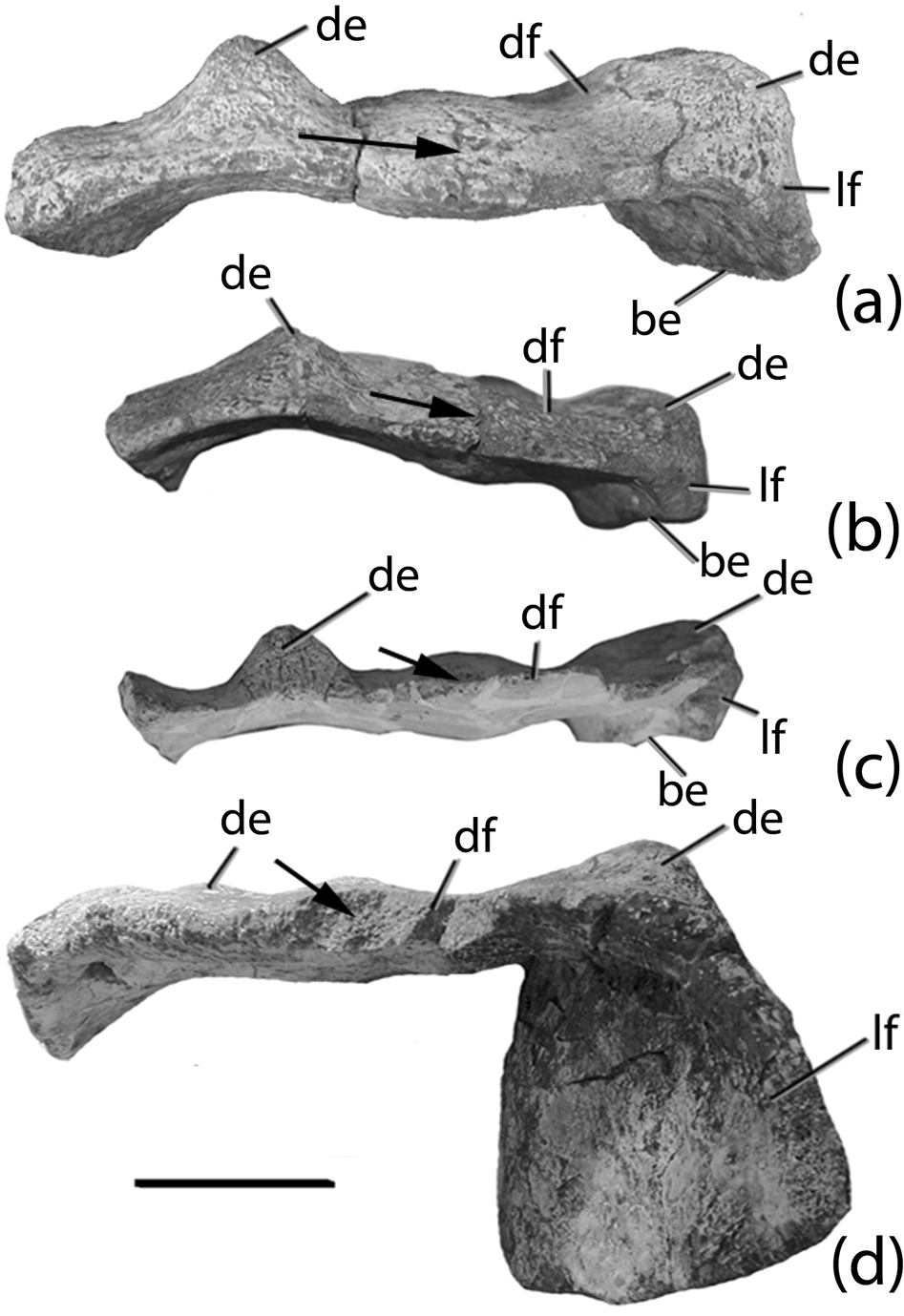
Desmatosuchus fused right lateral and paramedian osteoderms of the 1st dorsal series in posterior views: (a) ANSP 14688 (D. haplocerus); (b) TMM 31173-54; (c) TMM 31213-1; (d) MNA V9300 (D. spurensis). Scale bar=5 cm. Arrows indicate suture between paramedian and lateral osteoderms. Abbreviations: be=broken edge; de=dorsal eminence; df=dorsal flange; lf=lateral flange.

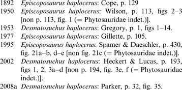
Lectotype. ANSP 14688, partial skeleton. Heckert & Lucas (Reference Heckert, Lucas, Heckert and Lucas2002) provided a lectotype designation, choosing a single lateral osteoderm to serve this role; however, Gregory's (Reference Gregory1953) referral to the material as the ‘type’ serves as an original lectotype designation (ICZN 1999, Article 74.6.1.1) and this encompasses the entire specimen ANSP 14688, minus the phytosaur material explicitly removed by Gregory.
Topotypes. TMM 40041-3 (originally TMM 18569-1), two partial cervical lateral osteoderms (Wilson Reference Wilson1950); TMM 40041-5, cervical osteoderm fragments.
ANSP 14688 consists of the fairly complete portion of the posterior cervical and anterior dorsal carapace from the right side of an aetosaur specimen. There is also a single cervical paramedian osteoderm from the left side. A partial scapula, a partial ilium, a sacral vertebral centrum and a dorsal vertebral centrum are included in ANSP 14688 which may or may not belong to an aetosaur (see discussion below). Based on comparison with known Desmatosuchus specimens (UMMP 7476, MNA V9300), ANSP 14688 contains osteoderms mostly from the anterior portion of the carapace, although one set of osteoderms has a morphology that is more consistent with the pelvic osteoderms (see below). In total, there are four lateral and at least eight paramedian osteoderms. In addition, there are boxes with more than 70 osteoderm fragments. A quick examination showed that at least a few fit together; therefore, it is possible that much of this armour could be put back together with a few hours of preparation time.
Cope (Reference Cope1892) provided the initial description of this material, but did not figure any specimens. Gregory (Reference Gregory1953) redescribed the material and was the first to provide figures. Later figures were provided by Spamer & Daeshler (Reference Spamer and Daeschler1995) and Gillette (Reference Gillette1977). The specimen was subsequently figured and briefly redescribed by Heckert & Lucas (Reference Heckert, Lucas, Heckert and Lucas2002); however, this description is difficult to follow because the same osteoderms are often figured as different elements (e.g. Heckert & Lucas Reference Heckert, Lucas, Heckert and Lucas2002, fig. 2), and in many cases elements are described as oriented on the wrong side of the body (see full discussion by Parker Reference Parker2008a). Parker (Reference Parker2008a) also proposed in detail a revision of the taxonomic status of the materials, but did not provide any description.
6. Description
6.1. Cervical and anterior dorsal osteoderms
Following Long & Ballew (Reference Long and Ballew1985), aetosaur osteoderms are assigned to different body regions based upon the type of vertebrae they cover (e.g. cervical, dorsal, caudal). However, in many forms, including Desmatosuchus, although the vertebral transition is somewhat clear (Case Reference Case1922; Parker Reference Parker2008a), paramedian osteoderms from the posterior cervical and anterior dorsal regions can be difficult to distinguish without the presence of associated lateral osteoderms (Parker Reference Parker2008a; contra Long & Ballew Reference Long and Ballew1985).
The most prominent osteoderm is a large, recurved lateral of the right (not left as stated by Heckert & Lucas Reference Heckert, Lucas, Heckert and Lucas2002) side (Fig. 12a–b; Gregory Reference Gregory1953, figs 1–3; Heckert & Lucas Reference Heckert, Lucas, Heckert and Lucas2002, fig. 1a–e). Although broken, the spine measures 16.3 cm in length from the base of the lateral flange to the broken spine tip. When complete, it probably had a length of around 20 cm. The length and recurvature of the spine suggest that this osteoderm is from the fourth or fifth cervical series. The spine is dorsoventrally flattened, with a tapering anterior edge and a more rounded posterior surface. The lateral flange is nearly twice as long as the dorsal flange, a character diagnostic of the cervical series in Desmatosuchus (Parker Reference Parker2008a). This osteoderm differs slightly in that the spine is directed more strongly dorsally and is less recurved than in D. spurensis (MNA V9300; UMMP 7476), and is more like those in the holotype of D. smalli (TTU P-9024; Gregory Reference Gregory1953, p. 21; Parker Reference Parker2008a).
A cervical paramedian osteoderm articulates perfectly with this lateral and thus was almost certainly associated in life (Fig. 12b; Gregory Reference Gregory1953, figs 1, 3; Heckert & Lucas Reference Heckert, Lucas, Heckert and Lucas2002, figs 1a, 2h, k). It is roughly quadrangular in dimension (Table 5). The osteoderm is dorsoventrally thickened (3 cm) with well developed tongue-and-groove (sensu Long & Ballew Reference Long and Ballew1985) articular surfaces, which are typical for desmatosuchines (Heckert & Lucas Reference Heckert and Lucas2000; Parker Reference Parker2007). A prominent dorsal boss rises from the centre of the osteoderm and the ventral surface is smooth and strongly mediolaterally concave. These dimensions and characteristics are consistent with placement as a posterior cervical paramedian based on more complete specimens (UMMP 7476).
A slightly smaller cervical paramedian from the left side (Fig. 12c; Heckert & Lucas Reference Heckert, Lucas, Heckert and Lucas2002, fig. 2e, g, j) has similar dimensions with the right side paramedian just described (Table 5), suggesting that they may correspond in position or that this osteoderm could fall into the position immediately anterior. Alternatively, Gregory (Reference Gregory1953, figs 9–10) figured it articulating with an anterior dorsal osteoderm series from the right side (described below). This is equally plausible given the similarity in the overall morphology of the posterior cervical and anterior dorsal, paramedians. The anterior lamina is broken away, but the rest of the osteoderm is complete. There is a prominent dorsal eminence that is situated centrally on the plate and oriented obliquely from posterolateral to anteromedial. The anterior and lateral portions of the dorsal surface of the osteoderm possess the faint pattern of pits and ridges characteristic of Desmatosuchus (Long & Ballew Reference Long and Ballew1985).
Conjoined paramedian and lateral osteoderms of the right (not left as identified by Heckert & Lucas Reference Heckert, Lucas, Heckert and Lucas2002) side (Fig. 12d–f; Gregory Reference Gregory1953, figs 9–11; Heckert & Lucas Reference Heckert, Lucas, Heckert and Lucas2002, fig. 2c) are from the anterior dorsal series (Gregory Reference Gregory1953), probably from the first or second series. Both osteoderms are incomplete, the lateral flange is missing from the lateral osteoderm and the anterior laminae are broken off from both. The dorsal flange of the lateral osteoderm is 5 cm wide and at least 9.2 cm long. The associated paramedian is similar to the others described previously in possessing a well developed dorsal boss and having a smooth strongly convex ventral surface. The paramedian is strongly sutured to the associated lateral osteoderm (Fig. 12e). The lateral plate is flexed obtusely at approximately 130 degrees from the horizontal (Fig. 12f). The lateral flange is missing, so its dimensions cannot be compared to the dorsal flange. Gregory (Reference Gregory1953) considered the dorsal eminence to be a broken off spine, yet reduced in comparison with spines in the cervical series. However, comparison of this osteoderm with those of other Desmatosuchus specimens (TMM 31173-54, TMM 31213-1, MNA V9300; Fig. 13) demonstrates that not much of the eminence is missing and that, overall, the morphology of the lateral osteoderm is quite consistent between these specimens. Interestingly, the paramedian osteoderm eminence in ANSP 14688 and TMM 31213-1 (both from the Dockum Group of Texas) is much more pronounced than in MNA V9300 (from the Chinle Formation of Arizona); however, an intermediate condition in TMM 31173-54 (also from the Dockum of Texas) suggests that this is simply a variable character and presently not taxonomically relevant.
6.2. More posterior osteoderms
Another lateral osteoderm of the right side in ANSP 14688 has a well-developed, but broken, spine (Fig. 14a–d; Gregory Reference Gregory1953, figs 12–13; Heckert & Lucas Reference Heckert, Lucas, Heckert and Lucas2002, fig. 1f, g). The dorsal flange is broad, but unfortunately the lateral flange is broken away. Heckert & Lucas (Reference Heckert, Lucas, Heckert and Lucas2002) considered the dorsal flange to represent a portion of a conjoined paramedian osteoderm; however, the proposed articulation between the elements is a large crack and only a single element is present. A broad dorsal flange and the development of the dorsal eminence into a well-developed spine suggest a more posterior placement in the dorsal carapace for this specimen (also see Gregory Reference Gregory1953). Parker (Reference Parker2008a) noted that the presence of well-developed spines in the anterior dorsal region of ANSP 14688 would be an autapomorphy for D. haplocerus as the dorsal lateral osteoderms of Desmatosuchus spurensis (e.g. MNA V9300) do not develop a strong spine until about the 10th row. However, a more posterior placement is further supported by articulation of one of these lateral osteoderms with a partial paramedian osteoderm (Fig. 14c–d). This paramedian is wider than long, so it belongs to at least the mid to posterior dorsal region (Parker Reference Parker2008a). Still, this placement is not unambiguous, because the dorsal flange of the lateral plate is thicker and shorter than mid to posterior dorsal laterals of MNA V9300. Indeed, these dimensions, as well as the robustness of the spine base, are more similar to the pelvic or anterior and mid caudal laterals of MNA V9300, and therefore these osteoderms are tentatively assigned to that region. Heckert & Lucas (Reference Heckert, Lucas, Heckert and Lucas2002) considered these to be cervical osteoderms, but the morphology of the articulating paramedian is not consistent with that assignment, as it is too thin and wide. Furthermore, the dorsal flange is too broad for osteoderms of the cervical lateral series.
“Episcoposaurus” haplocerus dorsal and caudal osteoderms (ANSP 14688): (a–b) right anterior caudal lateral osteoderm in dorsal (a) and anterior (b) views; (c–d) same right anterior caudal lateral osteoderm articulated with a paramedian osteoderm in posterior (c) and dorsal (d) views; (e–h) paramedian osteoderms from either the dorsal or caudal regions. Scale bar=5 cm. Abbreviations: al=anterior lamina; de=dorsal eminence; df=dorsal flange; lf=lateral flange.

Spines of the dorsal lateral series in D. smalli (TTU P-9024) are more gracile and elongate, and possess ventrally recurved tips, which differ from the shorter, more robust and straight spines of D. spurensis (UMMP 7476; MNA V9300). Unfortunately, the preserved spines in ANSP 14688 are too fragmentary to determine which morphotype they possess. This is the main reason why Parker (Reference Parker2008a) considered D. haplocerus to be a nomen dubium.
The majority of the paramedian osteoderm fragments in ANSP 14688 are thinner and consistent with the dorsal paramedian region. Several fairly complete and partial dorsal paramedians have been reconstructed from fragments (Fig. 14e–h; Gregory Reference Gregory1953, fig. 14; Heckert & Lucas Reference Heckert, Lucas, Heckert and Lucas2002, fig. 2a–d, i). These osteoderms are wider than long, possess a randomly arranged dorsal surface ornamentation of pits and grooves, a thin anterior lamina and a centered, raised dorsal eminence. The surface ornament is more “honeycombed” and thus more similar to D. smalli (TTU P-9024) than D. spurensis (UMMP 7476) (Parker Reference Parker2008a). In addition, as previously mentioned, there are boxes of osteoderm fragments in ANSP 14688, which with formal preparation may form more elements.
6.3. Other material
The dorsal vertebra (Fig. 15a) mentioned by Gregory (Reference Gregory1953, p. 17) belongs to a phytosaur and should be excluded from the lectotype materials.
Non-osteoderm, postcranial material from ANSP 14688: (a) phytosaur dorsal vertebral centrum in lateral view; (b–c) phytosaur sacral vertebra in ventral (b) and posterior or anterior (c) views; (d) aetosaur mid-caudal vertebral centrum in ventral view; (e) left phytosaur ilium in lateral view; (f–g) proximal portion of a right aetosaur scapula in lateral (f) and medial (g) views. Scale bar=5 cm. Abbreviations: gl=glenoid; sr=sacral rib; t=tuber; trt=triceps tubercle.

An isolated sacral vertebra (Fig. 15b–c; Gregory Reference Gregory1953, figs 6–8; Heckert & Lucas Reference Heckert, Lucas, Heckert and Lucas2002, fig. 3c–d) is 5.8 cm high, 7 cm wide and 6.5 cm long. The neural arches are broken away and the proximal portion of the right sacral rib is fused to the centrum. The ventral surface is convex, not flattened as in aetosaurs. Overall, the morphology compares more favourably with a phytosaur and thus this specimen should be excluded from the lectotype (Heckert & Lucas Reference Heckert, Lucas, Heckert and Lucas2002).
The caudal centrum (Fig. 15d) described by Gregory (Reference Gregory1953, p. 17) is a badly weathered anterior caudal centrum that is 5.6 cm high, 5 cm wide and 5 cm long. The neural arch is broken away, as are the apparently very short transverse processes. The ventral surface is grooved, with the anterior portion separating the ventrally extending articular surfaces for the chevron bones. This morphology is consistent with the anterior caudal vertebrae (∼8th position, Parker Reference Parker2008a) of aetosaurs and, therefore, probably belongs with the specimen.
A nearly complete left ilium is also present in ANSP 14688; however, it was not mentioned by Cope (Reference Cope1892) and a field tag with the specimen states that it was collected from near the same site as the armour, but on a return visit (Gregory Reference Gregory1953). Spamer & Daeschler (Reference Spamer and Daeschler1995, fig. 21c) and Heckert & Lucas (Reference Heckert, Lucas, Heckert and Lucas2002, fig. 3e, f) figured only the anterior portion of the element, but the posterior portion is present as well (see Fig. 15e; Gregory Reference Gregory1953, fig. 15). This specimen differs significantly from the ilia of aetosaurs (e.g. UMMP 7322) in having an extremely short anterior iliac blade and a low iliac blade with a nearly horizontal dorsal margin, belongs to a phytosaur, and should be removed from the lectotype (Gregory Reference Gregory1953, p. 23; Heckert & Lucas Reference Heckert, Lucas, Heckert and Lucas2002, pp. 200, 203; Parker Reference Parker2008a, p. 34; contra Spamer & Daeschler Reference Spamer and Daeschler1995, fig. 21c; Heckert & Lucas Reference Heckert, Lucas, Heckert and Lucas2002, p. 194, fig. 3e, f).
The proximal portion of a right (not left as identified by Heckert & Lucas Reference Heckert, Lucas, Heckert and Lucas2002) scapula is present (Fig. 15f–g; Gregory Reference Gregory1953, figs 4, 5; Heckert & Lucas Reference Heckert, Lucas, Heckert and Lucas2002, fig. 3 a, b). It measures 18 cm in length and is anteroposteriorly broad. The acromion ridge is concave, and not strongly convex as in phytosaurs (UCMP 26699; Camp Reference Camp1930; McGregor Reference McGregor1906, fig. 20). The supraglenoid area extends strongly posteriorly, and is kinked medially. There is a well developed triceps tubercle on the posterior surface approximately 8 cm above the glenoid lip (Fig. 15f–g). Overall, the element compares well with that of aetosaurs, including Desmatosuchus (TTU P-9024; Small Reference Small1985, fig. 7). A teardrop-shaped tuber is present on the posterior edge just dorsal to the glenoid fossa, which is consistent with this identification (Fig. 15f; Gregory Reference Gregory1953, fig. 4; Nesbitt Reference Nesbitt2011, character 219).
6.4. Topotypes
Parker (Reference Parker2008a, p. 34) provided a discussion of the status of topotypes assigned to E. haplocerus (also see Wilson Reference Wilson1950). Properly referred material includes two lateral osteoderms from TMM 40041-3 and 39 cervical and anterior dorsal plate fragments (TMM 40041-5), supposedly collected from the type locality by Cope in Reference Cope1892. These specimens share diagnostic characters with E. haplocerus and therefore are referable to that taxon (Parker Reference Parker2008a).
6.5. Summary
Reexamination of the type materials of Episcoposaurus haplocerus reveals that the majority of the material belongs to a single individual, including the osteoderms, caudal centrum and scapula. The ilium and dorsal vertebrae belong to phytosaurs and should be removed from the type materials. McGregor (Reference McGregor1906) states that the type of E. haplocerus was found associated with remains of the phytosaur “Phytosaurus superciliosus” (TMM 18562-1), which consists chiefly of disarticulated skull material. Thus, it is possible that these elements belong with that specimen. The sacral centrum is also more consistent with that of phytosaurs and should also be removed.
7. Conclusions
Reexamination of the historic type and referred materials of the aetosaurs Typothorax coccinarum, Episcoposaurus horridus and E. haplocerus, using an apomorphy based approach, allows for the removal of non-aetosaurian specimens from the material; a task that was once considered impossible (e.g. Gregory Reference Gregory1953). Typothorax coccinarum and Episcoposaurus horridus are still found to be synonymous following Gregory (Reference Gregory1953), and if the type materials of T. coccinarum (three osteoderm fragments in USNM 2865) are found to not be diagnostic, then T. coccinarum would be a nomen dubium and E. horridus would be a valid taxon containing all referred material of Typothorax coccinarum. Following Gregory (Reference Gregory1953), E. haplocerus is assigned to the genus Desmatosuchus, because it possesses autapomorphies of that taxon (e.g. Case Reference Case1922); however, the type materials are too incomplete to determine unambiguous apomorphic relationships with either D. spurensis or D. smalli. Therefore D. haplocerus is restricted to the type materials (Parker Reference Parker2008a).
This paper demonstrates the taxonomic and morphological importance of reexamining and redescribing historical specimens, especially type specimens. Despite the past common use of referred specimens as proxy “types” for many fossil vertebrates, the taxonomic names and specimen assignments rest exclusively on the apomorphies preserved in the original type specimens. For that reason, the case for the validity of Typothorax coccinarum is not clear cut. The holotype and paratype osteoderm fragments (USNM 2585; USNM 2586) are barely diagnosable, especially given the presence of similar ornamentation in paratypothoracin osteoderms. If Typothorax antiquum is valid (Heckert et al. Reference Heckert, Lucas, Rinehart, Celeskey, Spielmann and Hunt2010; contra Parker Reference Parker, Parker, Ash and Irmis2006; Parker & Martz Reference Parker and Martz2011), then the type materials are not sufficient to diagnose T. coccinarum to the exclusion of T. antiquum, and T. coccinarum must be considered a nomen dubium. This would leave aetosaur workers with two options: (1) use the next available name for diagnostic material which is Episcoposaurus horridus; or (2) petition the ICZN to make AMNH FR 2709 the neotype of T. coccinarum. Of course, the status quo option is to continue to use AMNH FR 2709 as a proxy holotype, on the basis that it is purportedly from the same locality and stratigraphic horizon as USNM 2585 (Huene Reference Huene1915; Lucas & Hunt Reference Lucas and Hunt1992). However, that assignment is based on the assumption that other typothoracisine aetosaurs are not present at the locality and, therefore, is based not on the fossils themselves but instead on biostratigraphic inferences, which is an inherently circular argument (Bell et al. Reference Bell, Gauthier and Bever2010). Whereas it may be desirable to retain these older names for simple convenience, historical, or even nostalgic reasons, simply ignoring undiagnostic type specimens and making further unsupported taxonomic assignments based on previously referred specimens can create serious confusion in future studies of these materials (Padian Reference Padian and Padian1986; Sullivan & Lucas Reference Sullivan and Lucas1999; Nesbitt & Stocker Reference Nesbitt and Stocker2008; Brochu Reference Brochu2013 (this volume)).
8. Acknowledgments
Thank you to Ernie Lundelius for suggesting this special volume be put together in honour of his longtime colleague the late Dr. Wann Langston Jr. and selecting me to contribute. This manuscript is dedicated to Dr. Langston, who regaled me with his stories as Dr. Charles Camp's graduate student during my time in Austin. Carl Mehling and Mark Norell facilitated the loan of the AMNH material. Ted Daeschler (ANSP), Lyndon Murray (TMM), Timothy Rowe (TMM) and Janet Gillette (MNA) provided access to specimens under their care. Discussions with Sterling Nesbitt and Jeff Martz helped clarify portions of the manuscript. Sterling Nesbitt photographed a portion of the AMNH material and Michael Brett-Surman provided photographs of the USNM material. Careful reviews by Julia Desojo, Bryan Small and handling Editor Michelle Stocker improved the final manuscript. This is Petrified Forest National Park Paleontological Contribution no. 31.