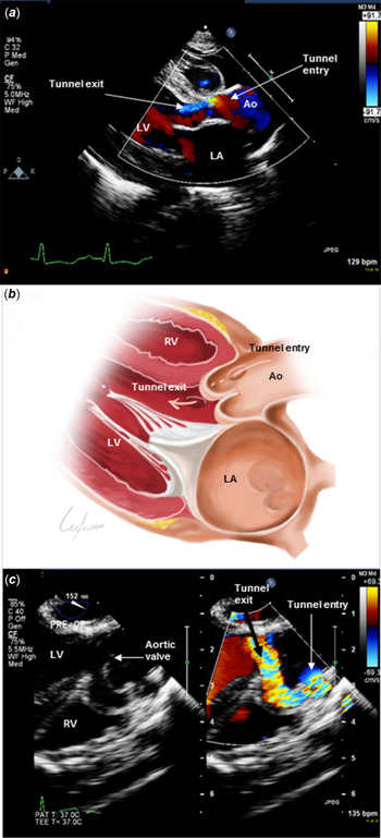
Aorto-left ventricular tunnel is an exceedingly rare CHD in which an additional tract is formed between the aorta and the left ventricle bypassing the usual pathway through the aortic valve (Fig 1b). The earliest description of this rare anomaly was indicated in the “Atlas of Acquired Diseases of the Heart and Great Vessels” by Edwards, J in 1961.Reference Edwards1 Levy et al have described this lesion in more detail in 1963.Reference Levy, Lillehei, Anderson, Amplatz and Edwards2 Their original description referred to this defect as “aortico-left ventricular tunnel.” However, the term “aorto-left ventricular tunnel” was introduced later and gained popularity.Reference McKay3 The exact incidence is unknown but estimated to represent less than 0.1% of congenital cardiac defects in pathological series.Reference McKay3 Patients with aorto-left ventricular tunnel may present with symptoms of heart failure due to volume overload; however, sometimes the only manifestation would be of an incidental murmur on auscultation or even with no apparent clinical findings. Prenatal diagnosis has been reported.Reference Greiber, Maroun, Vejlstrup and Jørgensen4 The lesion usually occurs as an isolated defect but some other associations have also been identified such as aortic valve stenosis, ventricular septal defect, valvular pulmonary stenosis, atrial septal defect, patent ductus arteriosus, and coronary abnormalities.Reference Martins, Sherwood, Mayer and Keane5,Reference Protopapas, Anderson and Backer6 Most affected patients need intervention in the neonatal period or early infancy due to progressive heart failure symptoms.Reference Kathare, Subramanyam, Dash, Muthuswamy, Raghu and Koneti7
(a) A still-frame of transthoracic echocardiogram at parasternal long-axis view for the patient number 3 in our series. Congenital ALVT with retrograde flow from aorta to left ventricle is clearly demonstrated. Left atrial dilatation is shown as well. (b) A schematic drawing demonstrating aorto-left ventricular tunnel with dotted line representing retrograde aortic flow from aorta towards the left ventricular cavity. (c) A still-frame of transesophageal mid-esophageal long-axis view for patient number 4 in the series, showing the entry point of ALVT just near the right coronary artery origin and the exit point to the left ventricular cavity just below the level of aortic valve annulus.

The repair of aorto-left ventricular tunnel is usually achieved by surgical patch closure. Long-term follow-up is recommended as there are number of possible complications that may ensue during follow-up. Progressive aortic valve insufficiency or development of aortic aneurysms are among the main long-term complications that have been attributed to multiple aetiologies such as lack of cusp support, aortic root dilation, leaflet perforation, or surgical distortion of the valve during the repair.Reference Greiber, Maroun, Vejlstrup and Jørgensen4,Reference Kathare, Subramanyam, Dash, Muthuswamy, Raghu and Koneti7,Reference Portelli Tremont, Kalra, Savarese and Lee8 Deterioration of the left ventricular function even years after intervention has been reported as well.
Due to paucity of knowledge about the long-term outcome of aorto-left ventricular tunnel, we report a case series of aorto-left ventricular tunnel patients in our centre. We describe the lesion before the repair and aim to evaluate the long-term outcome of this rare disease.
Methods
All patients that were diagnosed with aorto-left ventricular tunnel and underwent repair between 2001 and 2020 at King Abdulaziz Cardiac Center were identified and reviewed retrospectively using electronic patient records system of the institution.
All relevant clinical, echocardiographic, perioperative, and surgical details were reviewed. We included all patients who had aorto-left ventricular tunnel diagnosis pre-operatively and confirmed aorto-left ventricular tunnel intraoperatively.
The echocardiographic data of the patients were reviewed on digitally stored data in Xcelera system (Philips, Andover, USA). The following variables were obtained: diameter of the regurgitant tunnel jet at the narrowest point, presence and grade of aortic regurgitation, presence and grade of aortic stenosis, presence of aortic diastolic reversal flow on Doppler interrogation, left ventricular end-diastolic dimension and z-score by M-mode, and left ventricular systolic function assessment by fractional shortening. All echocardiographic parameters were compared pre-operatively and post-operatively at the last follow-up visit.
Results
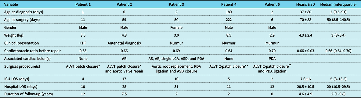
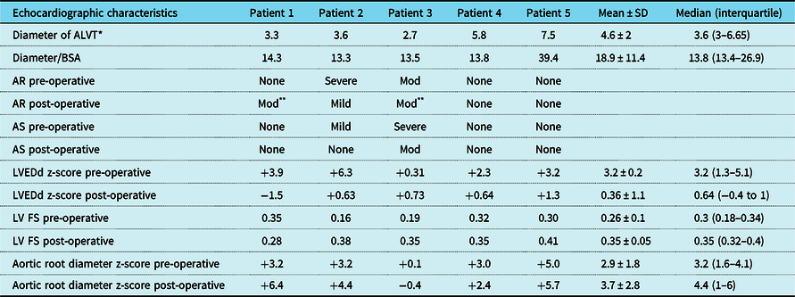
Our series included four male patients and one female. Their mean age at diagnosis was 37 days. Three out of five patients (60%) were diagnosed due to auscultatory murmur. Patient number 1 in the series presented early at day 7 of age with symptoms and signs of congestive heart failure. Patient number 2 in the series was diagnosed antenatally at 33 weeks of gestation due to dilated left ventricular seen during obstetric ultrasonography. His fetal echocardiogram showed dysplastic thickened aortic valve with eccentric regurgitant jet, which raised early suspicion of aorto-left ventricular tunnel. Postnatal echocardiogram confirmed a dysplastic regurgitant aortic valve with an additional severe paravalvular eccentric regurgitation jet, associated with dilated left ventricle correlating with early sign of impending heart failure. Patients 3, 4, and 5 were diagnosed relatively early in life after auscultating a heart murmur and before clinically significant symptoms of heart failure become recognisable. Although some of our aorto-left ventricular tunnel cases were clinically occult, all patients demonstrated radiologically variable degree of increased cardiothoracic ratios. The demographic and perioperative data of all five patients are summarised in Table 1. All patients were diagnosed by echocardiography (Fig 1, Table 2).
Demographic and perioperative data of five patients diagnosed with ALVT

ALVT = Aorto-left ventricular tunnel; AS = aortic valve stenosis; AR = aortic valve regurgitation; ASD = atrial septal defect; CHF = congestive heart failure; LCA = left coronary artery; LOS = length of stay; PDA = patent ductus arteriosus
* Synthetic (Gore-Tex) patch closure of ALVT from aortic side
** Patch closure of ALVT from aortic and ventricular sides with two separate synthetic (Gore-Tex) patches
Pre-operative and post-operative echocardiographic characteristics for five patients with ALVT

ALVT = Aorto-left ventricular tunnel; AS = aortic valve stenosis; BSA = body surface area; LVEDd = left ventricular end-diastolic dimension; LV FS = left ventricular fractional shortening; Mod = moderate
* Diameter of ALVT was measured in mm at the narrowest point on colour Doppler image
** Reversal flow noted on abdominal aortic Doppler interrogation
Three out of five patients had associated congenital heart lesions. Patient 2 had aortic regurgitation that was detected antenatally. Patient 3 had aortic stenosis and regurgitation, atrial septal defect, patent ductus arteriosus, and a single left coronary artery. Patient 5 had an associated patent ductus arteriosus (Table 1).
All five patients underwent surgical repair at our institution. Patients 1 and 2 had patch closure of aorto-left ventricular tunnel from aortic side only. In case of patient 2, surgical inspection revealed a thickened dysplastic aortic valve so aortic valve repair with commissuroplasty was carried out concomitantly with aorto-left ventricular tunnel repair. The aortic valve integrity was restored near normal and the regurgitation improved post-repair. Patient 3 had severely stenotic dysplastic aortic valve and also a thickened regurgitant pulmonic valve which precluded Ross procedure, and he ended with aortic root replacement using homograft size of 7 mm. This patient had an open sternum which was closed 3 days after the repair. All other patients had smooth post-operative course. Patients 4 and 5 had aorto-left ventricular tunnel double patch closure at both aortic and left ventricular ends, and aortic valve was intact by intraoperative transesophageal echocardiography assessment. Perioperative details are summarised in Table 1.
The median duration of ICU and hospital stay were 5 and 20 days, respectively. Median duration of follow-up was 4.6 years, ranging from 6 months to 12 years, (Table 1). Patient number 1 progressed from no pre-operative regurgitation to moderate post-operative aortic regurgitation at maximal follow-up of 12 years. Aortic regurgitation improved from severe to mild in patient number 2 who had concomitant aortic valve repair. Patient number 3 who underwent aortic root replacement had non-progressive moderate aortic regurgitation. Patients 4 and 5 had competent normal functioning aortic valve on follow-up.
Most patients in our series (80%) demonstrated dilated aortic root z-scores before surgery (Table 2). Three patients (1, 2, and 5) showed progression of aortic root dilation on last follow-up, while two patients (3 and 4) showed improvement of aortic root dilatation with age. All patients were asymptomatic at last follow-up visit with complete obliteration of aorto-left ventricular tunnel on echocardiography.
Discussion
Congenital aorto-left ventricular tunnel is an extremely rare condition characterised by an abnormal vascular channel that originates above sinus of Valsalva and tunnels towards ventricular cavity and enters the left ventricle inferior to the coronary cusp level. An anatomically similar lesion has been reported to connect to right ventricle.Reference Protopapas, Anderson and Backer6 The aetiology of this rare defect is unknown exactly but thought to be due to failure of development of the extracardiac tissue plane that separates cushions that develop into facing aortic and pulmonary sinuses and their respective leaflets. This tissue plane usually develops due to regression of surrounding muscle.Reference McKay3 An anatomical classification for different variants of aorto-left ventricular tunnel has been described and attributed to Hovaguimian et al.Reference Hovaguimian, Cobanoglu and Starr9
The largest publication up to date of aorto-left ventricular tunnel cases was published recently by the European Congenital Heart Surgeons Association – World Society for Pediatric and Congenital Heart Surgery. It was a multi-institutional retrospective analysis and comprehensive review, with 15 participant centres and a total number of 42 patients with aorta-ventricular tunnels, including left and right ventricular tunnels. Their analysis showed that aortic insufficiency tends to remain stable or improved, in contrast to aortic stenosis that might require aggressive timely management. Most of the patients in their series underwent surgical repair in form of patch closure.Reference Protopapas, Anderson and Backer6
Clinically, the presentation of aorto-left ventricular tunnel most commonly involves congestive heart failure symptoms, or less commonly, an incidental murmur discovered in neonates or young infant.Reference Martins, Sherwood, Mayer and Keane5 Transthoracic echocardiography represents an excellent modality for diagnosing and assessing aorto-left ventricular tunnel, putting in mind the various other lesions which cause aortic regurgitation and heart failure such as sinus of Valsalva fistula, truncus arteriosus with regurgitant valve, aortopulmonary window, aortic cusp prolapse into a ventricular septal defect, large patent ductus arteriosus, coronary fistula, and congenital aortic valvular lesions with aortic regurgitation.Reference McKay3 Of note, the aortic origin of aorto-left ventricular tunnel is always above the sinotubular junction, while the ruptured aneurysmal sinus of Valsalva courses below the sinotubular level.Reference Protopapas, Anderson and Backer6 A special feature which helps to differentiate aorto-left ventricular tunnel from coronary cameral fistula is that aorto-left ventricular tunnel never passes through cardiac myocardium to reach the ventricular cavity, in contrast to coronary fistulas.Reference Kathare, Subramanyam, Dash, Muthuswamy, Raghu and Koneti7
Although spontaneous closure of aorto-left ventricular tunnel has been documented in single report,Reference Martins, Sherwood, Mayer and Keane5 the vast majority of reported cases were repaired by surgery, preferably before 6 months of age.Reference Li, Yan and Shen10 Less commonly, catheter-based intervention has been reported as well.Reference Chessa, Chaudhari and De Giovanni11 Multiple surgical techniques were described in the literature for approaching aorto-left ventricular tunnel defects, including direct suture or patch closure either at the aortic end or at the ventricular end of the tunnel, with some reports favouring patch closure especially in smaller patients to avoid aortic valve distortion.Reference Martins, Sherwood, Mayer and Keane5,Reference Protopapas, Anderson and Backer6 A particular attention should be paid to coronary artery ostium, especially the right as it is the most common site for tunnel origin, to avoid distortion or compression during surgical repair.Reference Protopapas, Anderson and Backer6
Data from our centre’s experience demonstrate good overall surgical outcome of aorto-left ventricular tunnel. All patients in our series had successful repair with no mortality, no recurrence, and no re-intervention during 12 years of follow-up. Most patients in our series showed variable degree of aortic roots dilatation prior to surgery, which is consistent with published reports. Reference Kathare, Subramanyam, Dash, Muthuswamy, Raghu and Koneti7 Post-repair, on last follow-up, 60% of cases showed progression of aortic root dilatation and that could play a role in aortic regurgitation development after aorto-left ventricular tunnel repair. However, a larger sample is needed to confirm this observation. All patients were in sinus rhythm at the last follow-up with no evidence of heart block.
Conclusion
Aorto-left ventricular tunnel represents an unusual cause of left heart failure in neonates and young infants. Early surgical repair of aorto-left ventricular tunnel has good outcome achieving complete obliteration with no residual aorto-left ventricular tunnel in all of our case series. We highlight the importance of long-term follow-up as some patients may exhibit progression of aortic regurgitation (20%), residual regurgitation (40%), or aortic stenosis (20%) post-aorto-left ventricular tunnel repair. Diastolic reversal showed good correlation with the presence of significant aortic regurgitation in our series. Long-term follow-up is recommended for aortic root dilatation and aortic valve competency, as valve function needs to be addressed in a timely manner to avoid further complications.
Acknowledgements
The authors thank Dr. Alaa Muhammad Alorabie (@Ala2muh_art) for artwork.
Financial Support
This research received no specific grant from any funding agency, commercial or not-for-profit sectors.
Conflict of Interest
None.
Ethical Standards
The study was approved by King Abdullah International Medical Research Center (KAIMRC) with IRB NCBE registration no. H-01-R-005.