Introduction
Oocyte quality is one of the most important factors that affects the outcome of porcine embryo development (Antosik et al., Reference Antosik, Kempisty, Jackowska, Bukowska, Lianeri, Brussow, Wozna and Jaskowski2010). Although many kinds of cloning animals have been produced during recent decades, objective criteria for selecting competent oocytes remain elusive. Oocyte quality is reflected by characteristics such as degree of expansion of the cumulus mass, presence of the first polar body (PB1), and anomalies in the absence of cytoplasm and zona pellucida (Segers et al., Reference Segers, Adriaenssens, Coucke, Cortvrindt and Smitz2008). However, these features are insufficient to confer the ability of oocytes to support full-term pregnancy for transgenic or somatic nuclear transfer research (Coticchio et al., Reference Coticchio, Sereni, Serrao, Mazzone, Iadarola and Borini2004). Therefore, establishing creative and reliable criteria to select oocytes with good quality and high competence for further embryo development is necessary. Acquisition of oocyte competence remains an unclear process revealed throughout oogenesis (Albertini et al., Reference Albertini, Sanfins and Combelles2003). Immediately prior to ovulation, both nuclear and cytoplasmic compartments interact and exchange materials (Funahashi et al., Reference Funahashi, Koike and Sakai2008). Germinal vesicle breakdown (GVBD), which signifies meiotic resumption and extrusion of PB1 are indicators of meiotic processes. However, emissions of GVBD and PB1 do not validate the complete or correct progression of oocyte meiotic apparatus to metaphase II in preparation for in vitro fertilization (De Vos et al., Reference De Vos, Van de Velde, Joris and Van Steirteghem1999). Various factors can affect spindle and cytoskeletal organization (Vanhoutte et al., Reference Vanhoutte, Sutter, Nogueira, Gerris, Dhont and Van der Elst2007), thereby compromising both meiosis II segregation and broader spindle-mediated events. Simultaneously, various events take place among oocytes in a cytoplasmic environment during final maturation, which enable the oocytes to support further embryo development.
Several researchers have questioned the possible correlation between the normal development of PB1 morphology to fertilization rate, cleavage rate, embryo quality, and blastocyst formation (Scott et al., Reference Scott, Finn, O’Leary, McLellan and Hill2007; Younis et al., Reference Younis, Radin, Izhaki and Ben-Ami2009; Navarro et al., Reference Navarro, de Araújo, de Araújo, Rocha, Reis and Martins2009; Rose & Laky, Reference Rose and Laky2013). Therefore, these indicators cannot ensure full-term development of matured oocytes. In this study, the grades of PB1 are compared with those of cortical granule (CG) distribution, the ratio of embryo cleavage and development to blastocyst stage, and the acetylation level of porcine embryos to enrich the methods and evaluate the quality of porcine matured oocytes. Thus, oocytes with good quality are produced for further experimental research and efficiency improvement.
Materials and methods
Chemicals and culture media
All chemicals were purchased from Sigma Chemical Company (St. Louis, MO, USA) unless otherwise specified. Tissue culture medium 199 with Earle's salts (TCM199, Lot No. 12340) was sourced from the Invitrogen Corporation (Carlsbad, CA, USA). Insulin–transferrin–selenium (ITS) and epidermal growth factor (EGF) was sourced from GIBCO (Logan Town, Utah, USA); pregnant mare's serum gonadotropin, human chorionic gonadotropin, and follicle-stimulating hormone was sourced from Ni Bo Sangsheng Pharmaceutical Co., Ltd (Ni Bo City, Zhejiang Province, China). The basic in vitro maturation (IVM) medium was a modified TCM199 supplemented with 0.1% polyvinyl alcohol, 0.57 mmol/l cysteine, 3.05 mmol/l glucose, and 0.91 mmol/l pyruvic acid Na salt. The medium used for collecting and washing cumulus–oocyte complexes (COCs) comprised of 10% newborn bovine serum (NBS) and Dulbecco's phosphate buffered saline (DPBS; Gibco 11500–030, Grand Island, NY, USA). The porcine zygote medium (PZM-3) was based on the formula in a previously published article (Hu et al., Reference Hu, Ma, Bao, Li, Cheng, Gao, Lei, Yang and Wang2011). The first antibodies for H3K9 acetylation were purchased from Abcam (lot number: ab10812), and the Cy3-conjugated goat anti-rabbit second antibody was purchased from Beijing Biosynthesis Biotechnology Co., Ltd (Lot: bs-0369P-Cy3).
Collection of ovaries
Experiments were based on the guidelines of the Association for Research in Embryo Transplantation, and were approved by the Animal Research Committee of Hunan University of Humanities, Science and Technology. Ovaries were collected at a local pig slaughterhouse from peripubertal gilts without retrieving information of age and breeding. Ovaries were stored in 0.9% (w/v) NaCl that contained 100 mg/l penicillin and streptomycin at 30–37°C, and were transported to the laboratory within 2 h after slaughter.
Preparation and culture of COCs
The follicular contents, including COCs, were collected by aspirating the visible antral follicles (around 2–6 mm in diameter) with a 10-ml syringe equipped with an 18-gauge needle. Only COCs with uniform ooplasms and compact cumulus cell masses were collected and washed three times with Ca2+-/Mg2+-free PBS plus 10% NBS. In this study, porcine oocytes were separately washed three times and cultured in the modified TCM199 adding these materials (10 IU/ml PMSG, 10 IU/ml hCG, 2.5 IU/ml FSH, 10 ng/ml EGF and 1% ITS) under 38.5°C temperature, 5% CO2 in air with saturated humidity (Hu et al., Reference Hu, Ma, Bao, Li, Cheng, Gao, Lei, Yang and Wang2011). After 42 h of in vitro maturation (IVM), cumulus cells were removed by gently pipetting with a fine-bore pipette in saline supplemented with 0.3% hyaluronidase (GIBCO, Logan town, Utah, USA) for 3–5 min to analyze the matured oocytes. The signs of maturation oocytes depended on protruding PB1 combined with cumulus cells surrounding modality of oocytes. Classification of PB1 morphology was performed using an inverted microscope (1×70 Olympus, Hamburg, Germany). Then, the polar bodies were evaluated (43 h after maturation) and oocytes were assigned to different groups depending on whether they showed a PB1 with fragmented (grade A), smooth or intact surface (grade B), rough surface (grade C) (Van de Velde et al., Reference Van de Velde, Nagy, Joris, De Vos and Van Steirteghem1997).
Immunocytochemistry staining of cortical granules
Following 42 h IVM, cumulus cell-free oocytes were washed twice in PBS, fixed with 3.7% paraformaldehyde for 30 min, and washed again three times in PBS. Oocytes were permeabilized in PBS with 0.1% Triton X-100 for 5 min, and then rinsed four times in PBS. Oocytes were incubated in PBS with 100 g/ml of fluorescent lens culinaris agglutinin–fluorescein complex for 30 min at 37°C. Finally, oocytes were rinsed and mounted on histology slides, and the localization of the CGs was evaluated with a Nikon fluorescence microscope (wavelength 488 nm). The distribution of CGs in porcine oocyte IVM was evaluated based on three categories: type I (CGs distributed in cytoplasm, but not on plasma membrane), type II (CGs distributed in the cortex and forming a fluorescent halo around the plasma membrane), and type III (CGs distributed partially on the plasma membrane and partially in the cytoplasm) (Hosoe & Shioya, Reference Hosoe and Shioya1997; Hu et al., Reference Hu, Ma, Bao, Li, Cheng, Gao, Lei, Yang and Wang2011).
Evaluation of level of acetylation of porcine embryos
To obtain parthenogenetically activated oocytes, the denuded oocytes were incubated in an activation medium from Cyto Pulse Sciences Company (Cytofusion™ Medium, Cyto Pulse Sciences, Inc., Holliston, MA, USA) for 5 min. The oocytes were then transferred into two 0.2-mm diameter platinum electrodes with a 1-mm gap and covered with the activation medium in a chamber connected to an electrical pulsing machine (PA-4000S Laboratory PulseAgile_Electroporation System; Cyto Pulse Sciences, Inc.). Similar to described in a previously published paper (Gupta et al., Reference Gupta, Uhm and Lee2008), activation was conducted with a single direct current (DC) pulse of 1.5 kV⁄cm for 30 ms. After culturing for 3 h in TCM199 medium that contained 2 mM 6-dimethylaminopurine (Sigma, St. Louis, MO, USA), embryos were washed at least three times in PZM-3 and cultured in the medium further.
The method used for indirect immunofluorescence was adapted from a modified work from Santos and colleagues (Santos et al., Reference Santos, Peters, Otte, Reik and Dean2005; Wang et al., Reference Wang, Kou, Zhang and Gao2007). All steps were performed at room temperature, unless otherwise stated. The collected embryos were fixed with 4% paraformaldehyde for at least 30 min, and then permeabilized for 30 min with 0.2% Triton X-100 in PBS. After three washings, all samples were incubated in a blocking solution (1% BSA and 0.05% Tween-20 in PBS) overnight at 4°C. Embryos were incubated with the first antibodies to H3K9 acetylation (1 μg/ml). After three washings, the embryos were incubated with a Cy3-conjugated goat anti-rabbit second antibody for 1 h. DNA was visualised with Hoechst 33342 stain, and all samples were mounted in anti-fade solution. A digital camera attached to the microscope was used to acquire images, and the number of pixels was measured using the MetaMorph imaging software (ImageJ 5.0; http://imagej.softpedia.com/). Background intensities were measured and subtracted from the final values. Experiments were performed independently three times, and the order of processing was randomized each time.
Experimental design
To analyse the effects of different polar body grades on porcine oocytes and further embryo development in vitro, they were first compared with the ratios of the three categories of CG distribution in porcine matured oocytes (protruding PB1). The effects of these grades on further embryo development in vitro were also analysed, including the rate of embryo cleavage, development to blastocyst stage, and mean cell number in the blastocyst. Furthermore, the effects of these grades on the acetylation level of porcine blastocyst were evaluated by analysing the immunofluorescence intensity in vitro. The entire experiment was repeated at least three times independently.
Statistical analysis
Statistical analyses of different experimental groups in different grades of PB1 were conducted by analysis of variance and t-test with the Statistical Package for Social Science 10.0 software. A P-value < 0.05 was considered to be significant.
Results
The type of CG distributions in porcine oocytes with different PB1 grades
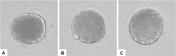
We examined the effects of polar body type (grades A, B, and C; Fig. 1) on CG distribution, divided into three types (types I, II, and III) adapted from a previous work (Hu et al., Reference Hu, Ma, Bao, Li, Cheng, Gao, Lei, Yang and Wang2011). Table 1 shows the type II CG distribution of oocytes with grade B PB1, which is significantly greater than those with grades A C (71.43% versus 52.46% and 50%; P < 0.05). However, types I and III CG distributions of oocytes with grade B PB1 are significantly lower compared with those of grades A and C (P < 0.05). Therefore, porcine oocytes with grade B PB1 may be better in terms of cytoplasmic maturation because the CGs are distributed below the zona pellucida.
Effects of different PB1 grades on CG distribution in porcine oocytes

a,bValues with different superscripts indicate significant difference (P < 0.05; n = 3). CG, cortical granules; PB1, first polar body; S.E., standard error.
Morphology of the first polar body (PB1). (A) Fragmented first polar body (grade A); (B) smooth or intact first polar body (grade B); (C) rough first polar body (grade C). Images are obtained using inverted light microscopy at original magnification ×400.

Development of porcine embryos with different first polar body types in vitro
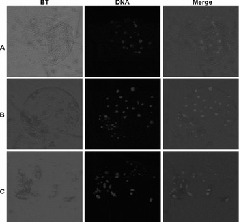
To compare the development of porcine embryos from activated oocytes with different PB1 grades, three different groups of oocytes in three kinds of grades PB1 were activated and cultured in vitro. Table 1 and Fig. 2 show that although no significant difference exists in the rate of porcine embryo cleavage among the three groups, the rate of porcine embryo development to blastocyst stage in the group in grade B PB1 is significantly greater compared with that of cells with grades A to C PB1 (30.81% versus 19.02% and 15.15%; P < 0.05). To evaluate the effects of different groups on the quality of porcine embryos, the mean cell numbers in the blastocyst were compared. Results indicate that the cell numbers in the blastocyst of the group in grade B PB1 are significantly greater than those in grade A or C PB1 (36.37% versus 24.67% and 28.67%; P < 0.05).
Number of cell nuclei in porcine blastocyst. (A) Group of oocytes with grade A first polar body (PB1); (B) group of oocytes with grade B PB1; (C) group of oocytes with grade C PB1. Images obtained by inverted light microscopy at original magnification ×400. BT, Bright field.

Level of H3K9 acetylation in porcine embryos with different first polar body types in vitro
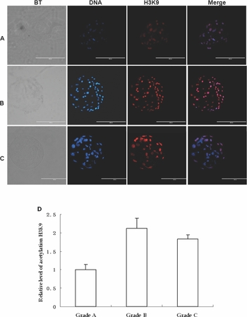
We compared the acetylation level of histone H3 lysine 9 in porcine blastocysts among the experimental groups. Figure 3A–C show that this level in porcine embryos from oocytes with grade B PB1 is greater than those in the embryos of other groups, which also agrees with the immunofluorescence intensity analysis results obtained using ImageJ 5.0 software (Fig. 3D).
Confocal micrographs of immunolocalization of the H3K9 dimethyl epitope in blastocysts from porcine oocytes with different first polar body (PB1) grades. (A) Embryos from parthenogenetic oocytes in grade A PB1; (B) embryos from parthenogenetic oocytes in grade B PB1; (C) embryos from parthenogenetic oocytes in grade C PB1. The embryos were labelled for acetylation of histone H3K9 (red) and Hoechst 33342 (green). Merged images of labelled histone H3K9 and Hoechst 33342 appear in orange. BT, Bright field.

Discussion
This study aims to firstly clarify whether different grades of PB1 (as shown in Fig. 1) in oocytes affect further porcine embryo development, in order to find the suitable methods to choose more competitive oocytes for further research in producing transgenic animals.
Previous studies have demonstrated that PB1 morphology is related to mature oocyte viability, and have the potential to predict oocyte performance and pregnancy achievement in infertile women undergoing intracytoplasmic sperm injection (ICSI) treatment (Younis et al., Reference Younis, Radin, Izhaki and Ben-Ami2009). Recently, it has also been found that PB1 morphology may reflect the development competence of oocytes in vitro (Rose & Laky, Reference Rose and Laky2013). In contrast, it has also been reported that irregular shape or fragmentation of the first polar body (PB1) is not related to subsequent embryo quality and blastocyst development (De Santis et al., Reference De Santis, Cino, Rabellotti, Calzi, Persico, Borini and Coticchio2005; Ten et al., Reference Ten, Mendiola, Vioque, de Juan and Bernabeu2007), The reason for this may be there is not a unified standard for testing further embryo quality, development and epigenetic events, so further research is still needed to understand the relationship between PB1 and embryo quality and et al. It has been reported that CG distribution may indicate the cytoplasmic maturation of oocytes (Wang et al., Reference Wang, Sun, Hosoe, Shioya and Day1997; Hu et al., Reference Hu, Ma, Bao, Li, Cheng, Gao, Lei, Yang and Wang2011). The results show that the distribution of type II CGs in oocytes with grade B PB1 are significantly greater compared with grades A and C (as shown in Table 1). Therefore, porcine oocytes with grade B PB1 are better in terms of cytoplasmic maturation (with CGs distributed below zona).
Oocytes with intact or smooth PB1 can generate better second-day embryos, greater blastocyst yields, and increased pregnancy and implantation rates (Younis et al., Reference Younis, Radin, Izhaki and Ben-Ami2009; Rose & Laky, Reference Rose and Laky2013). It has also been found that embryos in the intact or smooth PB1 group show increased rate of blastocyst formation compared with the fragmented PB1 group (P < 0.05) (Navarro et al., Reference Navarro, de Araújo, de Araújo, Rocha, Reis and Martins2009; Rose & Laky, Reference Rose and Laky2013). In this paper, the rate of porcine embryo development to blastocyst stage in the group with grade B PB1 is significantly greater than in the other groups (as shown in Table 2). The mean number of cells in each blastocyst is also significantly greater than in the other groups (as shown in Table 2 and Fig. 2). All of the results are similar to the previous reports. Therefore, previous data were supported by our results, suggesting an important prognostic function of PB1 morphology in embryo development and quality.
Development of porcine embryos with different types of PB1 in vitro

a,bValues with different superscripts indicate significant difference (P < 0.05; n = 3). PB1, first polar body; S.E., standard error; SEM, standard error of the mean.
It was found that the acetylation level of histone H3 at the early stages of the porcine embryo is positively correlated with the subsequent development of somatic cell nuclear transfer (Das et al., Reference Das, Gupta, Uhm and Lee2010). It also has been reported that the acetylation level of this histone H3K9 was crucial for gene activation of early embryos development, because the tri- and dimethylations of H3K9 were gradually demethylated in the cloned embryos, and (Wang et al., Reference Wang, Kou, Zhang and Gao2007). The acetylation level of histone H3 at the early stages of the porcine embryo is positively correlated with the subsequent development of somatic cell nuclear transfer (SCNT) embryos, which are important for the vital development of SCNT embryos in miniature pigs (Yamanaka et al., Reference Yamanaka, Sugimura, Wakai, Kawahara and Sato2009). To clarify the reason for the increase in the ratio of porcine embryos developing to blastocyst stage and the quality of porcine embryos, acetylation levels of histone H3 lysine 9 were analysed in the three groups.
The level of acetylated histone H3 lysine 9 in porcine embryos from oocytes with grade B PB1 is greater than those in the embryos in the other groups (as shown in the Fig. 3), indicating that porcine embryo development in group B is correlated with the competing oocytes with good quality by affecting the acetylation of histone H3 in porcine embryos in vitro. Therefore, porcine oocytes with grade B PB1 are predicted to further develop and exhibit higher acetylation level expression of histone H3.
Therefore, it could be speculated that porcine oocytes with grade II PB1 are more competitive for embryo quality and development, but there is still a need for further research in many of the fields.
Acknowledgements
The current work was supported by the Chinese National Natural Science Foundation (#31301978), the Youth Research Program of Hunan Province Education Department in 2013 (13B056) and the highly educated project of Hunan University of Humanities, Science and Technology (HUHST).