INTRODUCTION
Improved understanding of the occurrence and spatio-temporal distribution of bivalve larvae during their planktonic stage necessitates the development of techniques for rapid larval identification. Such knowledge holds significant benefits for marine ecological studies targeting supply-side assessments, biodiversity estimates, bio-indicators and the introduction and spread of invasive species; for better management of shellfisheries focused on issues such as stock management, fisheries recruitment and population dynamics; and for improved aquaculture practices through efficient site selection and forecasting of the date for installation of equipment to optimize spat collection (Kanno, Reference Kanno1970; Ito, Reference Ito1977; Slater, Reference Slater2006).
Despite these benefits, morphological identification of bivalve larvae is extremely difficult to perform because of the small larval size, similarities in their shape and colour, and the absence of any protruding parts to aid in their identification (Caceres-Martinez & Figueras, Reference Caceres-Martinez and Figueras1998; De Vooys, Reference De Vooys1999; Slater, Reference Slater2005). Though morphological methods are technically feasible with some species, they are inappropriate for the rapid processing of plankton samples from multiple sites because of their time consuming nature. Advances in marine biotechnology have led to the development of molecular methods of identification based on DNA extraction such as dot blot hybridizations, PCR–RFLP, nested PCR or SSCP (Bell & Grassle, Reference Bell and Grassle1998; Frischer et al., Reference Frischer, Danforth, Tyner, Leverone, Marelli, Arnold and Blake2000; Hare et al., Reference Hare, Palumbi and Butman2000; Lopez-Pinon et al., Reference Lopez-Pinon, Insua and Mendez2002; Bendezu et al., Reference Bendezu, Slater and Carney2005; Hansen & Larsen, Reference Hansen and Larsen2005; Larsen et al., Reference Larsen, Frischer, Rasmussen and Hansen2005; Livi et al., Reference Livi, Cordisco, Damiani, Romanelli and Crosetti2006). Although such methods have considerable potential for the rapid analysis of plankton samples, bivalve larval tracking can only occur if the quantification of larvae can be facilitated. With larval destructive methods requiring DNA extraction, larval quantification is possible only by multiple repetitions of the technique with individual larvae (Larsen et al., Reference Larsen, Frischer, Rasmussen and Hansen2005) or from a standard graph prepared using real-time PCR which assumes that all of the larvae in a sample are of a standard size and contribute an equal quantity of DNA (Vadopalas et al., Reference Vadopalas, Bouma, Jackels and Friedman2006; Dias et al., Reference Dias, Batista, Shanks, Beaumont, Davies and Snow2009). Neither of these methods are capable of providing an estimate of the larval growth rate and a hence a prediction of the date of larval settlement.
In contrast, in-situ hybridization (ISH) techniques are non-destructive. Fluorescence in situ hybridization (FISH) has been widely used with single-celled microorganisms leading to significant advances (Amann et al., Reference Amann, Krumholz and Stahl1990; Scholin et al., Reference Scholin, Miller, Buck, Chavez, Harris, Haydock, Howard and Cangelosi1997; Miller & Schonlin, 1998; Amann & Ludwig Reference Amann and Ludwig2000; Simon et al., Reference Simon, Campbell, Örnolfsdottir, Groben, Guillou, Lange and Medlin2000; Wagner et al., Reference Wagner, Horn and Daims2003; Hosoi-Tanabe & Sako, Reference Hosoi-Tanabe and Sako2006; Tujula et al., Reference Tujula, Holmström, Mußmann, Amann, Kjelleberg and Crocetti2006); however, applications with multicellular organisms have been restricted to only a few larval species (Goffredi et al., Reference Goffredi, Jones, Scholin, Marin III and Vrijenhoek2006; Henzler et al., Reference Henzler, Hoaglund and Gaines2010). Since this technique relies on the ability to distinguish and detect specifically bound fluorescent molecules and resolve them from non-specific background signals, it is implicit that anything which obscures visualization of the change in fluorescence intensity will be problematic. With marine samples in particular, autofluorescence caused by endogenous fluorophores such as chlorophyll, lipofuscin, collagen, elastin, NADH, riboflavins and flavin coenzymes, is a universal and well-recognized problem (Mosiman et al., Reference Mosiman, Patterson, Canterero and Goolsby1997; Webster et al., Reference Webster, Wilson, Blackall and Hill2001; Hosoi-Tanabe & Sako Reference Hosoi-Tanabe and Sako2006; Tujula et al., Reference Tujula, Holmström, Mußmann, Amann, Kjelleberg and Crocetti2006; McCarthy et al., Reference McCarthy, Urquhart and Bricknell2008). Strong autofluorescence has been attributed as the major constraint limiting the application of FISH techniques with marine bivalve larvae (Pradillon et al., Reference Pradillon, Schmidt, Peplies and Dubilier2007) and has led to the development in some instances of more complex ISH colorimetric assays (Le Goff-Vitry et al., Reference Le Goff-Vitry, Chipman and Comtet2007a, Reference Le Goff-Vitry, Jacquelin and Comtetb; Pradillon et al., Reference Pradillon, Schmidt, Peplies and Dubilier2007).
Laboratory protocols for performing FISH comprise several principal steps: sample fixation and permeablization; probe hybridization; post-hybridization washing to flush unbound probe from cells; and finally data capture and analysis. Sample fixation treatments have generally been developed to preserve morphological and/or genetic characteristics for long-term storage (Sawada et al., Reference Sawada, Saito, Hosoi and Toyohara2008). However, for FISH, the ideal fixative not only functions as a preservative but also permeabilizes cell membranes to facilitate penetration of labelled probes into cells and may also reduce autofluorescence of the sample matrix (Miller & Scholin, Reference Miller and Scholin2000). A wide range of fixation treatments are cited in the literature, several of which report enhanced autofluorescence due to the fixative(s) used during the sample preservation step (Del Castillo et al., Reference Del Castillo, Llorente and Stockert1989; Baschong et al., Reference Baschong, Suetterlin and Laeng2001; Viegas et al., Reference Viegas, Martins, Seco and do Carmo2007).
Given the limited knowledge regarding autofluorescence in marine planktonic organisms generally and the absence of any comparative studies of autofluorescence in marine bivalve larvae, we have investigated the effect of a range of fixatives on autofluorescence as a practical step towards the development of a FISH technique for bivalve larval identification. Specifically, in this study we compared the autofluorescence intensity of fresh bivalve larvae (control) to that of samples fixed by physical means, namely freezing at −80°C, and by different chemical fixatives: 70% ethanol; 95% ethanol; modified saline ethanol (MSE); Carnoy's fixative; formalin followed by 70% ethanol; formalin followed by 90% methanol; formalin followed by MSE and phosphate buffered saline (PBS) and glycerol (50:50).
In addition to investigating the effect of the fixative on autofluorescence intensity, we also compared a range of treatments cited in the literature for autofluorescence reduction. The rationale for this study was that a dual approach to minimizing autofluorescence comprising firstly, selection of the optimum fixative and secondly, an autofluorescence reduction treatment was more likely to achieve the low intensity autofluorescence required for FISH studies. Identification of a suitable autofluorescence reduction treatment for bivalve larvae would also support investigations on archived material. Given the widespread use of MSE as a preservative in the marine field, we compared the autofluorescence intensity of bivalve larvae stored in this fixative to that of samples subjected to the following chemical treatments to reduce autofluorescence: Sudan Black B; Trypan Blue; proteinase K digestion; sodium borohydride in PBS; Chemicon™ Autofluorescence Eliminating Reagent (Millipore); ammonia–ethanol; ammonium chloride; acetone (80%); and cupric sulphate solution (10 mM copper sulphate in 50 mM ammonium acetate).
MATERIALS AND METHODS
Bivalve larval samples
Approximately 300,000 larvae of the blue mussel, Mytilus edulis (Linnaeus) at 15–21 days post-fertilization were transferred from a commercial hatchery (Cartron Point Shellfish Ltd., County Clare, Ireland) to the laboratory in 1000 ml of filtered and UV sterilized seawater at approximately 4°C. Upon arrival at the laboratory, larvae were aliquotted into samples of approximately 1000 larvae in 1.5 ml micro-centrifuge tubes and, with the exception of a sample of fresh larvae (control), fixed according to the protocols below.
Fixatives
For freezing at −80°C, larvae were immersed in 1 ml of filtered and UV sterilized seawater from the hatchery. Chemical fixatives were prepared as follows: 70% ethanol was prepared from 95% ethanol (Sigma Aldrich) using de-ionized water (Millipore); modified saline ethanol (MSE) was prepared by mixing 22 ml of 95% ethanol, 5 ml deionized water and 3 ml of 25X SET buffer (3.75 M NaCl, 25 mM EDTA, 0.5 M Tris HCl adjusted to pH 7.8); Carnoy's fixative was prepared by combining 60 ml of 99% ethanol (BDH Chemicals), 30 ml of chloroform (BDH Chemicals) and 10 ml glacial acetic acid (Sigma Aldrich); formalin was prepared by diluting formaldehyde (37%) (BDH Chemicals) with de-ionized water; PBS and glycerol (50:50) fixative was prepared by mixing 50 ml PBS (137 mM NaCl, 2.7 mM KCl, 10 mM sodium phosphate dibasic, 2 mM potassium phosphate monobasic adjusted to pH of 7.4) and 50 ml of glycerol (BDH Chemicals). For each of the chemical fixatives used, excess seawater was removed from the larval aliquot by pipette, 1 ml of fixative added, the tube gently agitated to ensure all of the larvae were completely immersed and the volume of fixative made up to 1.5 ml. Chemically-fixed larvae were stored in the dark at room temperature until required. Where initial fixation with formalin or dilute formalin was performed, this fixation step continued for 1 hour after which larvae were washed three times in the second fixative before storage as above.
Autofluorescence reduction
The effect of fifteen treatments for autofluorescence reduction was investigated using approximately 50 Mytilus edulis larvae which had previously been fixed and stored for 10 months in modified MSE. Treatments for autofluorescence reduction were prepared as follows: Sudan Black B (0.1%) and Sudan Black B (saturated) were prepared by dissolving 50 mg and approximately 4 g of Sudan Black B (Sigma Aldrich) respectively in 50 ml of either 70% or 95% ethanol; Trypan Blue solution (10%) and Trypan Blue (250 µg/ml) were prepared by dissolving Trypan Blue (BDH Chemicals) in de-ionized water; the two concentrations of the enzyme proteinase K (Sigma Aldrich) were prepared by dissolving the respective weight of enzyme in PBS; sodium borohydride (1 mg/ml) was prepared by dissolving sodium borohydride (Sigma Aldrich) in PBS; Chemicon™ autofluorescence eliminating reagent (Millipore) was used as per the manufacturer's guidelines; ammonium ethanol solution was prepared by dissolving 0.667 g of ammonium chloride in 35.7 ml of 95% ethanol and made up to 50 ml with de-ionized water; ammonium chloride solution (50 mM) was prepared by dissolving 0.13375 g of ammonium chloride in 50 ml of de-ionized water; acetone (80%) was prepared by dilution with de-ionized water and cupric sulphate solution (10 mM) was prepared by mixing 0.5 g of copper sulphate in 200 ml of ammonium acetate (50 mM).
For evaluation of each autofluorescence reduction treatment, excess MSE was removed from a larval aliquot by pipette and larvae treated according to protocols cited in the literature (Beisker et al., Reference Beisker, Dolbeare and Gray1987; Mosiman et al., Reference Mosiman, Patterson, Canterero and Goolsby1997; Schnell et al., Reference Schnell, Staines and Wessendorf1999; Baschong et al., Reference Baschong, Suetterlin and Laeng2001; Hosoi-Tanabe & Sako, Reference Hosoi-Tanabe and Sako2005; Viegas et al., Reference Viegas, Martins, Seco and do Carmo2007).
Optimum treatment duration for the two most effective autofluorescence reduction treatments, saturated Sudan Black B in 95% ethanol and Chemicon™ were investigated by comparing the autofluorescence intensity of larvae after treatment durations ranging from 2–60 minutes. Suitability of the two most effective autofluorescence reduction treatments for subsequent use in a FISH protocol was assessed using a fluorescein isothiocyanate (FITC)-labelled Mytilus-specific probe (FITC-CCGACGCAAATGGGGATCGG) (Le Goff-Vitry et al., Reference Le Goff-Vitry, Chipman and Comtet2007a). Samples of MSE fixed Mytilus edulis larvae were treated with either saturated Sudan Black B in 95% ethanol or Chemicon™ for the respective optimum treatment duration followed by hybridization with FITC-labelled probe (probe concentration 2 µM, hybridization temperature 46°C for 3 hours, formamide concentration 40%).
Data capture
Subsamples of larvae (N = 25 ± 11) from each of the different fixatives were examined for autofluorescence after 1 hour, 24 hours, 7 days, 10 months and 3 years. Images of autofluorescent larvae were recorded in both the FITC and Cy3 channels using an Olympus BX 50 epifluorescent microscope fitted with CCCD camera DP-70 using Cell-F software. Filters used were Olympus MNIBA3 for FITC (excitation 470–495 nm, emission 510-550 nm, dichromatic mirror 505 nm) and Olympus MWIGA3 for Cy3 (excitation 530–550 nm, emission 575–625 nm and dichromatic mirror 570 nm). Digital images were captured using 100× magnification and 20 milliseconds exposure time. Autofluorescence intensity of larvae was determined using the ‘daime’ software program (Daims et al., Reference Daims, Lücker and Wagner2005) and a minimum threshold value of 20 relative fluorescence units (RFU). Mean autofluorescence intensities of larvae preserved by each fixation method and viewed through the FITC and Cy3 filters were compared to that of the fresh larvae.
For each of the autofluorescence reduction treatments, subsamples of larvae (N = 11 ± 5) were examined in both the FITC and Cy3 channels and autofluorescence intensity determined as above. For the two most successful autofluorescence reduction treatments, optimization of the duration of the treatment was performed. Using these optimized protocols, the FITC-labelled probe was used to determine if either autofluorescence reduction treatment interfered with subsequent FISH.
Data analysis
For the fixative trial, statistical analysis performed using Minitab 15 (Minitab Inc., USA) consisted of a two-way analysis of variance (ANOVA) followed by Dunnett's multiple comparison test to a control. Mean autofluorescence intensities of fresh larvae for each respective filter set were used as the control values. Factor levels used in the analysis were fixative type and storage time. For the autofluorescence reduction trial, statistical analysis consisted of a one-way ANOVA followed by Dunnett's multiple comparison test. The control values were the autofluorescence intensities of MSE-fixed larvae stored for 10 months for each respective filter set. To assess the compatibility of the autofluorescence reduction treatments with FISH, a two-way ANOVA followed by Tukey's multiple comparison tests was used.
RESULTS
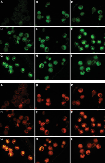
Autofluorescence intensity was not uniform within individual larvae. Some parts exhibited intense autofluorescence while other parts often exhibited little or none (Figure 1). Relative fluorescence values recorded with the FITC filter set were generally less than those recorded with the Cy3 filter set. Autofluorescence intensities with the different fixatives and autofluorescence reduction treatments used in this investigation exhibited relatively high coefficients of variation (CVs) ranging from 8% to 45%, regardless of the filter set used. Similar high CVs with FISH studies have been reported in the literature (Wallner et al., Reference Wallner, Amann and Beisker1993).
Autofluorescence of 21 days post-fertilization Mytilus edulis larvae viewed through the FITC filter (top) and Cy3 filter (bottom) with 20 milliseconds exposure after preservation for 10 months by: (A) freezing at −80°C; (B) 70% ethanol; (C) 95% ethanol; (D) modified saline ethanol (MSE); (E) Carnoy's fixative; (F) formalin and 70% ethanol; (G) formalin and 90% methanol; (H) formalin and MSE; (I) phosphate buffered saline and glycerol.

Fixatives
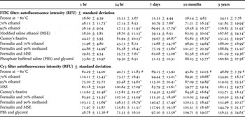
Mean autofluorescence intensities varied considerably between different fixation methods, different storage durations and with the filter set through which larvae were examined (Table 1). Fixative induced autofluorescence was apparent within 1 to 24 hours for several of the chemical fixatives. With one exception, all of the chemical fixatives evaluated significantly increased the autofluorescence intensity of larvae when stored for between 10 months and 3 years. High levels of fixative induced autofluorescence were observed in both the FITC and Cy3 filters. The lowest autofluorescence intensities were recorded in fresh larvae and those frozen at −80°C when examined through the FITC filter. Autofluorescence intensities of some of these larvae were below the minimum threshold (20 RFU) for detection using the standard parameters of the ‘daime’ software. Larvae frozen at −80°C exhibited no significant increase in autofluorescence intensities compared to the fresh larvae (P > 0.05).
Mean autofluorescence intensities (RFU) of Mytilus edulis larvae in different fixatives after 1 hour, 24 hours, 7 days, 10 months and 3 years examined through the FITC filter (top) and Cy3 filter (bottom) with 20 milliseconds exposure. Statistical analysis was performed using Dunnett's multiple comparison to control values for fresh larvae (29.93 ± 5.14 RFU with the FITC filter, 65.33 ± 15.97 RFU with the Cy3 filter). Values significantly increased and decreased at 95% CI and are shown with a* and a # respectively (N = 25 ± 11).

Autofluorescence reduction
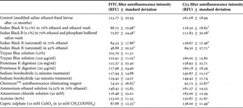
Mean autofluorescence intensities of Mytilus edulis larvae after 10 months preservation in MSE (control) and following fifteen different autofluorescence reduction treatments examined through the FITC and Cy3 filters are provided in Table 2. Treatments resulting in significant reduction in autofluorescence intensity at the 95% confidence interval are marked by an asterisk in Table 2. Larvae in the untreated MSE control after 10 months fixation (Table 2) exhibited significantly higher autofluorescence intensities than corresponding values reported in the fixative treatment trial (Table 1) because of the increased exposure time from 20 milliseconds to 50 milliseconds used during image capture.
Mean autofluorescence intensities (RFU) of Mytilus edulis larvae after 10 months storage in modified saline ethanol (control) following a range of autofluorescence reduction treatments examined through the FITC and Cy3 filters with 50 milliseconds exposure. Statistical analysis using Dunnett's multiple comparison. Values significantly lower than the control larvae at 95% CI are marked with* (N = 11 ± 5).
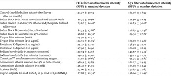
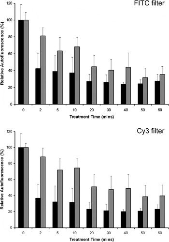
Of the fifteen autofluorescence reduction treatments investigated, those using Trypan Blue, proteinase K, sodium borohydride, ammonium ethanol, ammonium chloride and acetone failed to show significant reduction in autofluorescence intensity in both FITC and Cy3 filters. The most effective treatments which exhibited significant reduction in autofluorescence intensity in both filters were those using Sudan Black B, Chemicon™ autofluorescence eliminating reagent and cupric sulphate solution. Treatment durations that resulted in the maximum reduction in autofluorescence intensity using the two most effective treatments were determined as 20 minutes with saturated Sudan Black B in 95% ethanol and 50 minutes with Chemicon™ autofluorescence eliminating reagent (Figure 2).
Relative autofluorescence intensity (%) of Mytilus edulis larvae preserved for 10 months in modified saline ethanol after autofluorescence reduction using a saturated solution of Sudan Black B in 95% ethanol (black) and Chemicon™ autofluorescence eliminating reagent (grey) to determine the optimum treatment duration. Error bars represent one standard deviation.

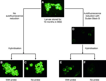
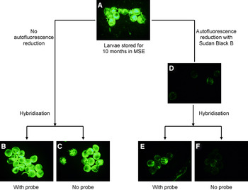
Using these optimized autofluorescence reduction treatments, their suitability for application in a FISH protocol was assessed using a FITC-labelled probe. Typical results with saturated Sudan Black B in 95% ethanol are shown in Figure 3. Mean autofluorescence intensity in the MSE-fixed larvae without autofluorescence reduction treatment (Figure 3A) was not significantly different after the hybridization protocol with or without FITC-labelled probe (Figure 3B, C) (P > 0.05), highlighting the difficulty in detection of probe-conferred fluorescence in the presence of strong autofluorescence. Significant reduction in the mean autofluorescence intensity was recorded following treatment with saturated Sudan Black B in 95% ethanol (Figure 3D) (P < 0.05). After hybridization with FITC-labelled probe, significant increase in the mean autofluorescence intensity of larvae previously treated with saturated Sudan Black B was recorded (Figure 3E) (P < 0.05). The control with no FITC probe (Figure 3F) confirmed that the FITC probe was responsible for the increased fluorescence and that the hybridization protocol used was not washing the autofluorescence reduction reagent from the larvae.
Compatibility of autofluorescence reduction treatment with Sudan Black B and fluorescence in situ hybridization using a fluorescein isothiocyanate (FITC)-labelled probe. Mytilus edulis larvae fixed in modified saline ethanol (MSE) for 10 months (A), MSE-fixed larvae without autofluorescence reduction and after the hybridization protocol with FITC probe (B) and without FITC probe (C), MSE-fixed larvae after autofluorescence reduction treatment with Sudan Black B (D), MSE fixed larvae after autofluorescence reduction treatment with Sudan Black B and the hybridization protocol with FITC probe (E) and without FITC probe (F).
DISCUSSION
Identification of bivalve larvae using FISH has significant advantages compared to other molecular methods. Since this is a non-destructive technique, counting larvae in plankton samples is straightforward, allowing spatial distribution to be mapped, supporting enhanced understanding of the fate of larvae in the plankton and the influence of oceanographic biophysical processes on larval dispersal (Goffredi et al., Reference Goffredi, Jones, Scholin, Marin III and Vrijenhoek2006). From a shellfish aquaculture perspective, such knowledge would support the selection of sites for optimal spat collection. Larval growth rate determination from shell measurements would support prediction of the date of the larval settlement (Slater, Reference Slater2006). Despite the clear benefits, the application of FISH techniques to bivalve larval identification is constrained by strong autofluorescence reported in marine specimens (Pradillon et al., Reference Pradillon, Schmidt, Peplies and Dubilier2007). A significant contributory factor to the intensity of this autofluorescence is the choice of sample fixative.
Fixatives
The initial objective of this study was to investigate the effect of different fixatives, the duration of fixation and the choice of filter set on the autofluorescence intensity of marine bivalve larvae. Physical fixation of larvae by freezing at −80°C has been widely reported in the literature (Demers et al., Reference Demers, Lagadeuc, Dodson and Lemieux1993; Bell & Grassle, Reference Bell and Grassle1998; Santaclara et al., Reference Santaclara, Espiñeira and Vieites2007; Phillips et al., Reference Phillips, Wood and Hamilton2008; Harvey et al., Reference Harvey, Hoy and Rodriguez2009). In this study, freezing at −80°C preserved the gross morphological structure of larvae and maintained autofluorescence intensity at the lowest levels. Autofluorescence intensities after freezing were comparable to those recorded with fresh larvae and in some instances were undetectable from the background threshold (20 RFU) using ‘daime’ image analysis software. Many chemical fixatives for the preservation of marine samples are reported in the literature. Ethanol has historically been used to preserve samples for a wide range of purposes. Pure ethanol (95% v/v) (Toro, Reference Toro1998a, Reference Torob; Andre et al., Reference Andre, Lindegarth, Jonsson and Sundberg1999; Launey & Hedgecock, Reference Launey and Hedgecock2001; Morgan & Rogers, Reference Morgan and Rogers2001; Larsen et al., Reference Larsen, Frischer, Rasmussen and Hansen2005; Toro et al., Reference Toro, Ojeda, Vergara, Castro and Alcapan2005; Vadopalas et al., Reference Vadopalas, Bouma, Jackels and Friedman2006, Pradillon et al., Reference Pradillon, Schmidt, Peplies and Dubilier2007; Santaclara et al., Reference Santaclara, Espiñeira and Vieites2007) can have a dehydrating effect on samples, possibly preventing the penetration of FISH probes, and is more frequently used at 70% (v/v) (Paugam et al., Reference Paugam, Le Pennec, Marhic and Andre-Fontaine2003; Wood et al., Reference Wood, Beaumont, Skibinski and Turner2003; Hansen & Larsen, Reference Hansen and Larsen2005; Taris et al., Reference Taris, Baron, Sharbel, Sauvage and Boudry2005; Pradillon et al., Reference Pradillon, Schmidt, Peplies and Dubilier2007). The use of MSE has been widely reported in the literature (Miller & Scholin, Reference Miller and Scholin1998; Peperzak et al., Reference Peperzak, Sandee, Scholin, Miller, Van Nieuwerburgh, Hallegraeff, Blackburn, Bolch and Lewis2000; Patil et al., Reference Patil, Gunasekera, Deagle and Bax2005) and this preservative has been described as the best fixative based on the preservation of cell integrity and optimal signal from fluorescent probes with phytoplankton stored for up to 1 month and barnacle larvae stored for between 2–6 weeks (Miller & Scholin, Reference Miller and Scholin2000; Goffredi et al., Reference Goffredi, Jones, Scholin, Marin III and Vrijenhoek2006). Ethanol-based fixatives (70% ethanol, 95% ethanol and MSE) evaluated in this investigation generally had less impact on autofluorescence intensity compared to other treatments, although values were elevated by comparison to the background level. Carnoy's fixative, a mixture of ethanol, chloroform and glacial acetic acid, has been reported as a suitable fixative for tissues from which DNA is to be extracted, although structural damage to tissues in histological investigations has been attributed to its use (Gerard et al., Reference Gerard, Naciri, Peignon, Ledu, Phelipot, Noiret, Peudenier and Grizel1994; Meithing et al., 2006; Koga et al., Reference Koga, Tsuchida and Fukatsu2009). Short fixation periods with paraformaldehyde and formalin are widely reported in protocols for FISH; longer fixation times have adverse effects on probe reactivity and on the intensity of the hybridization signal (Miller & Scholin, Reference Miller and Scholin2000; Simon et al., Reference Simon, Campbell, Örnolfsdottir, Groben, Guillou, Lange and Medlin2000; Staughton et al., Reference Staughton, McGillicuddy and Weinberg2001; Sako et al., Reference Sako, Hosoi-Tanabe and Uchida2004; Mikulski et al., Reference Mikulski, Morton and Doucette2005; Le Goff-Vitry et al., Reference Le Goff-Vitry, Chipman and Comtet2007a, Reference Le Goff-Vitry, Jacquelin and Comtetb; Pradillon et al., Reference Pradillon, Schmidt, Peplies and Dubilier2007). Both Carnoy's solution and the fixatives involving an initial formalin treatment generated the highest levels of autofluorescence in the larval samples used in this investigation. PBS and glycerol (50:50) more commonly used as a mounting medium has been suggested as a fixative for marine bivalve larvae (Stoecker K., personal Communication, 2006). In this study, PBS and glycerol (50:50) proved unsuitable for fixation purposes on the basis that autofluorescence intensity levels increased continuously over the 3 year period of investigation. Fixation induced autofluorescence can develop after periods as short as 1 hour and may affect one filter set more than another. In summary, in this comparative investigation of different fixation treatments, physical fixation by freezing at −80°C was the most suitable method of fixation for periods up to 3 years, with no significant increase in autofluorescence recorded.
Autofluorescence reduction
In developing a treatment to reduce or eliminate autofluorescence, one of three different approaches is generally used: extraction of the causative compound from the sample; alteration of the structure of the fluorescent component; or masking the autofluorescence by a suitable dye (Baschong et al., Reference Baschong, Suetterlin and Laeng2001). Extraction of the autofluorescent compound is most commonly performed where chlorophyll is the causative agent using an organic solvent such as ethanol, methanol or acetone (Hosoi-Tanabe & Sako, Reference Hosoi-Tanabe and Sako2005). Photobleaching, enzymatic or chemical treatments such as proteinase K digestion or sodium borohydride, can reduce autofluorescence by altering the structure and properties of the autofluorescent compound (Beisker et al., Reference Beisker, Dolbeare and Gray1987; Clancy & Cauller, Reference Clancy and Cauller1998; Baschien et al., Reference Baschien, Manz, Neu and Szewzyk2001; Neumann & Gabel, Reference Neumann and Gabel2002). Masking the autofluorescence can be accomplished by a range of different dyes such as Sudan Black B or Trypan Blue (Mosiman et al., Reference Mosiman, Patterson, Canterero and Goolsby1997; Schnell et al., Reference Schnell, Staines and Wessendorf1999). Each treatment may be used individually or in combination to improve the signal to noise ratio between background fluorescence and probe-conferred fluorescence and may be performed either before or after the FISH probe hybridization step (Baschong et al., Reference Baschong, Suetterlin and Laeng2001; Suetterlin et al., Reference Suetterlin, Baschong and Laeng2004; Viegas et al., Reference Viegas, Martins, Seco and do Carmo2007). In this study to compare autofluorescence reduction treatments, saturated Sudan Black B in 95% ethanol or Chemicon™ autofluorescence eliminating reagent resulted in significant and rapid reduction in autofluorescence to between 20–40% of the original intensity. Both treatments were compatible with subsequent FISH using a FITC-labelled probe.
In summary, as a practical step towards the development of a FISH technique for bivalve larval identification, we have confirmed that fixation by freezing at −80°C is the most suitable method of preservation whilst maintaining autofluorescence at background levels. With samples of bivalve larvae preserved in MSE, significant autofluorescence reduction can be achieved using the masking agents, saturated Sudan Black B in 95% ethanol or Chemicon™ autofluorescence eliminating reagent. Further work will be required to determine if these treatments are suitable for use with a wider range of planktonic organisms.
ACKNOWLEDGEMENTS
We wish to acknowledge the assistance of the research team in the Centre of Applied Marine Biotechnology at Letterkenny Institute of Technology. This work was supported by the Irish Research Council for Science Engineering and Technology (S.H., grant number RS/2005/79). Funding under the Higher Education Authority Technological Sector Research Strand III (grant number CRS/06/LYO1) and Enterprise Ireland Applied Research Enhancement programme (grant number RE-2004-0008) supported the contributions of A.M. and J.S. respectively.