Introduction
Cerium oxide nanoparticles (CNPs) are widely used in a variety of applications [Reference Rui, Zhang, Zhang, Ma, He, Gui, Li, Zhang, Zheng, Chu, Guo, Chai, Zhao and Zhang1, Reference Baalousha, Le Coustumer, Jones and Lead2], including those associated with catalysis [Reference Fu, Saltsburg and Flytzani-Stephanopoulos3, Reference Walkey, Das, Seal, Erlichman, Heckman, Ghibelli, Traversa, McGinnis and Self4], fuel cells [Reference Lashtabeg and Skinner5], solar energy [Reference Corma, Atienzar, Garcia and Chane-Ching6, Reference Krenzke and Davidson7], and protecting human health [Reference Walkey, Das, Seal, Erlichman, Heckman, Ghibelli, Traversa, McGinnis and Self4, Reference Chen, Patil, Seal and McGinnis8, Reference Asati, Santra, Kaittanis and Perez9]. However, there are also concerns about the fate and possible deleterious impacts of CNPs on the environment and their potentially toxic effects on human exposure [Reference Gagnon and Fromm10, Reference Karakoti, Munusamy, Hostetler, Kodali, Kuchibhatla, Orr, Pounds, Teeguarden, Thrall and Baer11]. Relevant to many applications is the ability of CNPs to transform between Ce3+ and Ce4+ oxidation states, alter structure, and change shape [Reference Kuchibhatla, Karakoti, Baer, Samudrala, Engelhard, Amonette, Thevuthasan and Seal12, Reference Zhang, Ma, Zhang, He, Zhang, Guo, Tai, Zhao and Chai13, Reference Zhang, Naka, Ohara, Kaneko, Trevethan, Shluger and Adschiri14]. Greater knowledge of the nature of the transformations of CNPs is important to understand the desirable and undesirable properties of CNPs and the observed inconsistencies in biological and chemical behaviors of CNPs in the literature [Reference Gagnon and Fromm10, Reference Karakoti, Munusamy, Hostetler, Kodali, Kuchibhatla, Orr, Pounds, Teeguarden, Thrall and Baer11, Reference Barkam, Ortiz, Saraf, Eliason, McCormack, Das, Gupta, Neal, Petrovici, Hanson, Sevilla, Adhikary and Seal15, Reference Kumar, Das, Munusamy, Self, Baer, Sayle and Seal16].
Bulk cerium oxide has two well-known stable stoichiometries: the cubic fluorite-type dioxide (CeO2) and the hexagonal cerium sesquioxide (Ce2O3). The close thermodynamic stability of these stoichiometries leads to a continuous range of partially reduced CeO2−x phases, where oxygen vacancies can be rapidly formed or eliminated [Reference Gangopadhyay, Frolov, Masunov and Seal17]. The capability to capture and release oxygen is important in a number of catalytic processes and is responsible for some of the useful biomedical applications. Several research groups have studied the physical, chemical, and structural changes in CNPs as a function of their size and reported intriguing observations and identified a variety of reasons for size-dependent behavior [Reference Grassian18, Reference Campbell, Parker and Starr19, Reference Candace20, Reference Billinge and Levin21]. These studies report chemical transformations of CNPs in different environments [Reference Tella, Auffan, Brousset, Issartel, Kieffer, Pailles, Morel, Santaella, Angeletti, Artells, Rose, Thiéry and Bottero22] and biological systems [Reference Zhang, Ma, Zhang, He, Zhang, Guo, Tai, Zhao and Chai13, Reference Kumar, Das, Munusamy, Self, Baer, Sayle and Seal16, Reference Szymanski, Munusamy, Mihai, Xie, Hu, Gilles, Tyliszczak, Thevuthasan, Baer and Orr23, Reference Ma, Zhang, Zhang, He, Zhang, Ding, Zhang, Zheng, Guo, Zhang, Chai and Zhao24]. The dopant and size dependence of the amounts of Ce3+ in CNPs has been studied, and higher concentrations of Ce3+ have been frequently reported in smaller size particles [Reference Wu, Wiesmann, Moodenbaugh, Klie, Zhu, Welch and Suenaga25]. Colvin [Reference Lee, Song, Cho, Puppala, Nguyen, Zhu, Segatori and Colvin26] and Seal [Reference Deshpande, Patil, Kuchibhatla and Seal27] research groups have shown the influence of particle size on the behavior of CNPs with reference to their use in biomedical applications. In a recent review examining the observed variation in CNP behaviors, Gagnon et al. [Reference Gagnon and Fromm10] found that biologically protective effects were more commonly, but not always, found for CNPs with diameter <20 nm.
Dynamic properties, specifically the regenerative oxidation state switching, of CNPs have been found to contribute to their biomedical applications. The switching behavior has been observed to be moderated by the presence of ligands and adsorbates in both engineered and natural environmental and biological systems [Reference Zhang, Ma, Zhang, He, Zhang, Guo, Tai, Zhao and Chai13, Reference Zhang, Naka, Ohara, Kaneko, Trevethan, Shluger and Adschiri14]. In a previous work, we reported a reversible transformation in chemical state of CNPs made up of ∼3 nm crystallites that agglomerated to particles of ≈12 nm [Reference Kuchibhatla, Karakoti, Baer, Samudrala, Engelhard, Amonette, Thevuthasan and Seal12] in response to the addition of H2O2 to an aqueous solution. Biological studies of the particles revealed that the particles made by this process were found to have beneficial antioxidant behaviors [Reference Zhou, Wong, Karakoti, Seal and McGinnis28, Reference Alili, Sack, Karakoti, Teuber, Puschmann, Hirst, Reilly, Zanger, Stahl, Das, Seal and Brenneisen29, Reference Karakoti, Singh, Dowding, Seal and Self30, Reference Dowding, Lubitz, Karakoti, Kim, Seal, Ellisman, Perkins, Bossy-Wetzel and Self31, Reference Colon, Herrera, Smith, Patil, Komanski, Kupelian, Seal, Jenkins and Baker32]. The detailed nature of environmentally driven transformations of CNPs is difficult to be fully characterized by ex situ methods because environmental changes can alter the particles [Reference Baer33]. Some of the useful in situ techniques cannot provide the level of direct molecular information important to understanding the details of the processes involved. Such challenges highlight the importance of semi in situ, close to in situ conditions but not absolutely in situ, characterization methods [Reference Zhang, Wang, Koberstein, Khalid and Chan34, Reference Baer, Amonette, Engelhard, Gaspar, Karakoti, Kuchibhatla, Nachimuthu, Nurmi, Qiang, Sarathy, Seal, Sharma, Tratnyek and Wang35, Reference Baer, Engelhard, Johnson, Laskin, Mueller, Munusamy, Thevuthasan, Wang, Washton, Elder, Baisch, Karakoti, Kuchibhatla and Moon36], and a combination of techniques is often required to understand the mechanisms driving the regenerative transformations and to minimize the unintended changes in the particles during analysis [Reference Kumar, Das, Munusamy, Self, Baer, Sayle and Seal16, Reference Grassian18, Reference Baer37].
Colleagues and reviewers of our earlier observations of chemical state transformations [Reference Kuchibhatla, Karakoti, Baer, Samudrala, Engelhard, Amonette, Thevuthasan and Seal12] challenged us to learn in more detail the nature of the transformations observed. Although changes in the chemical state of CNPs have been observed to be size dependent, other experimental and theoretical studies have found that the presence of surface water can also alter the state of the particles [Reference Inerbaev, Karakoti, Kuchibhatla, Kumar, Masunov and Seal38]. Our observations of the influence of changes in an aqueous solution environment potentially add an additional variable influencing the chemical state of CNPs. In this paper, we report a more detailed examination of the nature of this peroxide-driven transformation using (semi) in situ characterization. Structural and chemical changes during interaction of CNPs in solutions in response to the addition of H2O2 were monitored using micro-XRD (X-ray diffraction) and Raman spectroscopy.
We observed that in response to the environmental changes—adding H2O2 to the CNPs, as in the case of suspensions—the particles made up of ∼3 nm crystallites fully transformed from a ceria structure to an amorphous complex and returned to the crystalline cerium oxide phase upon solution aging. For comparison, after the addition of a solution containing H2O2, ceria nanoparticles of ∼40 nm were not observed to undergo this transformation and particles made up of crystallites of ∼8 nm appeared to partially transform during the time frame of our experiments. Observations from both micro-XRD and Raman studies indicate that CNPs of smaller size can transfer fully between a peroxide phase consisting of Ce, O, and H when in the Ce4+ state and a highly defected cerium oxide when in Ce3+ state. Larger sized CNPs do not readily undergo such transformation but partially transform or transform at a much slower rate.
Results and discussion
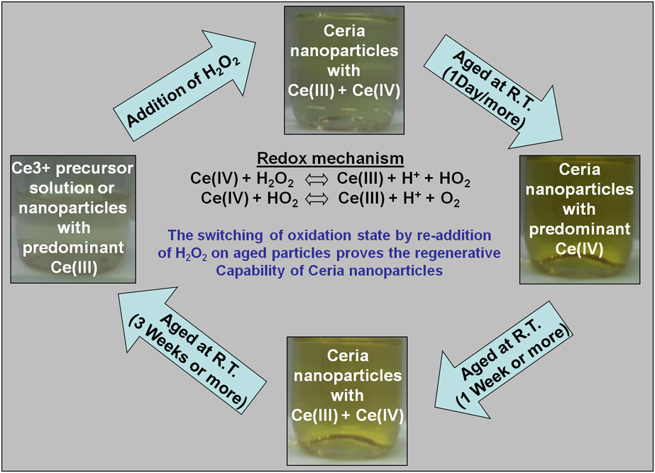
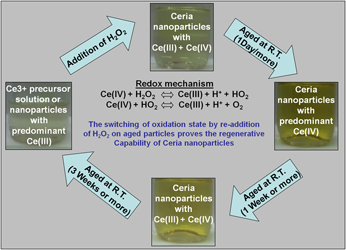
In our earlier work, as summarized in Fig. 1, it was shown that the wet chemical methods using hydrogen peroxide as an oxidizing agent initially produced, within about a day, CNPs with cerium in predominantly +4 oxidation state [Reference Kuchibhatla, Karakoti, Baer, Samudrala, Engelhard, Amonette, Thevuthasan and Seal12]. Aging CNPs in solution at room temperature leads to a slow transition of cerium from +4 to +3 oxidation state. Detailed ultraviolet–visible spectrophotometry, transmission electron microscopy (TEM), and X-ray photoelectron spectroscopy (XPS) analyses of the as-synthesized and aged CNPs were reported previously [Reference Kuchibhatla, Karakoti, Baer, Samudrala, Engelhard, Amonette, Thevuthasan and Seal12, Reference Karakoti, Kuchibhatla, Babu and Seal39, Reference Karakoti, Singh, Kumar, Malinska, Kuchibhatla, Wozniak, Self and Seal40, Reference Kuchibhatla, Karakoti and Seal41]. This cycle of oxidation and reduction of the CNPs sets the framework for the experiments described in this paper. As reported in earlier studies, TEM images of particles extracted from solution at different times and with different oxidation states did not appear to differ [Reference Inerbaev, Karakoti, Kuchibhatla, Kumar, Masunov and Seal38]. The Raman and XRD experiments described above were undertaken to learn more about what was happening to the particles during the transitions and in presence of H2O2.
Schematic representation of the regenerative capability and oxidation state switching of ceria NPs in an aqueous environment. The pictures of bottles containing the NP suspension in DI water show the color changes after different aging periods. Adapted from [Reference Kuchibhatla, Karakoti, Baer, Samudrala, Engelhard, Amonette, Thevuthasan and Seal12] and used with permission.

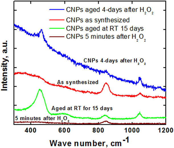
Raman spectroscopy data collected during CNP synthesis were collected at stages consistent with those given in Fig. 1: during synthesis, after particle aging, and upon the addition of H2O2 solution (Fig. 2). The discussion of the Raman spectra for each of the crystallite size focuses on the relative signal strengths of two Raman lines: (i) ∼460–470 cm−1 (cerium oxide) and (ii) ∼845 cm−1 (cerium oxyhydroxide/peroxide) [Reference Patil, Seal, Guo, Schulte and Norwood42]. A third Raman line at ∼877 cm−1 (surface or unreacted peroxide) could be observed soon after the addition of H2O2 solution to the dried or aged particles but is not essential to this discussion as it disappears within a few hours after adding the H2O2 solution to the particles. The presence, absence, and relative strengths of peaks (i) and (ii) provide information about the transformations.
Raman analysis of 3 nm CNPs at different stages of their oxidation–reduction cycle. Raman spectroscopy data collected immediately after synthesis, after particle aging and upon the addition of H2O2 solution to crystalline CNPs and aged, after addition of H2O2, at room temperature for 3–4 days. Variations in the wave numbers of 2–5 units and change in width of peaks may be ascribed to defects in the particles.

The as-synthesized 3 nm CNPs in the +4 oxidation state did not show any peak corresponding to cerium oxide (∼460–470 cm−1); however, a peak at ∼845 cm−1 was observed. After aging at room temperature for 15 days, the 845 cm−1 peak intensity decreased and a peak at ∼450–460 cm−1 evolved, indicating the transformation to nanocrystalline cerium oxide. For similar aging periods where this peak appeared, optical and XPS measurements indicated the presence of Ce3+ in CNPs [Reference Dowding, Lubitz, Karakoti, Kim, Seal, Ellisman, Perkins, Bossy-Wetzel and Self31], i.e., higher oxygen vacancy concentration [Reference Baer, Amonette, Engelhard, Gaspar, Karakoti, Kuchibhatla, Nachimuthu, Nurmi, Qiang, Sarathy, Seal, Sharma, Tratnyek and Wang35]. When these aged 3 nm CNPs were treated with H2O2, the peak at 450–460 cm−1 disappeared and the peak at 845 cm−1, as observed for the freshly synthesized 3 nm particles, reappeared. At roughly 3–4 days after the addition of H2O2 solution, the peak at 845 cm−1 started to disappear and the peak corresponding to crystalline cerium oxide reappeared. To test for reproducibility of the non-ceria peak, a subset of the studies were repeated at a later time on a different Raman system by a researcher not familiar with the earlier measurements. These measurements were consistent with the observations, particularly observing the presence of the ≈845 cm−1 peak upon the addition of H2O2 solution.
These cyclic observations of disappearance of the cerium oxide Raman peak upon addition of hydrogen peroxide and its reappearance upon aging in air suggest that the cycling of cerium oxide proceeds with a transformation between a defected ceria state and an oxyhydroxide state. This state is metastable and decomposes as the solution ages at room temperature and in ambient air to generate crystals of CNPs.
The presence of the CeO2 peak at a lower wave number (450–460 cm−1) than characteristic of bulk ceria (465 cm−1) corroborates well with the observations made by Scholes et al. [Reference Scholes, Hughes, Hardin, Lynch and Miller43]. A shift to the lower wave numbers for nanosized particles is attributed to contributions from phonon confinement, strain, size distribution, defects, and variations in phonon relaxation with particle size.
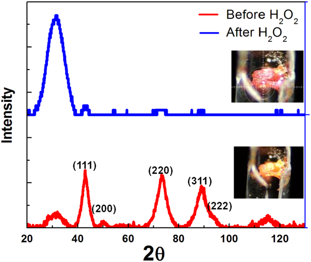
The presence of a Raman peak characteristic of an oxyhydroxide, ∼845 cm−1 peak, immediately raises questions of particle structure. Therefore, micro-XRD data also were collected on aged particles and after the addition of H2O2. Consistent with the cerium oxide peaks observed by Raman, aged (dry) 3 nm crystallites show clear diffraction peaks corresponding to the fluorite lattice of cerium oxide, as labeled in red in Fig. 3. A lower intensity broad peak corresponding to a 2θ value of ∼31° was also observed in the diffraction pattern along with fluorite structure cerium oxide peaks. The broad peak could not be matched with any known peaks of cerium oxide or related structures from the literature or the database. Addition of few drops of 30% H2O2 to this sample caused the fluorite structure to disappear and the relative intensity of the broad peak corresponding to a 2θ value of ∼31° increased. The inset digital images in Fig. 3 show the change from yellow-colored CNPs for the aged sample (Ce3+) to orange red (Ce4+) after the addition of H2O2. In this transition stage, it is not possible to ascertain whether these particles are pure CNPs or some other intermediate phase without further supporting data.
Micro-XRD data of cerium oxide samples (∼3 nm crystallite size) after aging (high Ce3+) and after the addition of H2O2. The insets show digital images of the nanoparticle and the color change with time, as captured on the micro-XRD instrument, and a broken capillary is also clearly visible in the images. The plots are overlaid for clarity and the intensities are not on the same scale and hence should not be compared quantitatively.

These results add a new dimension to previous work on the interaction of cerium with H2O2, which has been of significant interest to the researchers working on catalysis, conversion coatings, and biomedical applications. The cerium-H2O2 interaction resulted in the formation of cerium oxyhydroxide/peroxide; Ce(H2O2)3+ with further reactions expected to produce a  ${\rm{O}}_2^{ - 2}$ containing species CeIV (O2)(OH)2. Such peroxide species were reported to result in Raman peaks around 840–850 cm−1. In an earlier study [Reference Scholes, Hughes, Hardin, Lynch and Miller43] on conversion coatings, it was shown that the formation of peroxide species is enhanced with an increase in the amount of hydrogen peroxide. It was also demonstrated with the help of Raman, XPS, and XRD that the crystallite size of cerium oxide in the coatings was smaller with higher amount of peroxide [Reference Scholes, Hughes, Hardin, Lynch and Miller43]. While the Raman spectroscopy data reported previously indicated the formation of peroxide phase during the synthesis of cerium oxide, our study shows that CNPs can undergo this reactive transformation between cerium oxide and cerium oxyhydroxide/peroxide even after the formation of CNPs.
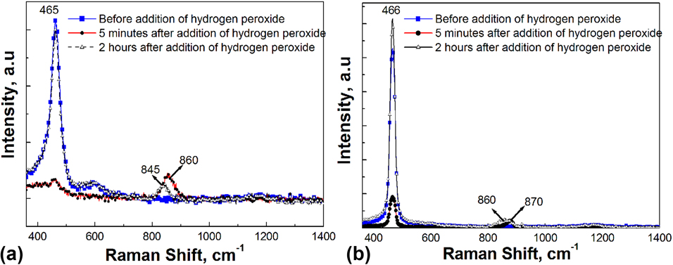
Because of previous studies suggesting size-dependent variations in the behaviors of CNPs, we undertook additional micro-XRD and Raman measurements on 8 and 40 nm CNPs. Comparison of the Raman data for 8 and 40 nm CNPs is shown in Fig. 4. The 3 nm particle data shown in Fig. 3 demonstrate the full cycle, including spectra from as synthesized, aged, after the addition of H2O2, and further aging after the addition. The spectra for the 8 and 40 nm CNPs are shown only up to 2 h after the addition of H2O2. At 5 min, the spectra show very low-intensity peak of crystalline cerium oxide, which is attributed to the masking effect of the residual hydrogen peroxide on the particles. Spectra collected 2 h after the addition of hydrogen peroxide show that both the ∼843 cm−1, oxyhydroxide/peroxide, and the ∼460–470 cm−1, cerium oxide, peaks are present for the 8 nm particles, but only 466 cm−1 cerium oxide peak is present for the 40 nm CNPs. It should be worth noting that along with the peroxide peak at ∼860–87 cm−1, the oxyhydroxide/peroxide peak is also present in case of 8 nm particles, whereas in case of 40 nm particles, the characteristic peak of oxyhydroxide (peroxide) peak is missing. This further strengthens the observation that 8 nm particle is partially transformed by the addition of H2O2, resulting in the intermediate phase, but the 40 nm CNPs show no evidence of a transformation. The peaks at 860–870 cm−1 are attributed to peroxide peaks due to the presence of residual hydrogen peroxide.
Raman spectra of (a) 8 nm and (b) 40 nm CNPs. The plots for the 8 and 40 nm CNPs show data of before addition of H2O2 and 5 min and 2 h after the addition of H2O2 solution, showing the absence of an oxyhydroxide/peroxide peak for the 40 nm particles and the presence of both the ceria and oxyhydroxide peak for the 8 nm particles. To ascertain the consistency and uniformity of information, data were collected from multiple locations. The peaks in the graph near 860–870 nm are the peroxide peaks—due to residual hydrogen peroxide left on the particles. Intensities of the Raman peaks shall not be used for quantitative comparison but for qualitative understanding alone.

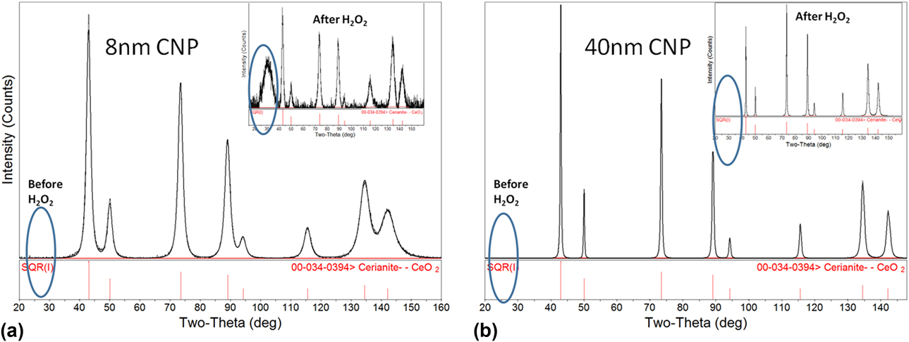
The micro-XRD data for the 8 nm and 40 nm particles indicate a similar behavior, as shown in Fig. 5. The diffraction peaks confirm mostly the structure of CeO2 after the addition of H2O2, with the presence of a low-intensity peak at a 2θ value of ∼31° for the 8 nm particles, and there was no evidence for changes in crystalline cerium oxide for the 40 nm particles. As might be expected, no major color changes were observed in 8 and 40 nm CNPs upon addition of H2O2 (data not shown).
Micro-XRD data from (a) 8 nm and (b) 40 nm CNPs before and after (inset) addition of H2O2 solution to the particles. The presence of an amorphous phase, as observed for the 3 nm particles is not observed for the 40 nm particles and present to a small degree for the 8 nm particles.

In the case of reaction between hydrogen peroxide and 3 nm/8 nm ceria crystallites, micro-XRD data confirm the presence of an amorphous-like phase (at a 2θ value of ∼31°), whose intensity varies with the CNP size. Hence, it is proposed that the amount of peroxide phase (amorphous in nature) is dependent on the extent of reaction or transformation of CNPs and decreases with increased crystallite size of CNPs. The presence of the low-intensity peak at 2θ value of ∼31° in the case of 3 nm crystallites before the addition of H2O2 can be attributed to the presence of this phase from the synthesis method involving H2O2, adopted by the authors. It is important to mention that irrespective of the method of synthesis, addition of H2O2 can produce the 31° peak, as evidenced in the case of 8 nm crystallites.
With aging or drying at room temperature after the addition of hydrogen peroxide, the crystalline cerium oxide peaks reappeared over time. Corresponding Raman and XRD data confirmed the time and crystallite size dependence of the transformation. Relatively smaller nanosized ceria particles with larger surface energy and defect concentration result in visible chemical transformation from crystalline cerium oxide to an amorphous peroxide phase in the presence of H2O2, as confirmed by the broadening and shift of the Raman peak [Reference Patil, Seal, Guo, Schulte and Norwood42, Reference Scholes, Hughes, Hardin, Lynch and Miller43, Reference Tarnuzzer, Colon, Patil and Seal44, Reference Tsunekawa, Fukuda and Kasuya45, Reference Tsunekawa, Sivamohan, Ohsuna, Kasuya, Takahashi and Tohji46]. Absence of cerium peroxide phases in the spectra obtained for 40 nm CNPs in the presence of H2O2 indicates greater stability of these CNPs (relative to 3 and 8 nm crystallites) at 40 nm size. The transformation tendency of the 8 nm CNPs is observed to be in-between the highly reactive 3 nm CNPs and relatively stable 40 nm CNPs.
As the peroxide phase is observed to be dependent on the extent of transformation or the extent of re-transformation, we propose that the regenerative oxidation state switching in CNPs with crystallite sizes below 10 nm may involve formation of molecular structures that do not appear to have a fluorite structure of crystalline cerium oxide. The environment-induced switching of Ce between the +3 and +4 oxidation state in solution [Reference Kuchibhatla, Karakoti, Baer, Samudrala, Engelhard, Amonette, Thevuthasan and Seal12] and in biological systems [Reference Zhang, Ma, Zhang, He, Zhang, Guo, Tai, Zhao and Chai13, Reference Szymanski, Munusamy, Mihai, Xie, Hu, Gilles, Tyliszczak, Thevuthasan, Baer and Orr23] has been observed by many research teams. However, the identification of a reversible phase with a non-ceria structure using optical absorption spectra was not reported and was likely not observable by the methods used. The use of semi in situ studies to monitor the transformation of nanoparticles with micro-XRD and Raman studies enabled the identification of the formation of a peroxide phase for small crystallites in a peroxide-rich environment.
The observed ability of surface species to induce phase transformations in smaller sized nanoparticles is consistent with other studies. For example, the adsorption of water on 3 nm ZnS altered both the surface and bulk structure [Reference Zhang, Gilbert, Huang and Banfield47]. Zhang et al. noted that the structure and reactivity of nanoparticles will depend on both particle size and the nature of surrounding molecules [Reference Zhang, Gilbert, Huang and Banfield47]. As in other examples, varying of surface ligands was found to control the crystal structure of ∼2 nm CdSe NPs [Reference Gao and Peng48] and ligands were observed to change the structure of nanosized Au square sheets from hcp to fcc [Reference Fan, Huang, Han, Bosman, Wang, Zhu, Liu, Li, Zeng, Wu, Shi, Li, Gan and Zhang49]. In the current study, it is proposed that the peroxide species react with surface cerium ions in 3 nm CNPs to transform the crystallites from a cerium oxide phase to a cerium oxyhydroxide/peroxide because of the relatively high number of surface cerium atoms and a generally higher number of structural defects due to particle instabilities [Reference Wu, Wiesmann, Moodenbaugh, Klie, Zhu, Welch and Suenaga25]. As the crystallite size grows, the reaction still takes place, but there is a lower relative number of surface cerium ions that undergo transformation to a cerium oxyhydroxide/peroxide as compared with the bulk crystalline cerium oxide and the larger particles have fewer defects and greater stability. At even higher crystallite sizes, the reaction is negligible compared with the bulk cerium ions, and thus, the particles do not show a reactive transformation from crystalline cerium oxide to a cerium oxyhydroxide/peroxide and its regeneration as highly defective cerium oxide.
The measurements reported primarily focused on observing transformations on a previously reported type of CNP but also involved the use of two additions, particles of different sizes from different sources. Although we have called out the importance of size, it is known that both size [Reference Gagnon and Fromm10, Reference Lee, Song, Cho, Puppala, Nguyen, Zhu, Segatori and Colvin26, Reference Deshpande, Patil, Kuchibhatla and Seal27] and history [Reference Gagnon and Fromm10, Reference Karakoti, Munusamy, Hostetler, Kodali, Kuchibhatla, Orr, Pounds, Teeguarden, Thrall and Baer11] can influence particle behaviors, but size, associated defective structure in smaller size, seems to be significant. The identification of a non-ceria transition phase for some types and sizes of crystallites may assist the efforts to understand the differences in biological impact of CNPs of different sizes and from different sources.
Conclusion
The authors started these experiments with the intention of obtaining greater molecular and structural understanding of the transformations previously observed for 3 nm CNPs. For these particles, a reversible transformation from a ceria to a non-ceria phase during the reaction of CNPs with hydrogen peroxide was found. For CNPs with crystallite size of 8 nm, a partial transformation was observed, and no transformation was observed for 40 nm particles. These observations are consistent with the frequently reported importance of surface ligands in CNP transformations [Reference Baalousha, Le Coustumer, Jones and Lead2, Reference Zhang, Ma, Zhang, He, Zhang, Guo, Tai, Zhao and Chai13, Reference Kumar, Das, Munusamy, Self, Baer, Sayle and Seal16] and the apparent benefits of small particle size in the regenerative behaviors of CNPs [Reference Gagnon and Fromm10]. The authors note that during the synthesis process for the 3–5 nm crystallite CNPs, the peroxide Ce4+ phase is intermediate between the initial solution and the final highly defected Ce3+ phase with a ceria structure. However, the smaller particles have the ability to transform between these two phases.
Considering cerium oxide’s ability to scavenge radicals and regenerative oxidation state switching ability, it appears that the ability of CNPs to transform between Ce4+ and Ce3+ rich phases is facilitated by small size but is not always constrained to be a transformation between defective and non-defective ceria phases. Likely, the tendency of small particles to have many vacancies in the ceria structure [Reference Wu, Wiesmann, Moodenbaugh, Klie, Zhu, Welch and Suenaga25], leading to the formation of the highly defective Ce3+ phase with a ceria structure, facilitates the transformation to other phases. The specific nature of the phases formed likely depends on the solution environment and the potential ligands contained [Reference Zhang, Ma, Zhang, He, Zhang, Guo, Tai, Zhao and Chai13, Reference Kumar, Das, Munusamy, Self, Baer, Sayle and Seal16]. The authors noted that in phosphate-containing environments, Ce phosphates can be formed [Reference Rui, Zhang, Zhang, Ma, He, Gui, Li, Zhang, Zheng, Chu, Guo, Chai, Zhao and Zhang1] and that the presence of polymers can alter the extent and rate of Ce +3 to +4 transformation [Reference Karakoti, Singh, Kumar, Malinska, Kuchibhatla, Wozniak, Self and Seal40].
It is also evident from this study that it is important to understand the size range in which nanoparticles will have the desired materials properties such as higher reactivity or stability and the ability to perform in various application environments [Reference Gagnon and Fromm10, Reference Baer, Amonette, Engelhard, Gaspar, Karakoti, Kuchibhatla, Nachimuthu, Nurmi, Qiang, Sarathy, Seal, Sharma, Tratnyek and Wang35]. Size-dependent reactive transitions, along with any influence of the synthesis methods used, may impact some of the desired behaviors and may be the reason behind the contradictory reports of CNP properties that appear in the literature [Reference Gagnon and Fromm10, Reference Karakoti, Munusamy, Hostetler, Kodali, Kuchibhatla, Orr, Pounds, Teeguarden, Thrall and Baer11, Reference Kuchibhatla, Karakoti, Baer, Samudrala, Engelhard, Amonette, Thevuthasan and Seal12]. It is essential to consolidate the observations reported in this article with additional studies, including first principle modelling, to ascertain the exact nature of the intermediate cerium peroxide species and the type of molecular bonding involved for the systems and environments involved. Such studies should be aimed at unequivocally establishing the chemistry and structure of this cerium-based peroxide intermediate phase (and potentially other similar phases in different environments) and provide the necessary impetus to move closer to practical use of CNPs in biomedical applications, including cancer treatment.
Materials and methods
Cerium oxide nanoparticles
CNPs primarily 3nm particles used in this study were synthesized in the laboratory, while larger particles used for comparison were purchased from different vendors. The sizes reported throughout the discussion are the average crystallite size, as it is an appropriate representation of the individual crystallite and particle reactivity. Therefore, in the entire article particles are referred to as 3, 8, and 40 nm particles, regardless of other measurements of particle size.
The CNPs with ∼3 nm crystallite size were synthesized by dissolving Ce(NO3)3·6H2O in De-ionized water (18.2 M Ω) at room temperature, and a stoichiometric amount of 30% H2O2 was added dropwise to yield a final 30 mM CNPs suspension, as reported previously [Reference Kuchibhatla, Karakoti, Baer, Samudrala, Engelhard, Amonette, Thevuthasan and Seal12]. The aging of this solution over a period of 15–20 days yields CNPs with predominantly Ce3+ oxidation state. Nanoparticles in as-synthesized or as-exposed to hydrogen peroxide in solution had a strong yellow/orange-red color due to the presence of cerium in Ce+4 oxidation state, while crystallites aged in solution were slightly yellow or clear due to the presence of cerium in predominantly Ce3+ state. The chemical state transformation of particles produced by this process was reported in some detail previously [Reference Kuchibhatla, Karakoti, Baer, Samudrala, Engelhard, Amonette, Thevuthasan and Seal12].
The particles of ∼10–20 nm were purchased as suspension from Alfa Aesar (Ward Hill, Massachusetts) (20% in solution) and used as is without any further purification or treatment. These particles were received as an acetate ion stabilized suspension at pH 3.0. The average crystallite size of these nominally 10–20 nm particles was found to be 8 nm.
Larger ∼50 nm particles were purchased as powder from Nanostructured and Amorphous Materials Inc. (Katy, Texas). The crystallite sizes of all three nanoparticles were determined using the Scherer equation from the XRD measurements. The crystallite size of these particles was observed to be ∼40 nm.
Preparation of particles for micro-XRD measurements
Suspensions of all the three sizes of nanoparticles were filtered using a 20 nm filter. A small agglomerate/chunk/portion of wet material was loaded into a capillary, carefully removed from the filter paper with a laboratory spatula (as indicated in the inset of Fig. 3) for the micro-XRD measurements. Particles loaded in the capillary were aged and were allowed to dry inside the capillary at laboratory conditions. Diffraction patterns were collected for as-loaded nanoparticles, after adding 30% hydrogen peroxide, and also after allowing further aging. To compare the structural changes that take place upon reaction with H2O2, the same amount of 30% hydrogen peroxide was added to the dried particles in capillary and allowed to react for 1 h.
Preparation of particles for Raman measurements
For Raman studies, the nanoparticles were collected over the filter paper before and after reaction with hydrogen peroxide. These particles were used for a variety of further studies looking at impacts of aging, drying, and peroxide additions. Some of the 3 nm particles were collected immediately after synthesis and, as observed in previous work, were equivalent to aged particles to which H2O2 was added.
X-ray diffraction
A Rigaku D/MAX RAPID II (Rigaku Americas, The Woodlands, Texas) microdiffractometer with a curved imaging plate and a rotating Cr anode operating at 35 kV and 25 mA was used for the XRD measurements. An optical prefix consisting of horizontal and vertical mirrors specific to Cr Kα radiation was employed in the incident beam to provide focused X-rays at the sample position. At the end of the incident beam path, a 0.3 mm collimator was used to optimize the resolution and signal-to-noise ratio of the diffraction patterns. The incident beam path from X-ray generator to the collimator is protected by O-ring sealed tubing and it is purged with He gas to improve the X-ray flux at the sample position. For the diffraction measurements of dried samples, thin film on Si substrate was mounted on a stainless steel sample holder. JADE 8.5 from Materials Data Inc. (Livermore, California) and PDF4+ database from ICSD (Inorganic Crystal Structure Database, FIZ Karlsruhe, Karlsruhe, Germany) were used to analyze XRD data.
Raman spectroscopy
Raman spectra were acquired on wet and dry powder samples directly from the filter paper in a backscattering configuration using a Spex Model 1877 Raman spectrometer (Edison, New Jersey). The 488 nm line of a Coherent (Santa Clara, California) Innova 307 Ar+ ion laser was used for excitation, and the detector was a Princeton Instruments’ (Trenton, New Jersey) LN/CCD detector. Laser power was attenuated to <10 mW at the sample to minimize decomposition. The slit width was 400 µm, and the exposure time was 100 s for all samples. Spectral acquisition and data analysis were performed using Winspec (Princeton Instruments) and Grams/32AI software (Galactic Industries, Salem, New Hampshire). The estimated uncertainty of the peak frequencies is ±1 cm−1. As described by Patil et al. [Reference Patil, Seal, Guo, Schulte and Norwood42], “cerium oxide has a cubic fluorite-type structure and belongs to the Oh 5 Fm3m space group. This structure has six optical-phonon branches, which yield three zone-center frequencies. These frequencies are 272, 465, and 595 cm−1, corresponding to the doubly degenerate TO mode, the triply degenerate Raman-active mode, and the nondegenerate LO mode, respectively. The triply degenerate Raman-active mode frequency can be directly detected by Raman measurements, whereas the TO and LO frequencies are given indirectly by fits to infrared reflectivity.”
Acknowledgments
A portion of the research reported here was performed in Environmental Molecular Sciences Laboratory (EMSL) a DOE user facility supported by the Office of Biological and Environmental Research and located at Pacific Northwest National Laboratory. Authors acknowledge the help from scientists in EMSL, specifically Dr. Nachimuthu Ponnusamy for his help with carrying out micro-XRD experiments and other researchers in Sudipta Seal (SS) group for their contributions to understanding CNP systems. CNP research by SS group was supported by NSF NIRT, NSF CMII and IREE, and NSF EEC (US Australia). Parts of the work are funded by NIH/NIEHS-U19 U19 ES019544 program.