Introduction
The fossil record of small mammals from the Miocene of the Iberian Peninsula is well known (Van der Meulen et al., Reference Van der Meulen, García-Paredes, Álvarez-Sierra, Van den Hoek Ostende, Kordijk, Oliver and Peláez-Campomanes2012; Casanovas-Vilar et al., Reference Casanovas-Vilar, Madern, Alba, Cabrera, García-Paredes, Van den Hoek Ostende, DeMiguel, Robles, Furió, Van Dam, Garcés, Angelone and Moyà-Solà2016; among others); but, most studies are focused on rodents. In addition, the distribution of insectivores sensu lato follows a latitudinal gradient; they are less abundant in southern Europe than in the northern parts of this continent (Furió et al., Reference Furió, Casanovas-Vilar and Van den Hoek Ostende2011; Van den Hoek Ostende et al., Reference Van den Hoek Ostende, Furió, Madern and Prieto2016), and for this reason, the remains of this group are more scarce in southern Europe than in the north.
The family Erinaceidae is an extant clade that includes hedgehogs and gymnures, also known as lunar rats. The subfamily Galericinae is nowadays confined to southestern Asia, having a widespread Holarctic distribution during the Paleogene and Neogene (Lopatin, Reference Lopatin2006). The genus Galerix Pomel, Reference Pomel1848 has a long record, ranging from the upper Oligocene to the lower Pliocene (Gould, Reference Gould1995; Zijlstra and Flynn, Reference Zijlstra and Flynn2015), and constitutes the best-represented erinaceid. The referal of Lantanotherium Filhol, Reference Filhol1888 to a specific tribe is uncertain, although Galerix and Lantanotherium have long been considered synonymous; some authors include Lantanotherium in the Galericini sensu lato or in the Echinosoricini (Viret, Reference Viret1940; Butler, Reference Butler1948; Storch and Qiu, Reference Storch and Qiu1991; Gould, Reference Gould1995; Lopatin, Reference Lopatin2006; Furió and Alba, Reference Furió, Alba, Pérez-García, Gascó, Gasulla and Escaso2011), but Engesser (Reference Engesser1979) and Bannikova et al. (Reference Bannikova, Lebedev, Abramov and Rozhnov2014) referred it to the Hylomyini, due to its resemblance to the genus Hylomys Müller, Reference Müller and Temminck1840. The subfamily Erinaceinae, which includes the hedgehogs, is represented in the Ribesalbes-Alcora Basin by a single tooth. Here, we have assigned this tooth to an indeterminate species of the current genus Atelerix Pomel, Reference Pomel1848. Although there is abundant literature regarding this genus and its synonymies, with some authors including it in Mioechinus Butler, Reference Butler1948 or in Postpalerinaceus Crusafont-Pairó and Villalta, Reference Crusafont-Pairó and Villalta1947 (Mein and Ginsburg, Reference Mein and Ginsburg2002; Prieto et al., Reference Prieto, Van den Hoek Ostende, Hír and Kordos2015; among others), that discussion is beyond the scope of the present work.
The family Herpetotheriidae is traditionally considered a group of marsupials close to, or within, the didelphimorphian opossums (Ziegler, Reference Ziegler, Rössner and Heissig1999). To other authors, it is a basal group of Metatheria, either related to the origin of marsupials, but distinct from them (Horovitz et al., Reference Horovitz, Martin, Bloch, Ladevèze, Kurz and Sánchez-Villagra2009), or included within the marsupialiforms (all of the basal groups of Metatheria, excluding deltatheroids) (Williamson et al., Reference Williamson, Brusatte and Wilson2014). Their initial adscription to Marsupialia (especially to the Didelphimorphia) was based on the study of the dental characters (Ladevèze et al., Reference Ladevèze, Smith and Smith2012). However, study of the cranium and complete skeletal remains showed that they cannot be considered true marsupials (Sánchez-Villagra et al., Reference Sánchez-Villagra, Ladevèze, Horovitz, Argot, Hooker, Macrini, Martin, Moore-Fay, de Muizon, Schmelzie and Asher2007; Horovitz et al., Reference Horovitz, Ladevèze, Argot, Macrini, Martin, Hooker, Kurz, de Muizon and Sánchez-Villagra2008). The record of the genus Amphiperatherium Filhol, Reference Filhol1879 spans from the lower Eocene to the middle Miocene (Crochet, Reference Crochet1980; Furió et al., Reference Furió, Ruiz-Sánchez, Crespo, Freudenthal and Montoya2012; Prieto and Rummel, Reference Prieto and Rummel2015). Its diversity reached a maximum during the Eocene and then progressively decreased until its extinction (Crochet, Reference Crochet1980; Furió et al., Reference Furió, Ruiz-Sánchez, Crespo, Freudenthal and Montoya2012). The species present in the Ribesalbes-Alcora Basin, Amphiperatherium frequens (von Meyer, Reference von Meyer1846), was divided by Koenigswald (Reference Koenigswald1970) into three subspecies, first rejected by Crochet (Reference Crochet1980), and subsequently accepted as chronosubspecies by Ziegler (Reference Ziegler1990). The validity of the genus Amphiperatherium or its synonymy with the genus Peratherium Aymard, Reference Aymard1850 have been well discussed in the literature (Ladevèze et al., Reference Ladevèze, Smith and Smith2012; and references therein).
This paper completes a series of works that deal with insectivores (Crespo et al., Reference Crespo, Furió, Ruiz-Sánchez and Montoya2018; Reference Crespo, Fagoaga, Montoya and Ruiz-Sánchez2019b, Reference Crespo, Marquina-Blasco, Ruiz-Sánchez and Montoyac) from the Ribesalbes-Alcora Basin. The material of Amphiperatherium frequens presented here was partially published by Furió et al. (Reference Furió, Ruiz-Sánchez, Crespo, Freudenthal and Montoya2012), but recovery of new material and new sites discovered have allowed us to revise the material and better understand the population from the Ribesalbes-Alcora Basin. Forthcoming papers will deal with the systematic palaeontology, palaeoecology, and biostratigraphy of rodents and other small mammals, as well as some of the more spectacular findings from the sites of this basin, thus completing our knowledge of the small mammal assemblage from the Ribesalbes-Alcora Basin.
Geographic and geologic setting
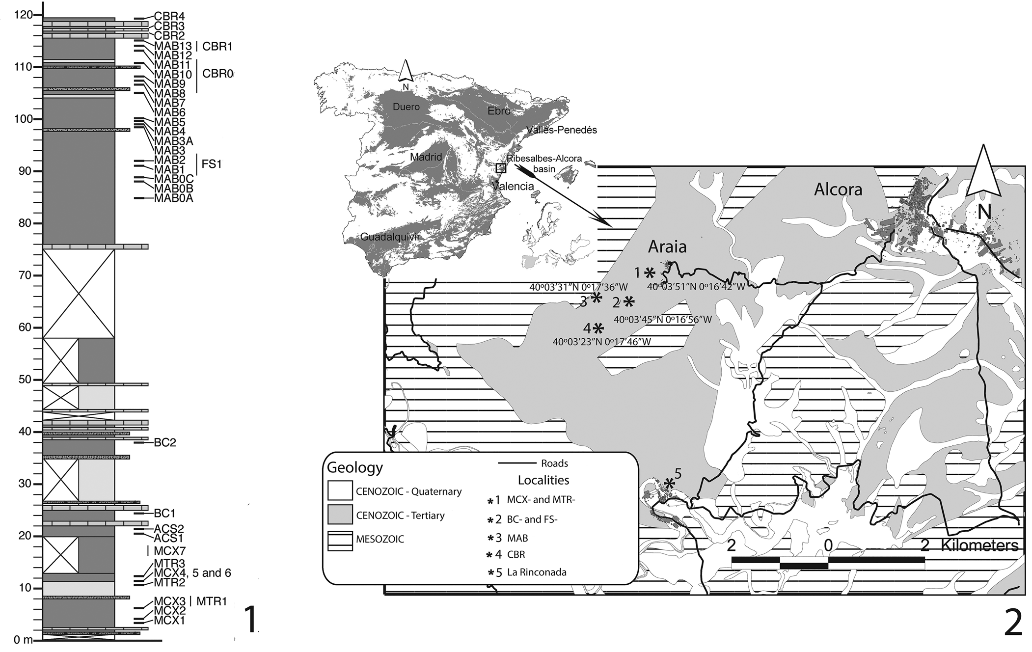
The stratigraphic column was performed in the Barranc de Campisano within the Ribesalbes-Alcora Basin, near the village of Araia d'Alcora (eastern Spain; Agustí et al., Reference Agustí, Anadón, Ginsburg, Mein and Moissenet1988; Crespo et al., Reference Crespo, Suárez-Hernando, Murelaga, Ruiz-Sánchez and Montoya2019a; Fig. 1.1). This side of the ravine is formed by gray and yellow mudstones, limestones, and sandstones, ~ 100 m thick, and comprises seven sections: Mas dels Coixos, Mas de Torner, Araia Cantera Sud, Barranc de Campisano, Foieta la Sarra, Mas d'Antolino B, and Corral de Brisca (Crespo et al., Reference Crespo, Suárez-Hernando, Murelaga, Ruiz-Sánchez and Montoya2019a). Agusti et al. (Reference Agustí, Anadón, Ginsburg, Mein and Moissenet1988) described the first sites, but this has been expanded to 45 new sites in this basin (Crespo et al., Reference Crespo, Suárez-Hernando, Murelaga, Ruiz-Sánchez and Montoya2019a). The sites studied here represent a similar stratigraphic range corresponding to the local Biozone C from the Calatayud-Daroca Basin (MN4, lower Aragonian, early Miocene) in the chronologic interval 16.5–16 Ma according to Van der Meulen et al. (Reference Van der Meulen, García-Paredes, Álvarez-Sierra, Van den Hoek Ostende, Kordijk, Oliver and Peláez-Campomanes2012; Crespo et al., Reference Crespo, Suárez-Hernando, Murelaga, Ruiz-Sánchez and Montoya2019a).
(1) Composite column of the Campisano ravine with positions of the studied sites. (2) Spanish Cenozoic basins, with location of the Ribesalbes-Alcora Basin, schematic distribution of sediments, and positions of the studied sites. ACS = Araia Cantera Sud; BC = Barranc de Campisano; CBR = Corral de Brisca; FS = Foieta la Sarra; MAB = Mas d'Antolino B; MCX = Mas dels Coixos; MTR = Mas de Torner. La Rinconada is the classic Fossil-Lagerstätte site with remains of insects, plants, and amphibians. Modified from Crespo et al. (Reference Crespo, Furió, Ruiz-Sánchez and Montoya2018, Reference Crespo, Suárez-Hernando, Murelaga, Ruiz-Sánchez and Montoya2019a).

Materials and methods
The photographic images of the specimens were taken with a SEM HITACHI 4800 at the Servei Central de Suport a la Investigació Experimental (SCSIE) of the Universitat de València Estudi General (UVEG).
For erinaceids, we used the nomenclature of Ziegler (Reference Ziegler1990) and Furió (Reference Furió2007) and the measurements of Doukas (Reference Doukas1986). For metatherians, we followed the nomenclature of Goin et al. (Reference Goin, Woodburne, Zimicz, Martin, Chornogubsky, Goin, Woodburne, Zimicz, Martin and Chornogubsky2016) and measurements used by Crochet (Reference Crochet1980). Measurements (length × width) are given in mm and were taken using a Leica MZ75 binocular microscope, by means of the displacement of a mechanical stage, connected to a Sony Magnescale measuring device. Lower teeth are designated with lower-case letters (lower molars: m1, m2, m3, and m4; lower premolars: p1, p2, p3, and p4; lower canine: c; lower incisor: i; lower decidual: dp) and upper teeth are written in upper-case letters (upper molars: M1, M2, M3, and M4; upper premolars: P1, P2, P3, and P4; upper canine: C; upper incisor: I; upper decidual: DP). Throughout the text, names of the localities often appear abbreviated as follows: BC = Barranc de Campisano; CBR = Corral de Brisca; FS = Foieta la Sarra; MAB = Mas d'Antolino B; MCX = Mas dels Coixos; MTR = Mas de Torner.
Repositories and institutional abbreviations
Figured and other specimens examined in this study are deposited in the Museu de la Universitat de Valencia d'Història Natural (MUVHN), Burjassot, Spain. Other cited institutions include: AL = Aristotle University of Thessaloniki, Greece; BHPSM = Bayerische Staatssammlung für Paläontologie und Historische Geologie, Munich, Germany.
Systematic paleontology
Class Mammalia Linnaeus, Reference Linnaeus1758
Order Eulipotyphla Waddell, Okada, and Hasegawa, Reference Waddell, Okada and Hasegawa1999
Family Erinaceidae Fischer, Reference Fischer1814
Subfamily Galericinae Pomel, Reference Pomel1848
Genus Galerix Pomel, Reference Pomel1848
Type species
Viverra exilis de Blainville, Reference de Blainville1839.
Galerix symeonidisi Doukas, Reference Doukas1986
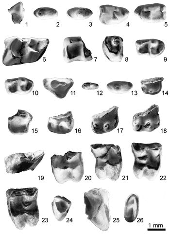
Figure 2.1–2.24
Holotype
AL 3036 (Doukas, Reference Doukas1986, pl. 1, fig. 6), M2 sin; Aliveri, Evia Island, Greece, early Miocene, MN4.
Teeth of the family Erinaceidae from the Ribesalbes-Alcora Basin. Galerix symeonidisi: (1) right p1 (lingual view, MAB5-165); (2) left p2 (MAB3-774); (3) right p2 (MAB5-757); (4) right p4 (MAB3-767); (5) left p4 (MAB5-411); (6) left m1 (MAB5-742); (7) right m2 (MAB3-730); (8) left m2 (MAB5-483); (9) right m3 (MAB4-4); (10) right m3 (MAB5-141); (11) left DP3 (MAB0A-83); (12) left P1 (MAB5-768); (13) left P2 (MAB11-121); (14) left P3 (MAB5-720); (15) right P3 (MAB5-756); (16) left P4 (MCX3-22); (17) left P4 (MCX3-121); (18) left P4 (MAB3-768-5); (19) left P4 (MCX3-22); (20) left M1 (MAB5-725); (21) right M1 (MAB13-4); (22) right M2 (MAB0A-84); (23) left M2 (MAB5-724); (24) left M3 (MAB5-729). cf. Lantanotherium sp. indet.: (25) left m1 (MAB3-52). cf. Atelerix sp. indet.: (26) right M3 (MAB3-799).

Occurrence in this basin
Mas dels Coixos 1, 3, and 4 (MCX1, MCX3, and MCX4); Mas de Torner 2 (MTR2); Barranc de Campisano 1 (BC1); Foieta la Sarra 1 (FS1); Mas d'Antolino B 0A, 0B, 3, 4, 5, 6, 11, and 13 (MAB0A, MAB0B, MAB3, MAB4, MAB5, MAB11, and MAB13); and Corral de Brisca 1 (CBR1).
Description
dp3.—The outline is subtriangular in occlusal view, with the posterior side being wider. The labial and lingual sides are straight. There is a small concavity near the protoconid. This cusp is conic and slightly elongated. The anterior cuspule is poorly developed and smaller than the posterior one. There is a small platform between the posterior cuspule and the protoconid.
dp4.—The outline is subrectangular, with two roots. There is a small lingual concavity, and the labial part is almost straight. The paraconid is well developed and it is isolated and lower than the protoconid and metaconid. The protoconid is the highest cusp, extending through nearly half of the tooth. The metaconid is slightly lower than the protoconid; it is only an accessory cuspid on the lingual side of the last cuspid. The cingulid extends around the posterior side of the tooth, including a small inclined valley on the posterolingual part. The posterolabial side is broken.
c.—The tooth is laterally compressed, and is triangular in anterior view. The root is well developed and the tooth is curved on the occlusal zone. On the labial side, there is a crest that extends from the apex to the base of the crown. The labial side is more curved than the lingual one. There are no cingulids.
p1.—This tooth has a single, well-developed root directed backward. The outline is ellipsoidal in occlusal view. The lingual side is protruding, whereas the posterior side is straight. There is only one cusp located toward the anterior part with two small cuspules on the anterior and posterior sides. There is no cingulid.
p2.—The tooth has two roots. Its outline is subrectangular in occlusal view. The protoconid is the main cusp and a small cuspule is present on the anterior side. The posterior side is rounded, except in the material from MAB5 and MCX3, which each have a small valley between the protoconid and the posterior crest.
p3.—The tooth has two roots. Its outline is almost elliptic in occlusal view, with a straight lingual side. The anterior cusp is small or absent. The protoconid is conic in shape. A small cingulid starts on the posterior side, and extends to the labial side, near the base of the protoconid. There is a small valley between the posterior cingulid and the protoconid.
p4.—The outline is subquadrate in occlusal view, except in the material from MAB3, in which it is rather subtriangular. The paraconid is well developed and somewhat lower than the metaconid. The latter is bulbous, and therefore the trigonid valley is poorly developed. The paraconid is connected to the protoconid. The metaconid is reduced and also connected to the protoconid. On the posterior part, there is a postcingulid that meets the entoconid near the labial side. This cusp is well developed in a third of the recovered material. Between the cingulid and the protoconid, there is a small valley with a well-developed entoconid or a wide valley with a less-developed entoconid. The posterior side is straight in two-thirds of the tooth length and is slightly concave in the remaining portion.
m1.—The trigonid and the talonid are similar in length and width. The anterior cingulid is narrow and extends from the base of the paraconid to the anterolingual base of the protoconid. The paraconid is slightly lower than metaconid and extends into a long, V-shaped paralophid in lateral view. The protoconid is higher than the metaconid; they contact each other by means of a protolophid, V-shaped in lateral view. The valley of the trigonid is deep. The metacristid is poorly developed and even absent in one-third of the molars. The entocristid is straight and descends abruptly. The entoconid is higher and larger than the hypoconid. The oblique cristid is low and ends near the base of the protoconid. The posterior cingulid slopes down toward the base of the hypoconid.
m2.—These are mostly broken teeth, each with the talonid narrower than the trigonid but similar in length. The anterior cingulid is relatively narrow. The paraconid is incorporated into the paralophid and near the straight valley of the trigonid. The protoconid is slightly higher than the metaconid; they are connected by a U-shaped protolophid. The metacristid is poorly developed in MAB3 material and absent in those from other sites. The talonid valley is wide. The oblique cristid is straight and connected to the protoconid. The entoconid is higher than the poorly developed hypoconid. The postcristid is straight and connected to the anterior cusps. The posterior cingulid is short and only present at the base of the hypoconid.
m3.—The talonid is narrower than the trigonid, but of similar length. The anterior cingulid is wide. The paraconid is included in the paralophid, and almost closes the narrow trigonid valley. The metaconid is as high as the protoconid; they are connected by a V-shaped protolophid. The metacristid is only a thickening, being absent in the MCX3 material. The talonid valley is wide. The oblique cristid is straight and not connected to the protoconid. The hypoflexid is connected to the valley of the talonid. The hypocone is poorly developed and as high as the entoconid. The postcristid is straight and connected to the hypoconid with the entoconid. The posterior cingulid is absent.
I3.—This is a half tooth with a curved principal cusp and a well-developed platform.
C.—The canine has two roots and only one cusp, and is subrectangular in occlusal view. The tip of the cusp is at the anterocentral part of the tooth, and is inclined toward the lingual side. There are no cinguli.
DP3.—This tooth is triangular in occlusal view. The paracone is the main cusp. A small secondary cusp is present in the posterocrista. The latter is slightly connected to the cingulum on the lingual side, but is not connected to the cuspule, which is located on the labial side. The parastyle is a small cuspule formed by the cingulum; there is a wide valley between the parastyle and the paracone. The protocone is small, and connected to the metastyle by a cingulum.
P1.—This is a small tooth, elliptical in occlusal view. The paracone is well developed and has a small, independent parastyle.
P2.—This tooth has two roots and is almost elliptical in occlusal view; the lingual side is straight. Its widest portion is at the middle of the tooth. The protocone is conical and the posterior side has a straight crest separated from the protocone by a small valley.
P3.—There are no complete specimens of this tooth. It is subquadrangular in occlusal view, with a poorly developed parastyle and a cingulum that connects the parastyle with the protocone. The paracone is well developed, being the highest cusp. The protocone and the hypocone are also well developed, although in the material from MAB5, the hypocone is somewhat smaller. The posterior valley is rounded and wide; in the material from MAB5, the posterior valley is subquadrangular. The tooth has three roots.
P4.—There are no complete specimens. The paracone is the highest and largest cusp; it is conical and directed backward. The posterior style is V-shaped and ends with an angular metastyle. The posterior cingulum is wide and well developed. The parastyle is straight. The protocone and the hypocone are well developed and independent from each other, although in the material from MCX3, each cusp is smaller. The paracone has a crest that connects with the parastyle. The posterior valley is wide and is closed by a straight posterior cingulum; it is straight on the preserved part.
M1.—The tooth is subquadrangular in occlusal view. The preparacrista is short and connected to a small parastyle. The anterior cingulum is narrow and starts from the anteromedial side of the protocone. The preprotocrista ends on the anterolingual side of the paracone. This cusp is connected to the metacone by the postparacrista and a short premetacrista. The metacone is higher than the paracone. The protoconule is well developed. The protocone is conical and its posterior crest is connected to the prehypocrista rather than the metaconule. The metaconule is large and semilunar in shape. Its anterior crest ends on the anterolingual side of the metacone; its posterior crest ends on its posterolingual side. The postmetacrista is short, strongly curved, and extends to the posterolabial side. The hypocone is conical and slightly narrow. The posterior side of the tooth is concave between the hypocone and the metacone, and its cingulum starts on the posterolabial side of the hypocone.
M2.—The outline of this tooth is subquadrangular in occlusal view. The parastyle is small and connects to the paracone by a short preparacrista. The anterior cingulum is short and narrow, and ends at the anteromedial side of the paracone. The paracone is lower than the metacone; the two cusps are connected by the postparacrista and by a short premetacrista. The protoconule is well developed. The protocone is conical. The posterior arm of the protocone is connected to the prehypocrista rather than the metaconule. The metaconule is well developed and crescent-shaped; its anterior arm ends near the anteriolingual side of the metacone, and its posterior arm ends on the posterolingual side of the metacone. The postmetacrista is short and strongly curved. The hypocone is conical and sligthly narrow. The posterior cingulum starts on the posterolabial side of the hypocone. The posterior side of the tooth is convex.
M3.—The outline of this tooth is subtriangular in occlusal view. The parastyle is feebly developed, being only a small thickening on the anterior cingulum, which is wide and well developed. The tooth has only three cusps, connected by crests, and they close a deep valley. The protocone is somewhat higher than the paracone and the metacone is the lowest cusp. There is a weak posterolingual cingulum.
Material
MCX3: 1 p1, 2 p2 (not complete), 1 p3, 1 m1 (not complete), 1 m3 (not complete), 4 P4 (not complete); MCX4: 1 p3, 1 M1/M2 (not complete); MTR2: 2 p1 (one not complete), 1 p2 (not complete), 1 m3 (not complete), 1 C; BC1: 1 m2 (not complete), 1 M3; FS1: 1 I (not complete), 1 M3; MAB0A: 1 DP3, 1 M2; MAB0B: 1 P2 (not complete), 1 M2; MAB3: 1 c, 1 p1, 2 p2 (one not complete), 1 p3, 2 p4 (one not complete), 1 m1, 1 m2 (not complete), 1 DP3 (not complete), 1 I3 (not complete), 1 P3 (not complete), 2 P4 (not complete), 1 M1 (not complete); MAB4: 1 p3 (not complete), 1 m3; MAB5: 1 dp4 (not complete), 2 p1, 4 p2 (one not complete), 2 p3, 4 p4 (one not complete), 5 m1 (three not complete), 3 m2 (two not complete), 3 m3 (one not complete), 2 P1 (one not complete), 2 P3 (not complete), 1 M1 (not complete), 2 M2 (one not complete), 1 M3; MAB6: 1 p4 (not complete); MAB11: 1 dp4, 1 c (not complete), 1 p2, 1 p4 (not complete), 1 m2 (not complete), 1 P2, 1 P4 (not complete); MAB13: 1 P2 (not complete), 1 M1 (not complete), 1 M2 (not complete); CBR1: 1 dp3, 1 P2 (not complete), 1 M3 (not complete).
Measurements
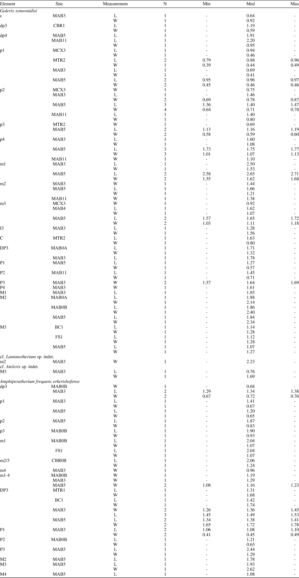
See Table 1.
Measurements of the teeth of erinaceids and metatherians from the Ribesalbes-Alcora Basin. L = length; Min = minimum; Med = median; Max = maximum; W = width.

Remarks
The most common erinaceid in the Ribesalbes-Alcora Basin is the small Galericini Galerix; it is also one of the most common insectivores in the lower-middle Miocene of Europe (Van den Hoek Ostende et al., Reference Van den Hoek Ostende, Furió, Madern and Prieto2016). The studied material is characterized by a M1/M2 with a semilunar metaconule, typical of the tribe Galericini. Among this tribe, it differs from members of Schizogalerix Engesser, Reference Engesser1980 for its undivided mesostyle in M1/2 and the posterior cingulum of the m1/2, usually connected to the entoconid. It differs from members of Parasorex von Meyer, Reference von Meyer1865 by the posterior arm of the metaconule, which is not extended to the posterolingual corner of the molar. Moreover, the p2 is larger than the p3. It differs from members of Riddleria Van den Hoek Ostende, Reference Van den Hoek Ostende2003b by the regular outline of the upper molars and less-developed cingulums. Finally, among other features, members of Deinogalerix Freudenthal, Reference Freudenthal1972 are larger (Van den Hoek Ostende, Reference Van den Hoek Ostende2001, Reference Van den Hoek Ostende2003b).
Within the genus Galerix, the species G. symeonidisi differs from other similar European species, e.g., G. aurelianensis Ziegler, Reference Ziegler1990, G. remmerti Van den Hoek Ostende, Reference Van den Hoek Ostende, López-Martínez, Peláez-Campomanes and Hernández-Fernández2003a, and G. exilis, in its smaller size, its metaconule disconnected from the postprotocrista, its short posterior crest on M1/M2, and a hypocone on P3. From G. kostakii Doukas and Van den Hoek Ostende, Reference Doukas and Van den Hoek Ostende2006, it differs in its small size (Van den Hoek Ostende, Reference Van den Hoek Ostende2001; Van den Hoek Ostende and Doukas, Reference Van den Hoek Ostende and Doukas2003; Doukas and Van den Hoek Ostende, Reference Doukas and Van den Hoek Ostende2006).
The presence of a poorly developed hypocone on P3 is a typical character of the Iberian populations of this taxon. Sometimes this cusp is so small that it consists only of a small enlargement on the posterior side of the protocone. In this feature, the Iberian populations differ from those of Central Europe (Van den Hoek Ostende and Doukas, Reference Van den Hoek Ostende and Doukas2003); the material from the Ribesalbes-Alcora Basin confirms this.
The oldest record of the genus Galerix is from the lowermost Miocene of Anatolia (de Bruijn et al., Reference de Bruijn, Ünay, Van den Hoek Ostende and Saraç1992; Van den Hoek Ostende, Reference Van den Hoek Ostende1992); this genus does not appear in Europe until MN2/3 at the sites of Stubersheim 3 and Wintershof West, with the species G. aurelianensis (Ziegler, Reference Ziegler1994; Van den Hoek Ostende, Reference Van den Hoek Ostende2003b). On the Iberian Peninsula, its first occurrence is the endemic species G. remmerti, reported in the Calatayud-Montalbán, Teruel, and Ebro basins (Van den Hoek Ostende, Reference Van den Hoek Ostende, López-Martínez, Peláez-Campomanes and Hernández-Fernández2003a; Van den Hoek Ostende and Furió, Reference Van den Hoek Ostende, Furió, Van den Hoek Ostende, Doukas and Reumer2005). Galerix symeonidisi is not found on the Iberian Peninsula until the end of MN3, in San Roque 4B from the Calatayud-Montalbán Basin, although this is uncertain (Van den Hoek Ostende, Reference Van den Hoek Ostende, López-Martínez, Peláez-Campomanes and Hernández-Fernández2003a).
At the beginning of MN4, Galerix remmerti was replaced by G. symeonidisi, which was itself gradually replaced by another species, G. exilis, which appeared shortly afterward. Eventually, G. symeonidisi became extinct in MN5 (Van den Hoek Ostende and Doukas, Reference Van den Hoek Ostende and Doukas2003). Discussing the co-occurrence of G. symeonidisi and G. exilis at the same site, Van den Hoek Ostende and Doukas (Reference Van den Hoek Ostende and Doukas2003) argued that in some sites of the Daroca-Calamocha Basin, their variability in size is too large to be attributed to a single species. Therefore, it is not possible to precisely identify the migration of G. exilis in Europe. Due to the overlap in size of the two species, G. exilis could have appeared earlier than can be inferred from the fossil record. At this time in Central Europe, Ziegler and Falhbusch (Reference Ziegler and Fahlbusch1986) found some intermediate populations; however, to other authors, e.g., Van den Hoek Ostende et al. (Reference Van den Hoek Ostende, Furió, Madern and Prieto2016), these represent a mixture of elements from each species.
The material studied here is characterized by its small size, which appears to be uniform, the lack of contact between the protocone and the metaconule, and the P3 with a small hypocone. These features suggest that Galerix symeonidisi is the species present in the Ribesalbes-Alcora Basin, although the presence of G. exilis cannot be completely excluded. The same conclusion can be made for the site of Montalvos 2 (Hordijk et al., Reference Hordijk, Bosma, de Bruijn, Van Dam, Geraedts, Van den Hoek Ostende, Reumer and Wessels2015). Galerix symeonidisi is also present in the nearby sites of Buñol (Van den Hoek Ostende and Furió, Reference Van den Hoek Ostende, Furió, Van den Hoek Ostende, Doukas and Reumer2005) in which it certainly might have co-occurred with G. exilis in Buñol. The Vallès-Penedès Basin is tentatively located in the site of Les Cases de la Valenciana (Jovèlls-Vaquè et al., Reference Jovells-Vaqué, García-Paredes, Furió, Angelone, Van den Hoek Ostende, Berrocal Barberà, DeMiguel, Madurell-Malapeira and Casanovas-Vilar2018)
Tribe Galericini sensu lato Lopatin, Reference Lopatin2006
or
Tribe Echinosoricini Butler, Reference Butler1948
Genus Lantanotherium Filhol, Reference Filhol1888
Type species
Erinaceus sansaniensis Lartet, Reference Lartet1851.
cf. Lantanotherium sp. indet.
Figure 2.25
Occurrence in this basin
Mas d'Antolino B 3 (MAB3).
Description
m1.—This tooth is a fragment of a large talonid, with well-developed entoconid and hypoconid of similar sizes, connected by a postcristid. The entocristid descends abruptly. The posterior cingulid is independent from the postcristid and extends toward the base of the hypoconid.
m2.—This tooth is a fragment of a large talonid, with an entoconid larger than the hypoconid. The entocristid descends abruptly. The postcristid connects the two cusps. The posterior cingulid is not connected to the postcristid and is parallel to the base of the hypoconid.
Material
MAB3: 1 m1 (not complete), 1 m2 (not complete).
Measurements
See Table 1.
Remarks
Two large fragments of erinaceid molars have been found in MAB3. The posterior side of the m1 is similar to the Lantanotherium material from Sansan, in having a postcristid with a weak connection to the posterior cingulid; this characteristic distinguishes Lantanotherium from Galerix (Engesser, Reference Engesser2009). In other genera, e.g., Atelerix, the posterior cingulid is connected to the labial cingulid, whereas in the material studied here, it is located below the hypoconid. In the genus Amphechinus Aymard, Reference Aymard1850, or at least in A. golpeae Gibert, Reference Gibert1974, the posterior cingulid seems connect to the hypocone; in other species, e.g., A. arvernensis (de Blainville, Reference de Blainville1839) or A. robustus (Lavocat, Reference Lavocat1951), there is no connection between the posterior cingulid and postcristid (Gibert, Reference Gibert1975; Sesé, Reference Sesé1980; Ziegler, Reference Ziegler1998). The oldest occurrence of Lantanotherium is in Central Europe in MN4 (Klietmann et al., Reference Klietmann, Nagel, Rummel and Van den Hoek Ostende2014). In the Iberian Peninsula, its record is poor and limited to L. sansaniensis (Lartet, Reference Lartet1851) from Buñol (Magro Basin) and L. piveteaui Crusafont-Pairó, Villalta, and Truyols, Reference Crusafont-Pairó, Villalta, Truyols, Crusafont, Villalta and Truyols1955 from Can Cerà (Vallès-Penedès Basin), its type locality (Van den Hoek Ostende and Furió, Reference Van den Hoek Ostende, Furió, Van den Hoek Ostende, Doukas and Reumer2005). Nevertheless, the type material of L. piveteaui was lost and it has been suggested that it might have belonged to another Galericini species (Van den Hoek Ostende et al., Reference Van den Hoek Ostende, Furió, Madern and Prieto2016). Therefore, we have provisionally assigned these teeth to cf. Lantanotherium sp. indet, awaiting new material collected in the Ribesalbes-Alcora Basin that might enable a more precise description and characterization of this taxon.
Subfamily Erinaceinae Fischer, Reference Fischer1814
Genus Atelerix Pomel, Reference Pomel1848
Type species
Erinaceus quadridactylus Pomel, Reference Pomel1848.
cf. Atelerix sp. indet.
Figure 2.26
Occurrence in this basin
Mas d'Antolino B 3 (MAB3).
Description
M3.—A much reduced tooth, elliptical in occlusal view. It has a well-developed protocone and a small paracone. There is a cingulum that almost surrounds the tooth and widens on the labial side; it has a small cusp on the posterolabial side. On the lingual side of the tooth, there is a small cusp.
Material
MAB3: 1 M3.
Measurements
see Table 1.
Remarks
The tooth found in MAB3 is a very reduced M3, typical of the family Erinaceidae. Atelerix is one of the smallest genera of this family. Other similar genera, e.g., Mioechinus or Postpalerinaceus, were considered junior synonyms by Mein and Ginsburg (Reference Mein and Ginsburg2002) and Prieto et al. (Reference Prieto, Van den Hoek Ostende, Hír and Kordos2015), respectively.
The record of Atelerix, which includes the current African hedgehog, is very scarce in the Iberian Peninsula, with a single species—the endemic A. butleri (Crusafont-Pairó, Villalta, and Truyols, Reference Crusafont-Pairó, Villalta, Truyols, Crusafont, Villalta and Truyols1955) (or ‘Mioechinus’ butleri after Prieto et al., Reference Prieto, Van den Hoek Ostende, Hír and Kordos2015) in its type locality of San Mamet (Crusafont-Pairó et al., Reference Crusafont-Pairó, Villalta, Truyols, Crusafont, Villalta and Truyols1955). However, this genus is more common in Central Europe and comprises A. rhodanicus Mein and Ginsburg, Reference Mein and Ginsburg2002 and A. depereti Mein and Ginsburg, Reference Mein and Ginsburg2002, two co-existing species from the middle Miocene of La Grive 5 (Mein and Ginsburg, Reference Mein and Ginsburg2002). The reduced morphology of the M3 and its small size allow us to assign it to the subfamily Erinaceinae. This specimen, found in the Ribesalbes-Alcora Basin, is one of the oldest remains of this genus in Europe. Due to these reasons and the scarcity of the studied material, we have left this material in open nomenclature as cf. Atelerix sp. indet.
Subclass Methateria Huxley, Reference Huxley1880
Family Herpetotheriidae Trouessart, Reference Trouessart1879
Genus Amphiperatherium Filhol, Reference Filhol1879
Type species
Oxygomphius frequens von Meyer, Reference von Meyer1846.
Amphiperatherium frequens erkertshofense (Koenigswald, Reference Koenigswald1970)
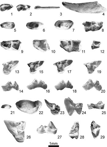
Figure 3
Holotype
BSPHM 1962 XIX 3174, M3 sin; Erkertshofen, Germany, early Miocene, MN4.
Teeth of the family Herpetotheriidae from the Ribesalbes-Alcora Basin. Amphiperatherium frequens erkertshofense: (1) right dp3 (MAB3-729); (2) right dp3 (MAB3-778); (3) i (lingual view, MAB0B-55); (4) c/C (MAB3-808); (5) left p1 (MAB3-866); (6) right p2 (MAB5-36); (7) left p3 (MAB0B-43); (8) right m1 (MAB0B-57); (9) left m1 (FS1-70); (10) left m2/3 (MAB3-741); (11) left m2/3 (CBR0B-60/61); (12) left m4 (MAB3-759); (13, 14) left DP3 (MTR1-4; modified from Furió et al., Reference Furió, Ruiz-Sánchez, Crespo, Freudenthal and Montoya2012): (13) occlusal view; (14) labial view; (15, 16) left DP3 (BC1-30; modified from Furió et al., Reference Furió, Ruiz-Sánchez, Crespo, Freudenthal and Montoya2012): (15) occlusal view; (16) labial view; (17, 18) left DP3 (MAB5-400; modified from Furió et al., Reference Furió, Ruiz-Sánchez, Crespo, Freudenthal and Montoya2012): (17) occlusal view; (18) labial view; (19, 20) left DP3 (MAB5-447; modified from Furió et al., Reference Furió, Ruiz-Sánchez, Crespo, Freudenthal and Montoya2012): (19) occlusal view; (20) labial view; (21) left P1 (MAB3-791); (22) left P3 (MAB5-776); (23) right M1 (MAB4-5; modified from Furió et al., Reference Furió, Ruiz-Sánchez, Crespo, Freudenthal and Montoya2012); (24, 25) left M2 (MAB5-395; modified from Furió et al., Reference Furió, Ruiz-Sánchez, Crespo, Freudenthal and Montoya2012): (24) occlusal view; (25) labial view; (26, 27) right M3 (MAB5-394; modified from Furió et al., Reference Furió, Ruiz-Sánchez, Crespo, Freudenthal and Montoya2012): (26) occlusal view; (27) labial view; (28) right M4 (MAB3-722); (29) left M4 (MAB5-730).

Occurrence in this basin
Mas dels Coixos 4 (MCX4); Mas de Torner 1 and 2 (MTR1 and MTR2); Barranc de Campisano 1 (BC1); Foieta la Sarra 1 (FS1); Mas d'Antolino B 0B, 3, 4, 5, 11, and 11B (MAB0B, MAB3, MAB4, MAB5, MAB11, and MAB11B); Corral de Brisca 0B, 0E, and 1 (CBR0B, CBR0E, and CBR1).
Description
i3.—This is a small tooth with a long and narrow root. The outline is subtriangular, and circular in anterior view. It has a large, single main cusp, with a small shelf on the anterior side.
c and C.—The upper and lower canines are simple and very similar; the crown is pointed and narrow and the root is thick. The occlusal and basal sides are curved and they diverge posteriorly; the lingual and labial sides are thickened. There is only one cusp.
dp3.—The talonid is wider than the trigonid, which is narrow with a small valley; in one of the teeth from MAB3, the trigonid is protoconid with a small metaconid. The paraconid and its cingulid are poorly developed in the other specimen. The metaconid and protoconid connect to each other by means of a short metacristid; this crest lies behind the metaconid; they are similar in size, but the protoconid is slightly higher. The entocristids are short, and the entoconid and the hypoconulid are poorly developed; the latter projects projects on the posterior side of the tooth. The hypoconid and the cristid obliqua are well developed. The talonid valley is deep and wide.
p1.—This tooth is elliptical in occlusal view, with two roots. It has a single main cusp, which is anteroposteriorly elongated with the highest part located on the anterior side. There are small cristids on the anterior and posterior sides. On the posterolabial side, there is a small cuspule that divides the posterior ridge.
p2.—This tooth is elliptical in occlusal view. The principal cusp is high and elongated along its central axis, with the tallest part in the central area. The anterior cristid is small; a second cristid is found at the posterior end, partly on the lingual side. A lingual valley separating this crest from a small cuspule is closed by the posterior cingulid. The posterior crest of the main cusp divides the posterior valley, which shows a well-developed lingual side and a poorly developed labial side.
p3.—This tooth is similar to the p2, but somewhat larger and more robust.
m1.—This tooth has two roots. The paraconid is the smallest cusp, and is located in a slightly more labial position than the metaconid. The paracristid is straight and V-shaped in lateral view. The protoconid is the highest cusp, and is pyramidal in shape. The metacristid is short and bears a notch. The metaconid is located near the protoconid. The trigonid valley is small and open. The talonid is wider and shorter than the trigonid, and its cusps are lower. The entoconid is well developed, lower in the material from FS1. The pre-entocristid ends below the metaconid. The hypoconid is located in a more labial position than the protoconid; its lingual side is concave. The cristid obliqua ends on the posterior side of the protoconid. The posthypocristid extends lingually toward the hypoconulid. The latter is the smallest cusp; it lies behind the entoconid and is backwardly projected. The talonid valley is deep and closed. There is only one cingulid, which is well developed and located on the anterolabial side of the trigonid. The material from FS1 has a small posterior cingulid.
m2/3.—The m2/3 are similar to the m1, but the trigonid is shorter and the talonid wider. There is a posterior cingulid.
m4.—This tooth is similar to the m2/3, but with a longer and narrower talonid. The entoconid and hypoconulid are connected; both the hypoconulid and the hypoconid are well developed. There is a posterior cingulid, except for a single specimen in MAB0B.
dP3.—The outline of this tooth is subtriangular in occlusal view. The metacone and the paracone are the highest cusps. The paracone is slightly lower than the metacone. The preparacrista is long. The ectoflexus is quite straight. The stylar cusp A is highly variable in shape and size: it can be well developed or just a small elevation on the labial edge; in BC1, the stylar cusp is lower and has three cuspules; in MAB0B material, it is small. The stylar cusp B is only an enlargement of the paracone. The size of the stylar cusp C is variable, from well developed to nearly absent; in MAB5 material, it is better developed than in specimens from other localities. The stylar cusp D is well developed, particularly in MTR1 and MAB3 material. The postparacrista and the premetacrista are straighter than in the permanent molars. The paraconule and the metaconule are poorly developed. The anterior cingulum can be connected to the preparaconular crest and the parastyle, or it can be absent. The development and connections of the postparaconular and premetaconular crests are highly variable. They can be well developed, with the postparaconular and the preparaconular crests connected and the premetaconular crest isolated; the postparaconular crest can be short and join the paraconule, whereas the premetaconular crest is well developed and isolated; alternatively, they can be well developed and isolated. There is a small cingulum on the posterolingual side of the protocone in MTR1 material.
P1.—This tooth appears subelliptical in occlusal view and has two roots. There is only one main cusp, with an anteroposterior enlargement. There are two crests, one each on the anterior and posterior sides, each ending in a small cuspule. The posterior crest extends to the labial side; it is curved in its middle part and divides the posterior valley into two parts; the lingual part is better developed. A small cingulum borders the posterior valley.
P2.—This tooth is similar to the P1, but larger.
P3.—This tooth is similar to the other premolars, but better developed.
M1.—This tooth is broken. The metacone is the highest cusp; the paracone and the labial side are broken. The protocone is lower than the other cusps. The protoconule and the metaconule are poorly developed. The preparaconular crest and the postmetaconular crest are isolated at their labial ends. The postparaconular and premetaconular crests are isolated on the lingual side and extend toward the protocone.
M2.—This tooth is fragmented; only the labial side is preserved. The more acute labial side is typical of the M2. The tooth is strongly worn. The stylar cusps A and C are small; the stylar cusp B is well developed, and stylar cusp D is intermediate in size. The metacone is larger than the paracone. The postparacrista and the premetacrista are less straight than on the anterior tooth.
M3.—This tooth is strongly worn and subtriangular in occlusal view. The anterior cingulum is short and isolated on the lingual side, and connects to a reduced stylar cusp A. The stylar cusp B is the best developed; stylar cusp D is also well developed, whereas stylar cusp C is absent. The metacone is the highest cusp; the protocone and the paracone are similar in height. The metaconule is small, and the postmetaconular crest is short.
M4.—The outline of this tooth is subtriangular in occlusal view. The anterior cingulum is short. The stylar cusp A is well developed. The preparacrista is long. The paracone is well developed, whereas the metacone is small. The postmetacrista is absent. The postparacrista and the premetacrista are straight. The paraconule and the metaconule are very small. The preparaconular crest ends on the labial side without any connection. The end of the postparaconular crest is also isolated in the middle of the trigonid valley. The postmetaconular crest is short and isolated on the labial side.
Material
MCX4: 1 m1-3 (not complete); MTR1: 1 dP3; MTR2: 1 M1 (not complete); BC1: 1 dP3; FS1: 1 m1, 1 m2-3 (not complete); MAB0B: 1 i, 2 c/C, 1 dp3 (not complete), 1 p3, 1 m1-4 (not complete), 1 m1-3 (not complete), 1 m1, 2 m2-3 (not complete), 2 m4 (not complete), 2 dP3 (not complete), 1 P2, 9 M1-3 (not complete), 2 M4 (not complete); MAB3: 2 dp3, 1 p1 (not complete), 1 p2, 1 m1-3 (not complete), 1 m2-3 (not complete), 2 m4 (not complete), 1 C, 3 dP3 (one not complete), 2 P1, 5 M1-3 (not complete), 2 M4 (not complete); MAB4: 1 M1 (not complete); MAB5: 1 p1, 1 p2, 4 m1-3 (not complete), 3 m2-3 (not complete), 4 dP3 (two not complete), 1 P3, 1 M2 (not complete), 1 M3, 2 M4 (not complete); MAB11: 1 M1 (not complete), 1 M2-3 (not complete); MAB11B: 1 dP3; CBR0B: 1 dp3, 1 m2-3 (not complete), 1dP3, 1 M1-2; CBR0F: 1 m1-4 (not complete); CBR1: 1 dP3.
Measurements
See Table 1.
Remarks
The presence of four molars in the hemimaxilla/hemimandible is typical of the metatherians, and distinguishes them from the rest of the small Miocene mammals in Europe (Ziegler, Reference Ziegler, Rössner and Heissig1999). The material from the Ribesalbes-Alcora Basin is characterized by the absence of a connection between the stylar cusps A and B in the upper molars, a typical feature of the genus Amphiperatherium (Furió et al., Reference Furió, Ruiz-Sánchez, Crespo, Freudenthal and Montoya2012), although Ladevèze et al. (Reference Ladevèze, Smith and Smith2012) raised doubts about the validity of this genus based on the nondiagnostic, highly variable differences with Peratherium. The great reduction of the stylar cusp C on the upper molars supports the specific adscription to A. frequens (see Furió et al., Reference Furió, Ruiz-Sánchez, Crespo, Freudenthal and Montoya2012). This species is subdivided into: A. f. frequens, the oldest subspecies, with the type locality in MN1 (Koenigswald, Reference Koenigswald1970); A. f. wintershofense (von Koenigswald, Reference von Koenigswald1970), with the type locality in MN3 (Koenigswald, Reference Koenigswald1970; Klietmann, Reference Klietmann2013), and A. f. erkertshofense, the youngest subspecies, with the type locality in MN4 (Koenigswald, Reference Koenigswald1970; Ziegler, Reference Ziegler1990, Reference Ziegler, Rössner and Heissig1999).
The studied fossils are small. The M3 better agrees with Amphiperatherium frequens erkertshofense, and is smaller than the M3 of the other two subspecies. The morphology of the upper dentition is generalized; all of the upper deciduals have a stylar cusp C, a typical character in the oldest Central Europe populations. The stylar cusp C can be present or not in the primitive population of A. f. erkertshofense from Petersbuch 28, but its absence in the permanent molars in Petersbuch 28, similar to the material from this site, has also been observed in our material (Klietmann et al., Reference Klietmann, Nagel, Rummel and Van den Hoek Ostende2014). This distinguishes it from A. f. frequens, in which this cusp is always present. The presence of a rounded protocone in MAB4 material has also been observed in A. f. wintershofense but not in the material from Petersbuch 28.
In the lower molars, the presence of the posterior cingulid on the m1-4 is typical of Amphiperatherium frequens frequens, and its occasional presence on the m4 is a derived character (Prieto and Rummel, Reference Prieto and Rummel2015). In our material and in the other subspecies, this cingulid can be present or not, and in the middle Miocene, it becomes rare or completely absent (Klietmann et al., Reference Klietmann, Nagel, Rummel and Van den Hoek Ostende2014; Prieto and Rummel, Reference Prieto and Rummel2015). In addition, the studied material lacks a crest closing the valley between the stylar cusps A and B, which is typical in A. f. wintershofense (see Koenigswald, Reference Koenigswald1970).
The variability of deciduous molars has already been remarked upon by Furió et al. (Reference Furió, Ruiz-Sánchez, Crespo, Freudenthal and Montoya2012), but the availability of additional upper deciduals and, for the first time in this basin, lower deciduals, allows us to further appreciate their variation. Among the upper deciduals, the stylar cusps are very variable in size, in morphology and in the occasional presence of a cingulum in the protocone (see the description of this element). The lower deciduals show extreme variability in the trigonid, i.e., the specimen MAB3-778 has a well-developed trigonid, but in MAB3-729, the valley of the trigonid is absent, the paraconid is only a crest of the protoconid, and the metaconid is very small.
Based on these characteristics and the similar morphology and size, the specimens from Araia d'Alcora have been ascribed to Amphiperatherium frequens erkertshofense. In addition, the studied fossils show different evolutionary stages in each character, an example of mosaic evolution, as it is the case in Petersbuch 28 material (Klietmann et al., Reference Klietmann, Nagel, Rummel and Van den Hoek Ostende2014).
Discussion
Biostratigraphy
The gymnure Galerix symeonidisi is a common Eulipothyplan in biozone MN4 (early Miocene), with its oldest record in the locality of Aliveri. This species would have been part of the migration wave that brought modern muroid rodents to Europe (Van den Hoek Ostende et al., Reference Van den Hoek Ostende, Furió, Madern and Prieto2016). Galerix exilis first appeared in Central Europe in MN4. However, due to the difficulty of recognising it when it co-occurred with G. symeonidisi, its first arrival to the Iberian Peninsula cannot be dated precisely. Galerix symeonidisi was gradually replaced by G. exilis, and eventually, only the latter is present in MN5 (Van den Hoek Ostende and Doukas, Reference Van den Hoek Ostende and Doukas2003). Occurrences of the gymnure Lantanotherium and the hedgehog Atelerix are very sparse in the early Miocene record but become more common in the middle Miocene (Engesser, Reference Engesser2009; Van den Hoek Ostende et al., Reference Van den Hoek Ostende, Furió, Madern and Prieto2016). A record of Lantanotherium in the Ribesalbes-Alcora Basin, if it is confirmed with more material, together with records in the sites of Buñol (Valencia, Spain) and the Vallès-Penedès Basin, would be one of the oldest known for the Iberian Peninsula (Furió and Alba, Reference Furió, Alba, Pérez-García, Gascó, Gasulla and Escaso2011), whereas in Central Europe it is more common and extends from biozone MN4 to MN11 (Furió et al., Reference Furió, Casanovas-Vilar and Van den Hoek Ostende2011). Scarce remains of Atelerix first appear in MN4; if confirmed with more material, this record would be one of the oldest in the European early Miocene and second oldest on the Iberian Peninsula, after the site of San Mamet from the Vallès-Penedès Basin (Van den Hoek Ostende and Furió, Reference Van den Hoek Ostende, Furió, Van den Hoek Ostende, Doukas and Reumer2005).
Amphiperatherium frequens has a very wide stratigraphic range from MN1 to MN6 (Furió et al., Reference Furió, Ruiz-Sánchez, Crespo, Freudenthal and Montoya2012; Prieto and Rummel, Reference Prieto and Rummel2015). Furió et al. (Reference Furió, Ruiz-Sánchez, Crespo, Freudenthal and Montoya2012) placed the last record of this metatherian in Europe in MN7+8, following age determination of the European sites Ergeten and Greuterschberg (Switzerland) by Kälin and Kempf (Reference Kälin and Kempf2009). Nevertheless, according to Prieto and Rummel (Reference Prieto and Rummel2015), this age determination is ill-founded; the Swiss sites should be considered as MN4 and the last occurrence of A. frequens actually occurs in MN6. This species is found at Cases de la Valenciana in the Vallès-Penedès Basin (Jovells-Vaqué et al., Reference Jovells-Vaqué, García-Paredes, Furió, Angelone, Van den Hoek Ostende, Berrocal Barberà, DeMiguel, Madurell-Malapeira and Casanovas-Vilar2018). The subspecies A. f. erkertshofense, present in the Ribesalbes-Alcora Basin, is typically from MN4 in Europe (Ziegler, Reference Ziegler, Rössner and Heissig1999).
Paleoecology
The extant gymnures often live in moist forests with abundant understory, always near water bodies (Ziegler, Reference Ziegler2005). According to Engesser (Reference Engesser2009), this assumption can be extended to the genera Lantanotherium and Galerix. However, to Furió et al. (Reference Furió, Casanovas-Vilar and Van den Hoek Ostende2011) and Prieto et al. (Reference Prieto, Van den Hoek Ostende and Böhme2011), only Lantanotherium can be ecologically compared to the extant gymnures, because it usually coexisted with the dimylid Plesiodimylus Gaillard, Reference Gaillard1897, an insectivore associated with moist environments (Crespo et al., Reference Crespo, Furió, Ruiz-Sánchez and Montoya2018). The absence of Lantanotherium in the fossil assemblages from the interior of the Iberian Peninsula supports this view (Furió and Alba, Reference Furió, Alba, Pérez-García, Gascó, Gasulla and Escaso2011). According to Van den Hoek Ostende and Doukas (Reference Van den Hoek Ostende and Doukas2003), the paleoecological requirements of G. symeonidisi were more moist than for G. exilis, which has been found in drier sites, e.g., Somosaguas (Luis and Hernando, Reference Luis and Hernando2000). The species found in the Ribesalbes-Alcora Basin is often recorded in lignite-rich sites, together with other wet taxa, although G. symeonidisi was a drier dweller than G. aurelianensis (see Van den Hoek Ostende and Doukas, Reference Van den Hoek Ostende and Doukas2003). Galerix remmerti, a small version of G. aurelianensis, might be an even drier dweller than G. symeonidisi, based on its absence from the only site with recorded beavers in the Calatayud-Montalbán Basin (Van den Hoek Ostende and Fejfar, Reference Van den Hoek Ostende, Fejfar, Van den Hoek Ostende, Nagel and Harzhauser2006). In addition, according to Van den Hoek Ostende (Reference Van den Hoek Ostende2001), the diet of G. symeonidisi might have been more herbivorous than that of the other Galerix species. Extant erinaceids live in a wide variety of habitats, including woods, meadows, bushlands, and dry zones (e.g., deserts). Therefore, this group is inadequate for making palaeoenvironmental inferences (Ziegler, Reference Ziegler2000).
In the sector of Araia d'Alcora, erinaceids are relatively abundant (Table 2) at both the bottom and the top of the synthetic stratigraphic column (Fig. 1.1). An abundance of specimens from this group is almost constant in remaining sites, although they are absent in CBR0B. Furthermore, the genus Galerix appears in MAB3, the only site with other species of erinaceids (Atelerix and Lantanotherium), which, unlike Galerix, were probably transient in the Ribesalbes-Alcora Basin. These changes in abundance and diversity probably do not reflect climatic changes.
Percentages (%) of the frequency of soricids (soricids + heterosoricids), dimylids, talpids, erinaceids, and herpetotheriids found in the Araia sites. Only intensively sampled sites (N > 50 teeth) are represented; the number in parentheses following the frequency is the percentage of that family versus the total number of mammalian teeth found in the sites studied at the Ribesalbes-Alcora Basin (modified from Crespo et al., Reference Crespo, Suárez-Hernando, Murelaga, Ruiz-Sánchez and Montoya2019a).

Amphiperatherium frequens has been interpreted as a forest dweller (Ziegler and Mörs, Reference Ziegler and Mörs2000). Kurz (Reference Kurz2005) proposed that this genus was a tree climber and more insectivorous than Peratherium. However, Sánchez-Villagra et al. (Reference Sánchez-Villagra, Ladevèze, Horovitz, Argot, Hooker, Macrini, Martin, Moore-Fay, de Muizon, Schmelzie and Asher2007) described this species as a runner and less arboreal than other metatherians. In addition, Furió et al. (Reference Furió, Ruiz-Sánchez, Crespo, Freudenthal and Montoya2012) emphasised that this species is often found in German sites that reflect humid conditions, and in Spain are located in sites with exceptional degrees of humidity. Nevertheless, the site (MAB0B) with more abundant remains of Amphiperatherium in the Ribesalbes-Alcora Basin and where this taxon is dominant is situated on a gypsum layer. Moreover, Klietmann et al. (Reference Klietmann, Nagel, Rummel and Van den Hoek Ostende2014) proposed that the different subspecies might actually reflect the local environmental conditions. This could also explain the more abundant presence of posterior cingulids in MAB0B when compared with other sites.
Conclusions
Systematic studies of metatherians and erinaceids from the Iberian Peninsula are very rare and often mostly limited to faunal lists. The Ribesalbes-Alcora Basin has a continental sequence similar in age to the local biozones Ca and Cb from the Calatayud-Montalbán area (MN4, early Miocene). These sites have yielded a relatively abundant fauna of these groups. Amphiperatherium frequens and Galerix symeonidisi are frequent in almost all studied assemblages, with a rich sample of mammal remains, and they can be considered permanent dwellers. On the other hand, Atelerix and Lantanotherium only appear in MAB3 and can be considered transient.
In this work, we refine the taxonomic position of Amphiperatherium frequens erkertshofense, partially published by Furió et al. (Reference Furió, Ruiz-Sánchez, Crespo, Freudenthal and Montoya2012). We describe more material of this species, including its lower dentition for the first time, which allows us to fully appreciate the high degree of variability of its decidual molars. Furthermore, we increase the stratigraphic record of this species in this basin. Galerix symeonidisi appears in almost all of the studied sites and is described for the first time from this basin. We also describe one of the first findings of the erinaceids Atelerix and Lantanotherium in Europe, if this can be confirmed.
The presence of Amphiperatherium is probably linked to relatively dry environments, as suggested by its abundance in MAB0B, a site located on a gypsum level. However, the more or less constant presence of erinaceids does not reflect their environmental preferences within the Ribesalbes-Alcora Basin, and for this reason, the eurytopics.
Acknowledgments
The survey and excavation campaigns in the area of Araia d'Alcora were funded by the Conselleria de Cultura i Esports of the Generalitat Valenciana for 2008–2011, through projects 2008/0433-CS, 2010/0528-CS, 2011/0230-CS, GV06/304, and GVPRE/2008/320. This research was also supported by the Spanish Ministry of Economy and Competitiveness (CGL2015-68333-P). Thanks are due to the helpful comments of J. Guillem, J. Klietmann, and L. Van den Hoek Ostende, a reviewer of the original manuscript. The support of the Consejo Nacional de Investigaciones Científicas y Técnicas (CONICET) toward this research for the senior author is hereby acknowledged.