INTRODUCTION
Dirofilaria immitis, also known as canine heartworm, has a worldwide distribution but as a general rule is most severe in tropical and warm/humid areas of the world. It is arguably the most important parasite of dogs in North America. As of 2013, heartworm-positive dogs had been reported in every state in the continental USA (Wang et al. Reference Wang, Bowman, Brown, Harrington, Kaufman, McKay, Nelson, Sharp and Lund2014; Yancey et al. Reference Yancey, Hegarty, Qurollo, Levy, Birkenheuer, Weber, Diniz and Breitschwerdt2014), and in multiple countries in South America (confirmed reports in Argentina, Colombia, Peru and Brazil, though recent data are sparse from the other countries) (Labarthe and Guerrero, Reference Labarthe and Guerrero2005; Vezzani et al. Reference Vezzani, Carbajo, Fontanarrosa, Scodellaro, Basabe, Cangiano and Eiras2011a , Reference Vezzani, Mesplet, Eiras, Fontanarrosa and Schnittger b ; Cuervo et al. Reference Cuervo, Sierra, Waisman, Gerbeno, Sidoti, Albonico, Mariconti, Mortarino, Pepe, Cringoli, Genchi and Rinaldi2013; Labarthe et al. Reference Labarthe, Paiva, Reifur, Mendes-de-Almeida, Merlo, Pinto, Juliani, de Almeida and Alves2014), as well as Southern Europe, Australia and parts of Africa (Simon et al. Reference Simon, Morchon, Gonzalez-Miguel, Marcos-Atxutegi and Siles-Lucas2009; Barker et al. Reference Barker, Langton, Helps, Brown, Malik, Shaw and Tasker2012; Otranto et al. Reference Otranto, Dantas-Torres, Brianti, Traversa, Petric, Genchi and Capelli2013; Maia et al. Reference Maia, Coimbra, Ramos, Cristovao, Cardoso and Campino2015). In Europe, the parasite seems to be spreading into previously non-endemic areas, especially in Central Europe (Penezic et al. Reference Penezic, Selakovic, Pavlovic and Cirovic2014; Sassnau et al. Reference Sassnau, Czajka, Kronefeld, Werner, Genchi, Tannich and Kampen2014; Tolnai et al. Reference Tolnai, Szell, Sproch, Szeredi and Sreter2014). In the USA, the prevalence of infection in domestic dogs (those that were tested by veterinarians) in 2010–2012 ranged from 2·9% in the Southeast to 0·4% in the Northeast, with an overall prevalence of 1·3% (Little et al. Reference Little, Beall, Bowman, Chandrashekar and Stamaris2014a ), lower than in a similar study in 2001–2007 (Bowman et al. Reference Bowman, Little, Lorentzen, Shields, Sullivan and Carlin2009). This prevalence is influenced by multiple factors, including climate, socio-economic factors, topography and the presence of vectors (Wang et al. Reference Wang, Bowman, Brown, Harrington, Kaufman, McKay, Nelson, Sharp and Lund2014). The parasite infects all canine species, and also cats, though in cats the infection and consequent disease is different when compared with the canid definitive hosts. On occasion humans become infected, so the parasite does have zoonotic potential. Dying worms in pulmonary artery branches can produce granulomas, a condition called pulmonary dirofilariosis, though most people with this condition will display no symptoms (Lee et al. Reference Lee, Montgomery, Theis, Blagburn and Eberhard2010). The granulomas appear as small, round lesions on radiographs. Due to the large number of dogs that are diagnosed every year, and the resulting expense of treatment, this disease is of huge economic importance in addition to its impact on the health and welfare of companion animals. In fact, millions of US dollars are spent on heartworm prevention, diagnosis and treatment each year. Because of the impact of disease both on the health of the animal and the expense of treatment, there exists a reasonable expectation that heartworm preventives will prevent the development of larval heartworms into adult worms. Despite the fact that there are many different products currently on the market to prevent heartworm disease, all of the active components are members of the same drug class, the macrocyclic lactones (MLs). Resistance against one of these drugs is extremely likely to manifest as resistance against all of them, and in the worst-case scenario could result in an increase in the incidence of heartworm disease in dogs and cats. Reports of loss-of-efficacy of the preventatives (Hampshire, Reference Hampshire2005) and frank resistance against them (Geary et al. Reference Geary, Bourguinat and Prichard2011; Bourguinat et al. Reference Bourguinat, Keller, Bhan, Peregrine, Geary and Prichard2011a ; Bowman, Reference Bowman2012; Pulaski et al. Reference Pulaski, Malone, Bourguinat, Prichard, Geary, Ward, Klei, Guidry, Smith, Delcambre, Bova, Pepping, Carmichael, Schenker and Pariaut2014) have understandably caused considerable concern. The purpose of this review is therefore to try to summarize the current position with regard to ML resistance in D. immitis as the authors understand it. It will become apparent that we do not understand very much about many aspects of this problem, not least how serious it may become, and therefore some aspects of the discussion may be somewhat speculative and based on personal opinion; we hope that this may stimulate additional research and contributions to resolve these issues. Resistance to the MLs is not a new phenomenon in parasitic nematodes, but has previously been documented mostly in gastro-intestinal parasites of livestock (Wolstenholme et al. Reference Wolstenholme, Fairweather, Prichard, von Samson-Himmelstjerna and Sangster2004; Wolstenholme and Kaplan, Reference Wolstenholme and Kaplan2012). Though it may be possible to learn some lessons from these systems and apply them to heartworm, there are significant differences in the parasite life-cycles and ecology that may limit this.
THE LIFE CYCLE
Adult D. immitis live in the pulmonary arteries, though in heavy infections the heart and other blood vessels are also colonized. Female worms can grow to 250–300 mm and males to a length of 100–150 mm. The adults can live up to 7 years or potentially longer in dogs. During this time, the adults will mate and produce microfilariae (Mf), which circulate in the blood. The Mf can then be ingested by a mosquito intermediate host. The mosquito is absolutely required for heartworm to develop. This is an important difference from the gastro-intestinal parasitic nematodes in which ML resistance has been described which have a direct life-cycle with no invertebrate vector. Once inside the mosquito, the Mf migrate for an average period of approximately 14 days, during which time, they develop to first-stage larvae (L1). Afterwards, the L1 molts twice to become an infectious third-stage larva (L3), which is in the head of the mosquito. Upon landing on a host, the mosquito will then take a blood meal. At this point, the larvae will ‘burst out’ of the proboscis. The larvae will then ‘surround’ the stylet (the piercing part of the mouthparts) in a pool of mosquito hemolymph. When the stylet is removed, the larvae then enter the host through the hole made by the stylet (Bowman and Atkins, Reference Bowman and Atkins2009). Once inside the definitive host, the L3s follow a complicated migration pathway. The L3 remain near to the site of entrance for approximately 3–4 days, during which time the L3 molt to fourth-stage larvae (L4). This is normally completed by 4 days, but may not occur until day 12 post infection (dpi). It is generally agreed that the molt to the last stage, known as the juvenile adult, occurs by day 58. By day 70 the first worms will arrive in their final location, the pulmonary artery. Most worms have reached this location by day 120. By 180 dpi, worms are sexually mature and have begun to produce Mf, thus completing the life cycle (Kotani and Powers, Reference Kotani and Powers1982). It is important to realize that while this is the generally accepted, ‘textbook’ form of life cycle, there could be variations in these times.
THE VECTOR AS PART OF THE LIFE CYCLE
Over 70 species of mosquitoes have the potential to act as vectors of heartworm, although 10 to 12 of them may be the most important. For example the mosquitoes in Tennessee, USA, that were positive for D. immitis were primarily Culex pipiens-complex (Fryxell et al. Reference Fryxell, Lewis, Peace, Hendricks and Paulsen2014), whereas D. immitis larvae were present in 9 of 16 species present in Arkansas (McKay et al. Reference McKay, Bianco, Rhodes and Barnett2013), with Aedes vexans and Anopheles quadrimaculatus as the most abundant. These authors also found that the prevalence of heartworm larvae in mosquitoes was ten-fold higher (73·7%) in insects collected from inside the kennel of an infected dog than from other sites (7·3%). Others have found that the presence of Culex quinquefasciatus, Aedes sierrensis, Anopheles punctipennis and Aedes (now Ochlerotatus) canadensis is also associated with higher heartworm prevalence (Wang et al. Reference Wang, Bowman, Brown, Harrington, Kaufman, McKay, Nelson, Sharp and Lund2014). While these are the most documented species, it is important to remember that there could be yet unidentified species, or newly introduced species, such as Aedes albopictus, which could also serve as vectors.
An important concept in relation to the role of the mosquito is seasonal transmission of heartworm (Knight and Lok, Reference Knight and Lok1998). Maturation of the L3 stage of the parasites in mosquitoes requires higher temperatures. Practically what this means is that the peak months of transmission are likely to be mid-summer in most places. For example, in the northern USA the transmission season is approximately 4 months, whereas in the southern USA it is approximately 6–8 months. In further southern climates, the transmission season would be extended even longer. Because of this variation and other factors, the current recommendation by the American Heartworm Society is for the administration of year-round heartworm prevention.
CURRENT METHODS OF PREVENTION AND TREATMENT OF INFECTION AND DISEASE
The only drug class currently in use for the prevention of heartworm disease is the MLs, which includes the avermectins (ivermectin, selamectin) and the milbemycins (moxidectin, milbemycin oxime). As normally administered, which for most products is monthly, the drugs do not prevent the initial infection by the L3 stage of the parasite, and are not rapidly effective at ‘curing’ the infection once adult worms have developed; they act by killing any L3 and L4 larvae present in the animal at the time they are given and thus prevent them from developing into the adult worms which cause disease. The length of time that the MLs persist in the animal after treatment, and will be able to kill any newly invading L3 larvae, varies with the individual drug and route of administration (McKellar and Gokbulut, Reference McKellar and Gokbulut2012). The plasma half-life of 200 μg kg−1 (which is much greater than the normal preventative dose) orally administered ivermectin in the dog is about 80 h (Gokbulut et al. Reference Gokbulut, Karademir, Boyacioglu and McKellar2006; Al-Azzam et al. Reference Al-Azzam, Fleckenstein, Cheng, Dzirnianski and McCall2007) but that of moxidectin is considerably longer (Al-Azzam et al. Reference Al-Azzam, Fleckenstein, Cheng, Dzirnianski and McCall2007; Lallemand et al. Reference Lallemand, Lespine, Alvinerie, Bousquet-Melou and Toutain2007). Topically applied selamectin has a plasma half-life of 250–300 h (Sarasola et al. Reference Sarasola, Jernigan, Walker, Castledine, Smith and Rowan2002; Dupuy et al. Reference Dupuy, Derlon, Sutra, Cadiergues, Franc and Alvinerie2004) Since the development of adult parasites from invading L3 takes several months, there is a fairly long window of opportunity (up to 2 months) after infection has taken place when the drugs can remove the larvae and prevent the development of disease (Guerrero et al. Reference Guerrero, McCall, Genchi, Vercruysse and Rew2002). Uniquely, any heartworm preventative is currently required to be demonstrated to be ‘100% effective’ in order to obtain a license from the Food and Drug Administration (FDA) (Hampshire, Reference Hampshire2005). To demonstrate this, dogs are normally inoculated with 50 infective D. immitis (L3) obtained from experimentally infected mosquitoes, and then 30 days later administered a single dose of the preventive being tested. No adult worms should be present in the infected and treated animals on euthanasia 7 months later. A control group is used to confirm that the parasites were capable of causing infection (Snyder et al. Reference Snyder, Wiseman, Bowman, McCall and Reinemeyer2011a ). To date, the majority of the monthly topical and oral preventive products have been tested by this method.
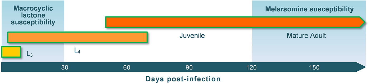
With the exception of ProHeart6®, an injectable formulation of moxidectin that confers protection for 6 months, the current preventives are designed to be administered monthly to prevent development of infecting L3 into adult worms that cause disease (Fig. 1). Only the L3 and L4 stages, but not adults, are susceptible to the drugs at the doses given in a single dose. The preventives kill the migrating stages of heartworm acquired during the previous month. They do not kill ‘forward’, that is, the drugs do not work by killing infective L3s acquired after administration of the preventive. In addition to their use to prevent disease, the MLs are administered to dogs that have become infected with heartworms and are undergoing treatment with melarsomine dihydrochloride (melaminylthioarsenate) to kill the adult worms (Bowman and Atkins, Reference Bowman and Atkins2009); the aim of this is to kill existing melarsomine-insensitive L3 and L4 that are potentially still in the animal. It is recommended to start a ML preventive (often ivermectin, which has been used in most studies) at the time of diagnosis, or up to 2–3 months prior to melarsomine treatment. Furthermore, the administration of a ML, though delaying treatment, also eliminates the ‘treatment/susceptibility’ gap (Fig. 1). By reducing the treatment gap, worms that have infected the dog within the past 2 months are eliminated, and those that infected within the past 2–4 months are allowed to mature to the point to where they can be killed using melarsomine. Melarsomine, an arsenical, is from a completely different drug class to the MLs and has a very different mode of action. There is no evidence that there would be any side-resistance between the MLs and melarsomine and so ML-resistant heartworms can still be removed using this regimen. Heartworm therapy therefore, unlike the preventives, is essentially a combination of different classes of drug, especially when the recent inclusion of tetracycline antibiotics, such as doxycycline (Bazzocchi et al. Reference Bazzocchi, Mortarino, Grandi, Kramer, Genchi, Bandi, Genchi, Sacchi and McCall2008), is considered. Combination therapies are common in antiviral (such as HIV) and antimalarial treatments, and are increasingly promoted for livestock use to reduce the risk of resistance. Theoretically, resistance to 2 different drug classes, such as melarsomine and the MLs, could be mediated via non-specific detoxification mechanisms, such as P-glycoprotein (P-gp) overexpression. However, although P-gps have been repeatedly implicated as mediators of ML-resistance, we can find no evidence in the literature that melarsomine can act as a P-gp substrate. In Trypanosoma bruceii, over-expression of a P-gp homolog does cause increased efflux of a melarsoprol adduct, but is not sufficient to cause actual resistance in vivo and was not detected in resistant parasites (Shahi et al. Reference Shahi, Krauth-Siegel and Clayton2002: Alibu et al. Reference Alibu, Richter, Voncken, Marti, Shahi, Renggli, Seebeck, Brun and Clayton2006).
Susceptibility of the mammalian stages of the D. immitis lifecycle to the macrocyclic lactones and melarsomine.

There has also been ‘off-label’ use of the ML to remove adult worms – so called ‘slow-kill’. This requires multiple applications of the drug, often combined with doxycycline (Venco et al. Reference Venco, McCall, Guerrero and Genchi2004; McCall, Reference McCall2005 ). Historically, this is less expensive than melarsomine treatment, but takes longer and fears have been expressed that widespread use of this regimen could contribute a selection pressure for drug resistance in D. immitis (Bowman, Reference Bowman2012).
ML MECHANISM OF ACTION
The MLs are potent activators of a family of inhibitory receptors found in nematodes and other organisms. In gastro-intestinal parasites and insects, the best-studied members of this family are the glutamate-gated chloride channels (GluCls), which are widely expressed in the nervous system and on pharyngeal muscle (Wolstenholme, Reference Wolstenholme2012). The MLs cause an essentially irreversible activation of these receptors, which has several deleterious consequences for the parasite, including paralysis together with inhibition of feeding and of egg-production (Wolstenholme and Rogers, Reference Wolstenholme and Rogers2005). In D. immitis, infertility caused by MLs has been attributed to effects on the male parasites (Lok et al. Reference Lok, Knight, Selavka, Eynard, Zhang and Bergman1995). Based on these phenotypes, in vitro assays for resistance have been developed for some species (Amarante et al. Reference Amarante, Pomroy, Charleston, Leathwick and Tornero1997; Kotze et al. Reference Kotze, Le Jambre and O'Grady2006; Kaplan et al. Reference Kaplan, Vidyashankar, Howell, Neiss, Williamson and Terrill2007; Demeler et al. Reference Demeler, Kuttler, El-Abdellati, Stafford, Rydzik, Varady, Kenyon, Coles, Hoglund, Jackson, Vercruysse and von Samson-Himmelstjerna2010, Reference Demeler, Gill, von Samson-Himmelstjerna and Sangster2013), and these are in routine use in some laboratories. An ivermectin-sensitive GluCl has been reported for D. immitis (Yates and Wolstenholme, Reference Yates and Wolstenholme2004) and 2 motility-based assays, a larval migration inhibition assay for L3 (Evans et al. Reference Evans, Moorhead, Storey, Wolstenholme and Kaplan2013) and a microfilarial motility assay (Bourguinat et al. Reference Bourguinat, Keller, Blagburn, Schenker, Geary and Prichard2011b ) have been described. However, in all cases the concentrations of drug that are required for any in vitro effect, either on the presumed ML receptor or on the motility of the worm, are far greater than those achieved in the host during prophylaxis or treatment and so it has been speculated that the effect of the ML may be to inhibit immunomodulation of the host by the parasite and hence permit innate immunity to remove the worms (Moreno et al. Reference Moreno, Nabhan, Solomon, Mackenzie and Geary2010; Vatta et al. Reference Vatta, Dzimianski, Storey, Camus, Moorhead, Kaplan and Wolstenholme2014). Other receptors can be activated or positively modulated by the MLs (Krause et al. Reference Krause, Buisson, Bertrand, Corringer, Galzi, Changeux and Bertrand1998; Rao et al. Reference Rao, Siddiqui, Prichard and Forrester2009; Accardi et al. Reference Accardi, Beech and Forrester2012; Hernando and Bouzat, Reference Hernando and Bouzat2014), but the importance of these to the mechanism of drug action in vivo is not known.
ML RESISTANCE IN HEARTWORM – REVIEW OF REPORTS
The potential emergence of ML resistance in D. immitis was first brought to public attention by Hampshire (Reference Hampshire2005) who reported that the US FDA had received complaints relating to lack of effectiveness of heartworm products starting in 1998 and that the numbers increased dramatically between 2000 and 2002, and rose again in 2002 and 2003 before dropping slightly in the last quarter of 2003. Perhaps unfortunately, this report coincided with the publication of a review arguing that the emergence of true anthelmintic resistance (a heritable change that allows a parasite to survive a previously lethal dose of the drug) in D. immitis was unlikely (Prichard, Reference Prichard2005). There was little disagreement with this view at the time, and it is interesting to revisit some of the arguments made in that review in order to try and establish which, if any, of the assumptions have proved to be inaccurate. The FDA report generated considerable interest and discussion and has stimulated a lot of research. Though it is clear that many, possibly most, ‘Lack of Efficacy’ (LOE) reports are due to failures of owner compliance, such as under-dosing or missing doses of preventative (Atkins et al. Reference Atkins, Murray, Olavessen, Burton, Marshall and Brooks2014) it is also now indisputable that genuinely ML-resistant heartworms are circulating, at least in the Mississippi delta region of the USA (Pulaski et al. Reference Pulaski, Malone, Bourguinat, Prichard, Geary, Ward, Klei, Guidry, Smith, Delcambre, Bova, Pepping, Carmichael, Schenker and Pariaut2014).
To date, much of the research on ML resistance in D. immitis has been reported in Conference Proceedings and there are relatively few peer-reviewed papers on the subject, though this number is likely to increase in the near future. Bourguinat et al. (Reference Bourguinat, Keller, Bhan, Peregrine, Geary and Prichard2011a ) produced the first peer-reviewed report demonstrating ML resistance in heartworms in vivo. In this case, the criterion for resistance was the failure of repeated treatments with ivermectin and milbemycin oxime to clear Mf from the blood of a dog that was born in New Orleans shortly after hurricane Katrina and transferred to Canada for adoption. The dog received 5 doses of melarsomine to kill adult worms and a total of 17 treatments with MLs, the last 2 of which were daily treatments for a week with high doses of milbemycin oxime. Despite this, the dog remained microfilaremic for 18 months although no antigen was detected following the adulticide treatment – implying that the adult worms had been removed. Previously, some of the same authors published a genetic study (Bourguinat et al. Reference Bourguinat, Keller, Blagburn, Schenker, Geary and Prichard2011b ) comparing three D. immitis isolates from LOE cases with a larger number of control isolates. The LOE isolates were judged to be potentially resistant based on an in vitro test on the Mf, where the number of motile worms in the presence of a single concentration of ivermectin (72 μg mL−1 – reported to be equivalent to 2X the EC95 concentration for a susceptible strain) was recorded by a human observer. The 2 papers clearly showed that parasites were circulating in the environment which produced Mf with dramatically reduced sensitivity to ML in vivo and in vitro. However, since the MLs are used prophylactically at very low doses to prevent heartworm disease, and not therapeutically to kill adult parasites or to block transmission by removing Mf (which requires higher doses, at least of ivermectin), it could be argued that a rigorous demonstration of resistance required that dogs would develop a patent infection despite being protected by administration of an approved ML preventative, especially in view of the very high concentrations needed to paralyze heartworm larvae in vitro (Bourguinat et al. Reference Bourguinat, Keller, Blagburn, Schenker, Geary and Prichard2011b ; Evans et al. Reference Evans, Moorhead, Storey, Wolstenholme and Kaplan2013). Although ‘surrogate’ assays using the larval stages of some species of GI parasite frequently do reflect the resistance status of the adult worms, this is not always the case (Tandon and Kaplan, Reference Tandon and Kaplan2004). At about the same time as the Bourguinat et al. papers, 2 reports described an isolate, designated MP3, that partially fulfilled this requirement; in one study (Snyder et al. Reference Snyder, Wiseman, Cruthers and Slone2011b ) 1 dog from a group of 14 each infected with 50 L3 contained a single adult worm at necropsy after treatment with a single dose of either ivermectin or milbemycin oxime; the drugs were therefore less than 100% effective. In a second study on the same isolate, groups of 8 dogs were infected with 100 L3 and then treated with a single dose of 1 of 4 commercial ML preparations (Blagburn et al. Reference Blagburn, Dillon, Arther, Butler and Newton2011). Only moxidectin (plus imidacloprid) was 100% effective in this case, with adult worms present in dogs treated with ivermectin, selamectin or milbemycin oxime. It is noteworthy than in these dogs, which were challenged with double the inoculum of L3 (100/dog) of the previous study, 7/8 of the animals ‘protected’ with the drugs other than moxidectin had at least 1 adult worm at necropsy and several from each group had both male and females present. However, although MP3 was originally isolated from a naturally-infected dog in Northeast Georgia with no known history of treatment with an ML (John McCall, personal communication), the strain had been passed since 2006 in a laboratory setting prior to these studies. In addition, the use of only a single dose of preventative was not sufficient to allow MP3 to be unequivocally designated as ‘resistant’, since monthly administration of the drugs is recommended to give complete protection against disease and this results in the worms being exposed to a minimum of 2 doses. A later study showed that 3 doses of milbemycin were completely effective at preventing the establishment of a patent infection with the MP3 isolate (Snyder et al. Reference Snyder, Wiseman, Bowman, McCall and Reinemeyer2011a ), confirming that it should not be considered to be genuinely ML-resistant.
More recently, 2 isolates from Louisiana dogs that fulfilled criteria, including persistence of circulating Mf following a microfilaricidal dose of ML (Geary et al. Reference Geary, Bourguinat and Prichard2011), for potential ML resistance (Pulaski et al. Reference Pulaski, Malone, Bourguinat, Prichard, Geary, Ward, Klei, Guidry, Smith, Delcambre, Bova, Pepping, Carmichael, Schenker and Pariaut2014) were used to infect mosquito cultures and the L3 that developed were used to experimentally infect 2 dogs each (69–144 L3/dog), which were then treated at monthly intervals with ivermectin. About 1 month post infection after infection, 1 dog in each group was treated with 12 μg kg−1 ivermectin by the subcutaneous (SC) route. The untreated control dog received a similar volume of only propylene glycol by the SC route. At monthly intervals thereafter, dogs received ivermectin (12 μg kg−1) or a propylene glycol sham treatment. The injection of ivermectin rather than oral administration of the commercial preventive formulation would have precluded any possibility that the dog might have vomited the treatment. Despite this, all the dogs developed patent infections, demonstrating that the inoculating parasites were resistant to the normal preventive regimen of ivermectin and that ML-resistant D. immitis are circulating in Louisiana. The heritability of the resistance was confirmed via the passage of the resistant parasites to a second dog.
Since there is now no doubt that ML-resistance is present in field D. immitis populations, the next question is how prevalent such parasites are. At the moment, we have very little information, largely due to the lack of a sensitive assay for resistance that has been validated by multiple laboratories. Two potential sources of information, which probably over-estimate the prevalence of true resistance but may be useful in tracking changes to it, are reports to the FDA of LOE cases and surveys of veterinarians . Both indicate that resistance may have increased at the end of the 20th century and the first few years of the 21st, but that this increase has levelled off in more recent years, in line with the overall decline in the prevalence of heartworm infections in the USA (Little et al. Reference Little, Beall, Bowman, Chandrashekar and Stamaris2014a ). A survey of Louisiana veterinarians found that 74% of respondents stated that they had seen cases of heartworm resistance and that 49% had seen cases where microfilaremia persisted and did not respond to ML therapy after adulticide treatment. In the majority of responses, only a few cases of apparent resistance were reported each year, with 1% or less of protected dogs developing heartworm disease (Pulaski et al. Reference Pulaski, Malone, Bourguinat, Prichard, Geary, Ward, Klei, Guidry, Smith, Delcambre, Bova, Pepping, Carmichael, Schenker and Pariaut2014). Geographic information system analysis of the distribution of those practices reporting higher numbers of resistance cases was interpreted to indicate they were not evenly distributed and that there were ‘hot spots’ of potential resistance emerging. More such surveys are urgently needed, since at the moment there is little evidence to indicate whether or not the incidence of resistance is increasing or spreading away from the Mississippi delta.
POTENTIAL MECHANISMS OF RESISTANCE
Macrocyclic lactone resistance emerged some time ago in gastro-intestinal parasitic nematodes of livestock and has been intensively studied in those species (Wolstenholme and Kaplan, Reference Wolstenholme and Kaplan2012). Despite this, no mechanism for ML resistance has been proven, though genetic changes that may be associated with resistance have been reported in a number of studies (Blackhall et al. Reference Blackhall, Liu, Xu, Prichard and Beech1998a , Reference Blackhall, Pouliot, Prichard and Beech b , Reference Blackhall, Prichard and Beech2003, Reference Blackhall, Prichard and Beech2008; Njue et al. Reference Njue, Hayashi, Kinne, Feng and Prichard2004; Ardelli et al. Reference Ardelli, Guerriero and Prichard2006; Eng et al. Reference Eng, Blackhall, Osei-Atweneboana, Bourguinat, Galazzo, Beech, Unnasch, Awadzi, Lubega and Prichard2006; Mottier and Prichard, Reference Mottier and Prichard2008; Williamson et al. Reference Williamson, Storey, Howell, Harper, Kaplan and Wolstenholme2011; Bourguinat et al. Reference Bourguinat, Keller, Blagburn, Schenker, Geary and Prichard2011b ). Many of these studies have focused on ABC transporters, in particular the ABCB (or P-glycoprotein (P-gp)) family, which transport xenobiotics out of cells. Possibly consistent with this idea, a polymorphism in a D. immitis P-gp gene was found in several LOE isolates and its frequency increased with the proportion of individual Mf that remained motile, in vitro, when exposed to a discriminating dose (defined as 2 × LD95) of ivermectin (Bourguinat et al. Reference Bourguinat, Keller, Blagburn, Schenker, Geary and Prichard2011b , Reference Bourguinat, Keller, Prichard and Geary c ). However, it is important to note that no causative effect of this polymorphism has been defined and more work urgently needs to be done in this area. It will be important to show that any ABC transporter or similar gene product that shows sequence or expression changes in resistant parasites is able to transport the MLs across cell membranes and that the genetic changes observed cause an increased efflux of the drugs from parasite tissues. Since there have been no reports that any of the MLs is able to overcome resistance to the others, such a resistance mechanism should also be able to transport all the drugs currently used as preventatives. Unfortunately, the costs and difficulties of maintaining and studying multiple resistant isolates may limit the experimental data that can be rapidly produced.
Other possible mechanisms of ML resistance that have been suggested for other species of parasitic nematode include changes to the GluCl target (Njue et al. Reference Njue, Hayashi, Kinne, Feng and Prichard2004; McCavera et al. Reference McCavera, Walsh and Wolstenholme2007), the possible involvement of additional ligand-gated chloride channels (Rao et al. Reference Rao, Siddiqui, Prichard and Forrester2009; Beech et al. Reference Beech, Levitt, Cambos, Zhou and Forrester2010) and changes in other genes, including those affecting the structure of the amphids (Freeman et al. Reference Freeman, Nghiem, Li, Ashton, Guerrero, Shoop and Schad2003; Urdaneta-Marquez et al. Reference Urdaneta-Marquez, Bae, Janukavicius, Beech, Dent and Prichard2014), chemosensory organs at the anterior end of the nematode with nerve endings exposed to the environment. The availability of a genome sequence for D. immitis (Godel et al. Reference Godel, Kumar, Koutsovoulos, Ludin, Nilsson, Comandatore, Wrobel, Thompson, Schmid, Goto, Bringaud, Wolstenholme, Bandi, Epe, Kaminsky, Blaxter and Maser2012) raises the prospect of comparative genomic studies that have the potential to reveal candidate resistance-associated polymorphisms, and these may eventually be developed into a genetic test for ML-resistance. Such a test is likely to require that specific polymorphisms are associated with resistance and can be detected and quantified by standard polymerase chain reaction (PCR) or Pyrosequencing-based methods, as measurement of relative transcript levels or the presence of multiple sequence polymorphisms, as reported for dyf-7 in H aemonchus contortus (Urdaneta-Marquez et al. Reference Urdaneta-Marquez, Bae, Janukavicius, Beech, Dent and Prichard2014), may make any genetic test complex and expensive.
THE DETECTION OF ML RESISTANCE IN HEARTWORM
Unfortunately, unequivocally demonstrating that a particular D. immitis isolate is or is not resistant to the ML preventatives is currently far from trivial. Candidate cases can be detected by the acquisition of a heartworm infection in a dog where it is likely that the owner has administered the preventative conscientiously, but other explanations than drug-resistant parasites can be put forward in the majority of cases (Atkins et al. Reference Atkins, Murray, Olavessen, Burton, Marshall and Brooks2014). The ‘gold’ standard for detection of genuine resistance is to take Mf from the blood of the infected animal, culture them through mosquitoes to the L3 stage and then use these to infect experimental animals that receive the appropriate dose of an ML preventative and then wait for the appearance of a patent infection, as recently described by Pulaski et al (Reference Pulaski, Malone, Bourguinat, Prichard, Geary, Ward, Klei, Guidry, Smith, Delcambre, Bova, Pepping, Carmichael, Schenker and Pariaut2014). In our facility the approximate cost of this procedure is US$15 000, and it takes about 8 months, which obviously rules it out as a practical diagnostic measure. Alternative methods have focused on the larval stages of the parasite life-cycle. The ML, as described above, target the development of migrating L3 and L4, but these are largely inaccessible for diagnostics as the L3 can only be isolated from mosquitoes and must be cultured in vitro to produce L4. Though a motility test for L3 has been described (Evans et al. Reference Evans, Moorhead, Storey, Wolstenholme and Kaplan2013), its practical use is therefore likely to be limited to the research environment. The Mf from the circulation of the infected animal is therefore by far the most convenient source of material, and motility based assays for resistance on this stage has been mentioned in some publications, though they have not yet been fully described and validated in multiple laboratories (Bourguinat et al. Reference Bourguinat, Keller, Blagburn, Schenker, Geary and Prichard2011b ; Storey et al. Reference Storey, Marcellino, Miller, Maclean, Mostafa, Howell, Sakanari, Wolstenholme and Kaplan2014). Such an assay would still require that blood from a suspected ML-resistant case be rapidly transported to the testing laboratory under conditions that maintain the viability of the Mf.
As an alternative, Geary et al. (Reference Geary, Bourguinat and Prichard2011) proposed an in vivo 7-day Mf suppression test. In this, a Knott's test would be carried out immediately before the infected dog is given a normally microfilaricidal dose of an ML (for ivermectin this would be 50 or 200 μg kg−1, depending on the breed and mdr1a genotype (Bowman and Mannella, Reference Bowman and Mannella2011)). The test would be repeated 7 days later, and a reduction in microfilaremia of <75% considered suggestive of infection with an ML-resistant parasite isolate. Such a test would not be absolutely quantitative, but cases in which the counts drop to 0 or close to it could be eliminated as potential resistance. If the dog has genuinely received regular preventative treatment, it should only be infected with resistant parasites and so a fairly high proportion of the circulating Mf should also be resistant in such a test, depending on the genetics of the trait. Any such test assumes that the resistance phenotype of the L3 and L4 larvae is reflected by the Mf, which is likely to be the case but yet to be formally proven.
LIKELY CLINICAL IMPACT OF ML RESISTANCE
On an individual level, the clinical impact on a dog infected with either susceptible or resistant strains of D.immitis will be the same in that the animal will require treatment using melarsomine dihydrochloride, with recommended daily administration of doxycycline for 4–6 weeks and restriction of exercise for up to 5 months. However, there could be a difference in the manifestation of clinical disease. It has been hypothesized that the number of adult worms present is correlated with the severity of disease, with more worms causing more severe disease, and most dogs infected with heartworms are essentially asymptomatic (Bowman and Atkins, Reference Bowman and Atkins2009). On animals not on preventive, presumably the number of worms would be correlated to the number of bites by D. immitis-infected mosquitoes. In animals on preventives, susceptible L3 and L4 would be killed, whereas resistant L3 and L4 would survive. If not every mosquito is infected with resistant heartworm larvae, then dogs infected with resistant heartworms and treated with preventive would have lower numbers of adults, since the susceptible worms will have been killed by the preventives. This could set up a scenario where asymptomatic animals infected with resistant heartworms would not be detected until heartworm testing at the animal's annual physical examination.
Up to this point, we have focused on the clinical impact on dogs, but we should also mention cats. While cats are normally amicrofilaremic when infected with heartworms, and therefore cannot serve as reservoirs for D. immitis, they still are affected by the presence of heartworms. A few adult worms can result in severe lung pathology, as well as cerebral and ocular disease, secondary to aberrant migration. Since a few worms can cause such severe sequelae, it is ideal if all of the infecting L3 and L4 are killed, but this would not be the case if the animal is infected with resistant worms. As stated above, it is likely that fewer worms are present in canine resistance cases, and that these cases are therefore more likely to be asymptomatic. Because cats can be severely affected by the presence of only a few heartworms, one could argue that infection with ML-resistant heartworms is likely to be more of a danger for cats than for dogs (Bowman and Atkins, Reference Bowman and Atkins2009).
THE POTENTIAL FOR RESISTANCE TO DEVELOP AND SPREAD
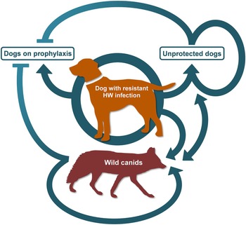
A very important question, now ML-resistance has been confirmed, is ‘How likely is it to spread, and how fast?’ At the moment, no answer can be convincingly given to this question. In some species of parasitic nematode, such as H. contortus, ML-resistance has spread rapidly and become nearly ubiquitous (Gilleard and Beech, Reference Gilleard and Beech2007; Gilleard, Reference Gilleard2013); in others, such as the small strongyles of horses, there have been sporadic reports of resistance, but it has yet to become a significant problem, despite intensive use of the drugs (Wolstenholme and Kaplan, Reference Wolstenholme and Kaplan2012). It is important to emphasize that there are important differences between heartworms and the GI parasitic nematodes that may have a major impact on how fast and how far ML-resistance will spread. The numerous heartworm infections in unprotected and feral dogs, together with wild canids, such as coyotes, wolves and foxes, mean that extensive refugia populations of parasites exist that are not exposed to drug selection. These refugia act as a source of drug-susceptibility alleles that dilute out those for resistance in the population as a whole and reduce the selection pressure for drug resistance (Van Wyk, Reference Van Wyk2001). Such refugia are often more limited in treatment of livestock parasites, as whole flocks were treated at a time when most of the worms were inside the hosts and ‘wild’ parasite populations are relatively small. The sizes of the heartworm refugia are, though, likely to vary locally, depending on the domestic and wildlife populations that are present (Fig. 2). Any loss of reproductive potential that may result from the acquisition of ML-resistance alleles, as observed in Onchocerca volvulus (Bourguinat et al. Reference Bourguinat, Pion, Kamgno, Gardon, Duke, Boussinesq and Prichard2007), will also tend to restrict their spread in the face of extensive refugia.
Populations of heartworms that might influence the rate of spread of drug resistance. The possible routes of transmission of resistant parasites from an infected dog are shown, along with those of susceptible populations that might act as refugium. These would not be able to infect protected animals on prophylaxis, though the resistant one would. All of the potential routes of spread are given equal weight in this figure, though in reality these would vary greatly. In particular, the motility of the parasite populations between wild, feral and domestic populations is unknown.

There is also an important difference in the sensitivity of our detection methods for resistance between GI parasites of livestock and heartworms. For livestock parasites, such as H. contortus, benzimidazole resistance-associated allele frequencies in some isolates can be as high as 30% and the isolate still be classed as ‘susceptible’ by a commonly used in vitro test, the egg-hatch assay (von-Samson Himmelstjerna et al. Reference von Samson-Himmelstjerna, Walsh, Donnan, Carriere, Jackson, Skuce, Rohn and Wolstenholme2009). None of the drugs used for livestock worm control is claimed to be ‘100% effective’ and observed efficacies of >95% are usually considered satisfactory (Coles et al. Reference Coles, Jackson, Pomroy, Prichard, von Samson-Himmelstjerna, Silvestre, Taylor and Vercruysse2006). The same drugs are used to both treat and prevent infections. In heartworms, by contrast, with compliant owners that treat regularly and have their dogs tested every year for circulating heartworm antigen, it is possible that infection with even a single resistant female worm could be detected by the current antigen test, especially if the sample is heated to inactivate antibody:antigen complexes (Little et al. Reference Little, Munzing, Heise, Allen, Starkey, Johnson, Meinkoth and Reichard2014b ). Those worms can then be killed using melarsomine. Widespread annual, or more frequent, testing together with proper therapy should therefore help to limit the spread of ML-resistant heartworms. Any resistant Mf will remain in the circulation of the treated dog and will be there before the antigen test is carried out, and this presents some risk of spread. The extent of that risk will depend on the exposure of the dogs to biting mosquitoes (dogs undergoing melarsomine therapy should be restrained from excessive exercise and so may not be outside as much) and the size and nature of the local refugium.
Some authors have worried that the use of MLs alone to remove adult worms (‘slow kill’) may increase the selection pressure for resistance (Bowman, Reference Bowman2012). Certainly, this method will fail to remove resistant adult worms from a dog that may be infected with both resistant and susceptible parasites and its use would not be a sensible course of action if the dog failed the Mf suppression test (Geary et al. Reference Geary, Bourguinat and Prichard2011). However, if the dog is only infected with susceptible adults, the situation will be different and depend on the genetics of resistance. For example, if resistance is due to changes in a single gene, which is unlikely based on experience with GI nematodes (Prichard, Reference Prichard2001; McCavera et al. Reference McCavera, Walsh and Wolstenholme2007; Wolstenholme and Kaplan, Reference Wolstenholme and Kaplan2012), and resistance is a recessive trait then infection with phenotypically susceptible heterozygous adults could result in the circulation of a proportion of Mf that are genetically and phenotypically resistant. Of course, such parasites could only be present if the dog had not been on prophylaxis. Since it is likely that the heterozygotes will be only a minority of the adults in an infected dog, it would be theoretically possible for such an animal to pass the suppression test, but still contain some resistant Mf. However, the numbers are likely to be small and transmission therefore less probable. The genetics of resistance are probably more complex than the single gene model which makes this analysis more difficult. If the dog is only infected with adults carrying ‘susceptible’ alleles then the only way that the ‘slow-kill’ procedure could select for resistance is if such Mf appeared as the result of spontaneous mutations in the mating adults. This is obviously possible, but such a mutation would need to be at least partially dominant to confer a resistance phenotype on the Mf and the number of such Mf in the circulation would be vanishingly small. It also worth pointing out that the melarsomine treatment protocol will also leave ML-resistant Mf in the circulation and that the inclusion of a tetracycline antibiotic in the treatment plan will slowly kill all the Mf and, make any survivors, even if they develop into L3, incapable of initiating a patent infection in a second dog (McCall et al. Reference McCall, Kramer, Genchi, Guerrero, Dzimianski, Mansour, McCall and Carson2014).
One interpretation of these factors, the large refugia and the efficacy of current drug combination therapies, might be that the risk of ML-resistance spreading in the heartworm populations is small. But none the less, it has emerged and in a geographical region, the Mississippi delta, where the refugium is undoubtedly very large. So, was the conclusion drawn by Prichard in 2005 (Prichard, Reference Prichard2005) that ‘it is unlikely that significant (emphasis added) avermectin/milbemycin resistance will be selected in D. immitis with current treatment strategies’ incorrect? It may not have been. This conclusion was based on ‘the proportion of the D. immitis population normally in refugia, the life cycle stage targeted, and the anthelmintic dosages used’, and one important consideration is the meaning of ‘significant’. Any infection in animals that would otherwise have been protected is of course very significant to those animals and their owners, but from a veterinary public health perspective it implies that a fairly large number of heartworm infections are being caused by resistant parasites, and to date that does not seem to have happened. The high reproductive rate of heartworms and the relatively low drug doses used as preventives will tend to favour the local selection of resistance alleles (Prichard, Reference Prichard2005) and the refugium in the insect vector may be small, but that in unprotected animals and wild canids is very large (Bowman and Atkins, Reference Bowman and Atkins2009). Given the very high infection rates of vectors that feed on dogs kept outdoors (McKay et al. Reference McKay, Bianco, Rhodes and Barnett2013) and possible limited overlap between these vectors and those feeding on the refugium of unprotected animals, it may be that there is a relatively high local selection pressure for resistance to emerge (Fig. 2). However, any spread of these resistance alleles beyond the limited area in which they have emerged will be limited by the large refugium they are then likely to encounter, which will dilute their impact and potentially lead to them disappearing (Van Wyk, Reference Van Wyk2001). This might explain why, despite the fact that almost half of the ‘Katrina’ dogs tested were positive for heartworm (Levy et al. Reference Levy, Lappin, Glaser, Birkenheuer, Anderson and Edinboro2011), at least some of these dogs are very likely to have been infected with ML-resistant parasites (Bourguinat et al. Reference Bourguinat, Keller, Bhan, Peregrine, Geary and Prichard2011a ; Pulaski et al. Reference Pulaski, Malone, Bourguinat, Prichard, Geary, Ward, Klei, Guidry, Smith, Delcambre, Bova, Pepping, Carmichael, Schenker and Pariaut2014) and they have been distributed widely through North America, the geographic spread of resistance seems to be limited.
The preceding paragraph might be seen as an optimistic interpretation and this optimism could easily be misplaced. It is important to note that we do not know the extent of resistance. If we compare the extent of resistance with the size of a fire, we do not know if we are dealing with a campfire or an acres-large wildfire. Most individuals agree that there is smoke on the horizon that shows resistance exists. If we are dealing with an uncontrolled wildfire, we might have expected to have seen a larger number of resistance cases. This does not seem to have been observed. More likely, the current state of resistance is a moderately-sized brush fire. However, it is important to remember that if a controlled fire is not monitored, it can quickly become much larger. This argues for an increase in activity to determine the ‘the size of the fire.’ Most importantly though, academics and veterinarians need to remember that, in the mind of the individual owner the size of the fire (i.e. extent of resistance) does not matter. What is important to the animal's owner is that their pet in now heartworm positive.
FINANCIAL SUPPORT
This research received no specific grant from any funding agency, commercial or not-for-profit sectors.