Introduction
Cardiovascular disease is one of the most severe and widespread global health problems of death and disability, due to its predominance. Cardiovascular diseases encompass diverse heart and blood vessel disorders through elaborate and sometimes unsteady pathological routes. Reference Guleria, Naga Srinivasu, Ahmed, Almusallam and Alarfaj1 If not recognised or poorly treated, cardiovascular diseases often cause acute myocardial infarction, chronic heart failure, and disabling strokes, as well as serious and life-threatening accidents. Reference Panis and Donal2 Early and reliable diagnosis is vital for effective cardiovascular disease management and enhancing patient outcomes.
MRI was widely used for evaluating the non-invasive cardiac due to the superior soft tissue contrast and functional image capability. Reference Backhaus, Schulz and Lange3 Cardiac MRI offers clinical information for diagnosing cardiomyopathies, heart failure, and myocardial abnormalities. Reference Singh, Singh, Kler and Sharma4 However, the manual interpretation of multi-parametric MRI scans was time-consuming due to inter-observer variability and reduced diagnostic consistency in the complex clinical cases. Reference Glessgen, Crowe and Wetzl5
Deep learning is a recent advancement of convolutional neural networks and vision transformers enhanced the automated cardiac MRI analysis. Reference Park, Kim, Ruan, Chow and Baskaran6 ResNet and EfficientNet frameworks have strong classification capability, and the transformer-based models enhance global contextual learning via a self-attention mechanism. Reference Muthulakshmi, Venkatesan, Prasanalakshmi, Bahiyah and Divya7 The hybrid convolutional neural network-transformer framework enhances the feature representation of local spatial information with long-range anatomical dependencies. Reference Jie, Chen and Lin8
Recent progress has several challenges to the clinical adoption of deep learning for cardiac MRI classification. However, convolutional neural network-based models suffer from capturing long-range anatomical dependency, and transformer-based approaches require high computational resources and large-scale training data. Reference Din, Qaraqe, Mourad, Qaraqe and Serpedin9,Reference Abdollahi, Nouri-Moghaddam, Mohajer, Mirzaei and Derakhshanfard10 Some studies lack robust preprocessing, an explainability mechanism, and reduce clinical trust, model transparency. Reference Bizimana, Zhang, Hounye, Asim, Hammad and El-Latif11,Reference Marey, Arjmand and Alerab12 To address these issues, a novel CardioFusion-XAI framework has been proposed and evaluated specifically for four-class cardiac MRI classification using the Sunnybrook Cardiac Dataset, consisting of heart failure with infarction, heart failure, hypertrophy, and normal cardiac categories. Therefore, the scope of this work is limited to automated classification within this dataset rather than comprehensive cardiovascular disease diagnosis.
Although the present study is evaluated using the Sunnybrook Cardiac Dataset, which primarily contains adult cardiac MRI cases, the proposed framework is designed as a general cardiac MRI analysis approach. The methodology may provide a foundation for future investigations involving paediatric and CHD populations, where accurate and interpretable cardiac image analysis is also clinically important. However, no paediatric or congenital cardiac datasets were included in the current study.
The contributions of the proposed CardioFusion-XAI framework are as follows:
-
• This study proposed a novel CardioFusion-XAI framework to enhance cardiac MRI quality via noise reduction, contrast enhancement, and structural normalisation.
-
• The proposed model combined with hybrid deep learning, like EfficientNet-B0, ResNet-50, and vision transformers, to capture both fine-grained local textures and long-range global anatomical dependency from cardiac MRI images.
-
• The proposed model used multi-scale convolutional neural network-transformer feature fusion to enhance discriminative cardiovascular disease classification performance and patient-level generalisation.
-
• The proposed model included explainable AI techniques like Grad-CAM and t-SNE visualisation to offer clinically interpretable decision support and enhance model transparency.
The organisation of the paper is structured as follows: Section 2 reviews various deep learning approaches for cardiovascular disease classification via a cardiac MRI. Section 3 explains in detail the proposed CardioFusion-XAI framework. Section 4 discuss about a result and analysis. Finally, Section 5 presents the conclusion and future work.
Literature survey
This section presents a deep learning and medical image, which enhances the automated cardiovascular disease diagnosis via a cardiac MRI. Some studies are focused on hybrid, machine learning, and transformer framework are enhanced diagnostic accuracy and reliability.
Sadr et al. Reference Sadr, Salari, Ashoobi and Nazari13 suggested a hybrid deep learning and machine learning framework for cardiovascular disease prediction via both the public and clinical datasets. This model integrated convolutional neural network, RNN, and classical classifiers for enhancing both the prediction accuracy and reliability. The suggested framework involves high architectural complexity but lacks validation across various demographic and acquisition settings.
Baghdadi et al. Reference Baghdadi, Farghaly Abdelaliem, Malki, Gad, Ewis and Atlam14 developed a gradient boosting-based cardiovascular disease prediction framework for early diagnosis via clinical records. This model attained high accuracy and the F1-score for effective asymptomatic case detection. However, the framework mainly focused on asymptomatic prediction and depended on the quality and completeness of clinical features.
Ogunpola et al. Reference Ogunpola, Saeed, Basurra, Albarrak and Qasem15 assessed seven machine learning models, which include convolutional neural network-based approaches for cardiovascular disease detection. The optimised gradient boosting model was achieved across various evaluation metrics for improving the diagnostic reliability. However, the suggested approach overfits risks and requires wide optimisation procedures, which lack practical reproducibility.
Liu et al. Reference Liu, Xu and Xu16 presented a neural classifier that utilises self-attention operations to model long-dependency for electrocardiography signal diagnosis. The suggested model was evaluated on the public Tianchi electrocardiography dataset, which integrates lead fusing blocks, transformer-XL encoder-based encoder modules. However, their study focuses on enhancing the interpretability, robustness, and expanding the clinical applications of AI-driven electrocardiography diagnosis.
Cockrum et a. Reference Cockrum, Nakashima and Ammoury17 advanced a deep learning algorithm that helped discriminate cardiac amyloidosis, both cine and late gadolinium enhancement of cardiac MRI. Their study, which was evaluated by the Ohio dataset, has the ability of the vision transformers model to differentiate the cardiac amyloidosis from hypertrophic cardiomyopathy. However, their study lacks the accuracy of disease detection to characterise the clinical decision support models.
Telangore et al. Reference Telangore, Azad, Sharma, Bhurane, San Tan and Acharya18 developed an artificial intelligence model of Sudden Cardiac Death (SCD) for early prediction via the electrocardiography signals. The suggested approach was evaluated by the MITBIH SCDs Holter Dataset, which enhances early SCD prediction as a valuable tool for continuous electrocardiography monitoring in high-risk patients. However, the suggested model predicts SCD a few minutes before onset, which limits the intervention time
Mirzaee Moghaddam Kasmaee et al. Reference Mirzaee Moghaddam Kasmaee, Ataei and Moravvej19 introduced a deep learning model called ELRLMD, which integrates ensemble learning and Reinforcement Learning for effective myocarditis diagnosis from cardiac magnetic resonance images. The suggested model was evaluated by the cardiac magnetic resonance dataset, which lacks risk convergence on local optima due to a suboptimal initial weight setting. The summary of existing cardiovascular disease classification approaches is provided in Supplementary Table S1.
Existing studies depend on a single-model framework or traditional machine learning methods with handcrafted features, which fail to capture both fine local textures and long-range contextual dependency in the complex cardiac MRI data. Moreover, some studies lack to integrates explainability mechanisms, which limits both the interpretability and clinical trust. To address these issues, this study proposes a novel framework that integrates enhanced preprocessing, hybrid convolutional neural network-transformer feature fusion, and explainable AI techniques for accurate and interpretable cardiac MRI classification.
Materials and methods
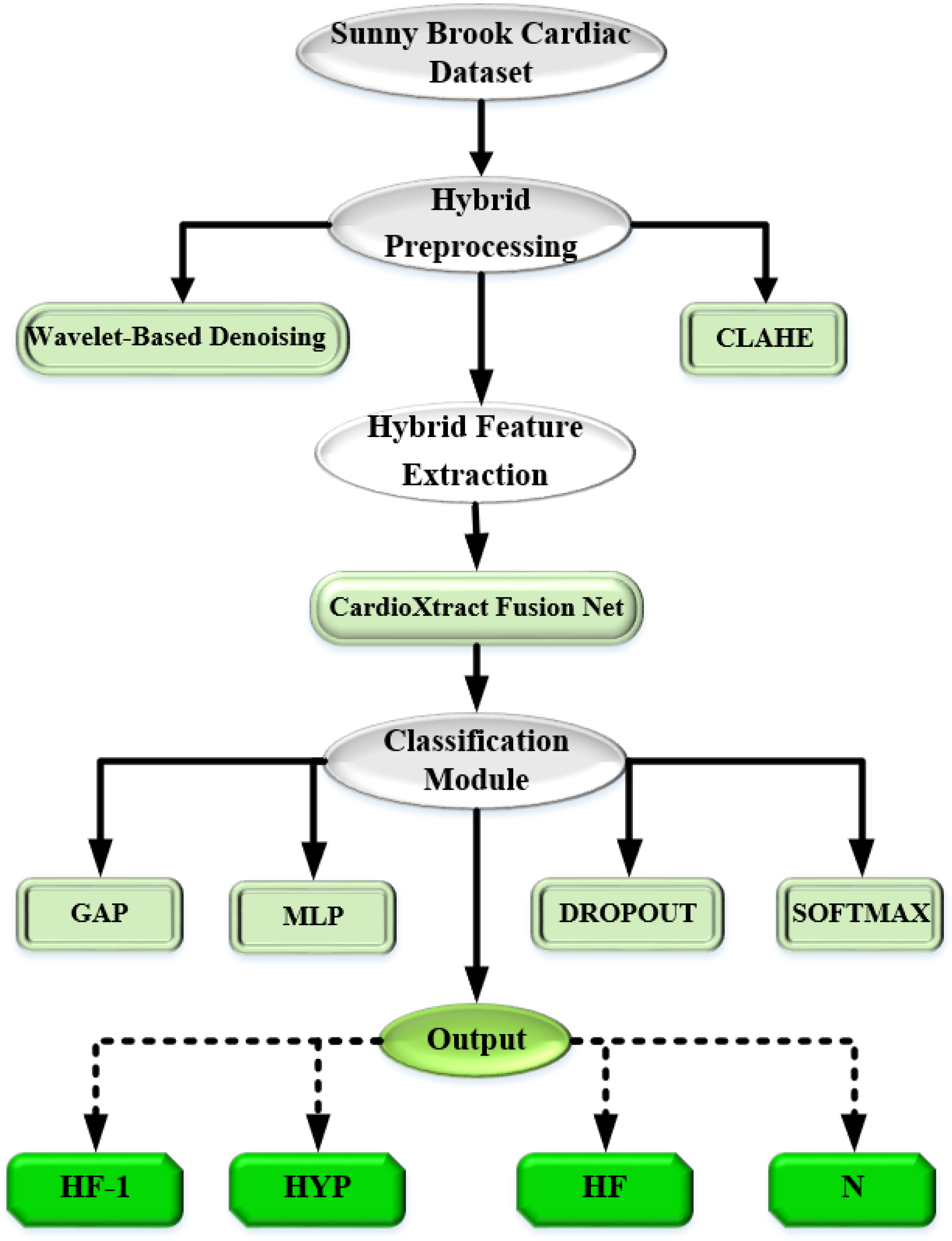
This study proposed a novel CardioFusion-XAI framework to enhance cardiac MRI quality via noise reduction, contrast enhancement, and structural normalisation. Initially, the Sunnybrook Cardiac Dataset (SBCD), which consists of fine cardiac MRIs, categorised into four like heart failure with infarction, hypertrophy, heart failure without infarction, and normal cardiac anatomy (N). The hybrid processing step includes wavelet-based denoising to remove the noise, whilst keeping the fine anatomical details and the structural boundaries intact. Next, contrast-limited adaptive histogram equalisation increases the local contrast cleverly, not exposing extremely subtle pathological features, but rather preventing the creation of any artificial artefacts.
The hybrid feature extraction step, supported by CardioXtract Fusion Net, forms the backbone of the proposed approach. The features that were extracted are fed into an intelligent classification for superior performance and generalisation. Global average pooling and dropout-regularised multilayer perceptron layers are applied for efficient classification and overfitting reduction. The properties that have been handled are then sent to a multilayer perceptron that employs dropout regularisation as a form of enhancement. Dropout is a method that works against overfitting by randomly turning off neurons during training. A detailed architectural framework and its flow are shown in Figure 1. Existing cardiac MRI classifications are dependent on convolutional neural network-only or transformer-only frameworks. In contrast, the proposed CardioFusion-XAI model integrates Wavelet-contrast-limited adaptive histogram equalisation preprocessing, complementary convolutional neural network feature extraction, transformer-based contextual fusion, and explainable AI mechanisms for robust and interpretable patient-level cardiac MRI classification.
Architectural framework for cardiac disease classification.

Image acquisition
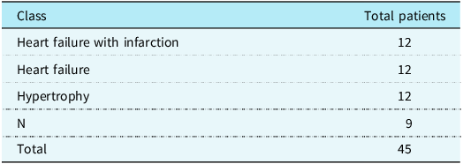
The proposed model was evaluated by an SBCD, which was publicly available as the 2009 Cardiac MR Left Ventricle Segmentation Challenge dataset. It contains 45 cine-MRI studies classified into four classes are shown in Table 1. It also includes 233 cine-MRI sequences, approximately 51,515 MRI slices with expert-annotated contours for reliable ground-truth validation. Five-fold cross-validation was performed using patient-level splitting to prevent inter-slice data leakage. All MRI slices belonging to a single patient were assigned exclusively to either the training or validation subset within a given fold. During evaluation, slice-level predictions generated by the proposed framework were aggregated into a single patient-level prediction using majority voting across all slices belonging to the same patient. Thus, the dataset was publicly available, fully anonymised, and no additional ethical approval was required.
Patient-level class distribution of the Sunnybrook Cardiac Dataset

Table 1. Long description
A table titled ‘Patient-level class distribution of the Sunnybrook Cardiac Dataset’ with two columns and five rows. The columns are labeled ‘Class’ and ‘Total patients’. The rows are labeled as follows: Row 1: Heart failure with infarction, 12; Row 2: Heart failure, 12; Row 3: Hypertrophy, 12; Row 4: N, 9; Row 5: Total, 45. The table shows the number of patients in each class, with the total number of patients being 45.
Preprocessing
Pre-processing plays a vital role in the hybrid WaveCLAHE Net to enhance the cardiac MRI quality before the model training. Initially, all the MRI images are resized into 256 × 256 pixels for input standardisation. Then, wavelet-based denoising was applied to suppress noise and maintain anatomical structures. Thus, the denoised image is reconstructed by inverse discrete wavelet transform after the soft-thresholding Reference Algolaib and Abdulshahed20 of wavelet coefficients was expressed as Equation (1):
 ${{\tilde w}_{j,k}} = {\rm{sign}}\left( {{w_{j,k}}} \right)\cdot\max \left( {\left| {{w_{j,k}}} \right| - {T_j},0} \right)$
where the wavelet coefficients are denoted by w j, k , At j, the threshold values are expressed by T j . Then contrast-limited adaptive histogram equalisation Reference Camargo, Figueiredo and Coelho21 was applied to improve both the local contrast and normalise intensity variations in cardiac MRI images. The histogram equalisation mapping function is expressed as Equation (2):
 ${{S_k} = T\left( {{r_k}} \right) = {{L - 1} \over {MN}}\sum _{j = 0}^k{n_j}}$
where the total number of pixels is denoted by MN, r j denotes the intensity of the number of pixels expressed by n j , and the number of intensity levels was represented by L. Finally, Z-score normalisation was performed to standardise intensity distributions across the dataset.
Feature extraction
After preprocessing, a hybrid feature extraction integrates EfficientNet-B0, Reference Muthulakshmi, Venkatesan, Prasanalakshmi, Bahiyah and Divya7 ResNet-50, Reference Anand, Lakshmi, Pandey and Pandey22, and vision transformers Reference Jacob, Borgohain, Chitiboi, Sharma, Comaniciu and Rueckert23 to capture both the local and global cardiac representations from MRI images. In that, EfficientNet-B0 and ResNet-50 extract discriminative spatial features, and the vision transformers performs global contextual fusion to model long-range anatomical dependency. The Vision Transformer utilised a patch size of 16 × 16, embedding dimension of 768, 12 transformer encoder layers, 12 attention heads, and a multilayer perceptron dimension of 3072. The detailed architecture of the proposed CardioXtract Fusion Net model was illustrated in Supplementary Figure S2.
EfficientNet-B0
The EfficientNet-B0 is used to extract both the fine-grained local features and myocardial texture representations from cardiac MRI images. From Block 7, the extracted high-level feature representations are expressed as Equation (3):
 $F={\rm EffB}0_{{\rm block\ }7}\left(I\right)$
where the input cardiac MRI images are denoted by I, from EfficientNet-B0, the extracted high-level feature map is represented by F. The extracted feature map is partitioned into non-overlapping patches and projected into D-dimensional embeddings, which are denoted as Equation (4):
 $P_{{\rm emb}}={\rm Conv}2{\rm D}\left(D,\, {\rm kernel\ size} =P,{\rm \ stride\ }=P\right)\left(F\right)$
where the patch size is denoted by P, the embedding dimensions are expressed by D, and the patch embedding is denoted by P emb . The extracted embeddings are a discriminative local cardiac information for consequent feature fusion. The EfficientNet-B0 framework was illustrated in Supplementary Figure S3.
ResNet-50
The proposed model integrates ResNet-50 to learn both the hierarchical spatial and anatomical representations from cardiac MRI images. The extracted deep features from the final residual stage were formulated as Equation (5):
 $F={\rm ResNet}50_{{\rm block\ }5}(I)$
where the input cardiac MRI images are expressed by I, via ResNet-50, the high-level feature maps are denoted by F. The extracted feature maps are converted into structured patch embeddings to maintain regional cardiac information for fusion learning. The architecture of ResNet-50 is shown in Supplementary Figure S4.
Vision transformer-based feature fusion in the cardioXtract fusion net
From the EfficientNet-B0 and ResNet-50, the features are extracted, and the vision transformers is used to perform a global contextual fusion. The synchronised convolutional neural network feature maps are represented as Equation (6):
 $F=\left[F_{{\rm ResNet}50 }\| F_{{\rm EffB}0 }\right]$
where the ResNet-50 and EfficientNet-B0 extract features are denoted by F ResNet50 and F EffB0. The fused feature map is partitioned into non-overlapping patches and projected into D-dimensional embeddings expressed as Equation (7):
 $z_{i}=W_{e}\cdot {\rm Flatten}\left(P_{i}\right)+b_{e}$
where the i th The image patch was denoted by P i , learnable projection parameters are expressed by W e ,b e , and the embedded token is represented by z i . Positional embeddings are added to maintain a spatial relationship among patches as indicated in Equation (8):
 $Z_{0}=\left[z_{1}+E_{1},z_{2}+E_{2},\ldots, z_{N}+E_{N}\right]$
where the i th patch tokens are associated with the positional embedding denoted by E i , and for the transformer encoder, the input token sequence is represented by Z 0. The multi-head self-attention Reference Jacob, Borgohain, Chitiboi, Sharma, Comaniciu and Rueckert23 mechanism is applied to model long-range feature dependency, denoted by Equation (9):
 ${\rm Attention}\left(Q,K,V\right)={\rm softmax}\left({QK^{T} \over \sqrt{d_{k}}}\right)V$
where the query, key, and value matrices are denoted by Q, K, V, and the key dimensions are represented by d k . The fused global representation is denoted as Equation (10):
 $F_{{\rm ViT}}={\rm MSA}\left(Z_{0}\right)$
where from the vision transformers, the globally contextualised features are denoted by F ViT . It enhances global contextual learning and discriminative cardiac disease representation.
Classification
After the CardioXtract Fusion Net model, the fused features are forwarded into classification for cardiovascular disease prediction. Initially, the global average pooling layer reduces spatial dimensions of the fused feature maps and maintains the most discriminative global representations. Then, the pooled features are fed into the multilayer perceptron with ReLU activation and dropout regularisation to enhance nonlinear feature learning and reduce overfitting. A dropout rate of 0.5 was employed within the multilayer perceptron classification head to reduce overfitting. Finally, the Softmax layer generates normalised probability scores for multi-class cardiac disease classification, such as heart failure, heart failure without infarction, hypertrophy, and normal classes. The detailed classification process is mathematically formulated in Supplementary Section S3.4.1–S3.4.4.
Visual analysis using Grad-CAM and t-SNE
This study employed Grad-CAM as a visual analysis tool to highlight image regions that contributed strongly to convolutional neural network-based feature extraction. Although Grad-CAM provides useful qualitative insights into model attention, it does not fully explain the final decision-making process of the integrated convolutional neural network–transformer architecture. Reference Gangadharaiah, Kavitha, Shafeeq and Karanth24 to improve clinical interpretability and generate visual heatmaps that highlight diagnostically relevant regions in cardiac MRI images. The generated activation maps help to identify anatomical structures, including myocardial walls and ventricular boundaries, which contribute to disease classification. Additionally, t-SNE was utilised to visualise the distribution and separability of learned feature representations across cardiac disease categories. These visualisations support qualitative assessment of feature discrimination but should not be interpreted as evidence of clinical explainability. Reference Ma, Chen and Ge25
Result and analysis
The proposed CardioFusion-XAI framework was evaluated by five-fold cross-validation for cardiovascular disease classification from cardiac MRI images. It was evaluated by several performance metrics like accuracy, precision, recall, specificity, F1-score, Cohen’s Kappa, and AUC metrics. Additionally, Grad-CAM visualisations and t-SNE feature projections are used to analyse the model interpretability and feature separability. The detailed preprocessing outcomes are provided in Supplementary Figure S5.
Performance metrics
The proposed CardioFusion-XAI framework was evaluated by five-fold level cross-validation and several metrics to ensure a reliable generalisation.
Implementation details and experimental configuration
The proposed CardioFusion-XAI framework was implemented on Python, TensorFlow, and Keras. ImageNet-pretrained EfficientNet-B0 and ResNet-50 models are integrated by the vision transformers fusion module. The model was trained by the Adam optimiser. Hyperparameters, including learning rate (0.0001), batch size (16), dropout rate, and training epochs, were selected based on preliminary validation experiments and prior literature on cardiac MRI classification. Early stopping and learning-rate scheduling were employed to reduce overfitting and improve model generalisation. Early stopping and learning rate reduction are applied to enhance convergence and reduce overfitting. Data augmentation included random rotation (±15°), horizontal flipping, zooming (0.9–1.1), width shifting (±10%), and brightness adjustment (0.8–1.2). Several data augmentations include random rotation, horizontal flipping, zooming, width shifting, and brightness adjustment. For all experiments, a fixed random speed of 42 was used for reproducibility. Thus, the framework was trained on an NVIDIA RTX 3080 GPU with 32 GB RAM and approximately 34.8 million trainable parameters. End-diastolic and end-systolic MRI phases are selected for the left ventricle for analysis. Model optimisation was performed using categorical cross-entropy loss and the Adam optimiser. Early stopping monitored validation loss with a patience value of 10 epochs, restoring the best-performing model weights. The learning rate was reduced by a factor of 0.5 when validation loss failed to improve for five consecutive epochs.
Quantitative evaluation metrics
The proposed CardioFusion-XAI framework was evaluated by several metrics, which were evaluated as follows:
-
• Accuracy represents the overall proportion of correctly classified instances among all evaluated samples and is computed as
 $\textit{Accuracy}={TP+TN \over TP+TN+FP+FN}$
-
• Precision indicates the reliability of positive predictions by measuring the ratio of correctly predicted positive cases to all predicted positives, expressed as
 $\textit{Precision}={TP \over TP+FP}$
-
• Sensitivity (Recall) assesses the model’s ability to correctly identify true positive cases from all actual positives, formulated as
 $\textit{Recall}={TP \over TP+FN}$
-
• Specificity reflects the capability of the model to recognise true negative cases among all actual negatives correctly and is calculated as
 $\textit{Specificity}={TN \over TN+FP}$
The proposed CardioFusion-XAI framework achieved high classification performance across all five cross-validation folds. To assess the stability of the proposed framework, performance metrics are reported as mean ± standard deviation across the five validation folds. The relatively low variability observed among folds indicates consistent model behaviour despite the limited dataset size. Nevertheless, the small number of available patients may lead to optimistic performance estimates, and therefore, the reported results should be interpreted with appropriate caution. The average metrics attained an accuracy of (0.9889 ± 0.0061), Precision (0.9893 ± 0.0058), Recall (0.9889 ± 0.0061), and F1-score (0.9890 ± 0.0060), showing a strong predictive capability with low variability across folds. Then, the model achieved high Specificity (0.9963 ± 0.0023), Cohen’s Kappa (0.9848 ± 0.0089), and AUC (0.9998 ± 0.0003), which represent excellent class separability and robust generalisation performance. The detailed fold-wise quantitative metrics are provided in Supplementary Table S3.
ROC and precision-recall curve analysis
The Receiver Operating Characteristic curve (ROC) and precision-recall analyses showed a strong discriminative performance across various cardiovascular diseases via a five-fold cross-validation. The detailed ROC curve is provided in Supplementary Figure S6.
Patient-level evaluation strategy
The class receiving the highest number of slice-level predictions was assigned as the final patient-level diagnosis. All quantitative metrics reported in this study were computed using patient-level predictions obtained from five-fold cross-validation.
Training and validation performance
The proposed CardioFusion-XAI framework has stable convergence and effective learning behaviour across all five folds with minimal overfitting. Both the training and validation curves have strong generalisation capability. Detailed training-validation accuracy and loss curves are provided in Supplementary Figures S7 and S8.
Confusion matrix analysis
The confusion matrix analysis has highly accurate classification across various cardiovascular diseases with less inter-class misclassification in five-fold cross-validation. Detailed confusion matrices are provided in Supplementary Figure S9. The confusion matrices presented in Supplementary Figure S9 correspond to patient-level predictions obtained after aggregation of slice-level outputs using majority voting. Each matrix, therefore, represents the final diagnostic classification of independent patients rather than individual MRI slices.
Interpretability analysis using Grad-CAM and t-SNE
The proposed framework employed Grad-CAM and t-SNE as supporting visual analysis techniques. Grad-CAM highlighted image regions that contributed strongly to convolutional neural network feature extraction, whereas t-SNE illustrated the separability of learned feature representations across disease categories. While these visualisations provide useful qualitative insights into model behaviour, they should not be interpreted as comprehensive explanations of the final convolutional neural network–transformer fused decision process or as definitive evidence of clinical interpretability. Detailed Grad-CAM visualisations are presented in Supplementary Figure S10.
Ablation study
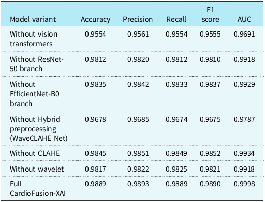
Table 2 displays the ablation study of individual modules and the proposed model. Standalone backbone architectures, including ResNet-50, EfficientNet-B0, and Vision Transformer, were evaluated under identical experimental settings. The results demonstrate that the proposed hybrid architecture consistently outperformed individual backbone models, confirming the complementary benefits of multi-scale convolutional neural network feature extraction and transformer-based contextual fusion. The proposed CardioFusion-XAI model attained 98.89% accuracy and 0.9998 AUC. Removing vision transformers, convolutional neural network branches, or preprocessing modules confirms that hybrid feature fusion and image enhancement are crucial for accurate cardiac MRI classification.
Ablation study of the proposed CardioFusion-XAI framework

Table 2. Long description
A table titled ‘Patient distribution across five cross-validation folds’ with five rows and three columns. The columns are labeled ‘Fold’, ‘Training patients’, and ‘Validation patients’. Each row represents a different fold, with the first column indicating the fold number from 1 to 5. The second column shows the number of training patients, which is consistently 36 for all folds. The third column shows the number of validation patients, which is consistently 9 for all folds. The table presents a clear and uniform distribution of patients across the five folds, with no variations in the number of training or validation patients.
Comparative analysis
The proposed framework was compared with several existing approaches. For fair comparison, all baseline models were reimplemented and evaluated using the same Sunnybrook Cardiac Dataset, patient-level five-fold cross-validation protocol, preprocessing pipeline, data augmentation strategy, optimiser settings, batch size, learning rate schedule, and early-stopping criteria employed by the proposed framework. This ensured that performance differences were attributable to model architecture rather than variations in experimental configuration, like convolutional neural network + LSTM, Reference Sadr, Salari, Ashoobi and Nazari13 VGG16, Reference Gurusubramani and Latha26, and conventional 2D convolutional neural network, Reference Mohonta, Motin and Kumar27 via the same dataset, preprocessing strategy, and patient-level data splitting for fair evaluation.
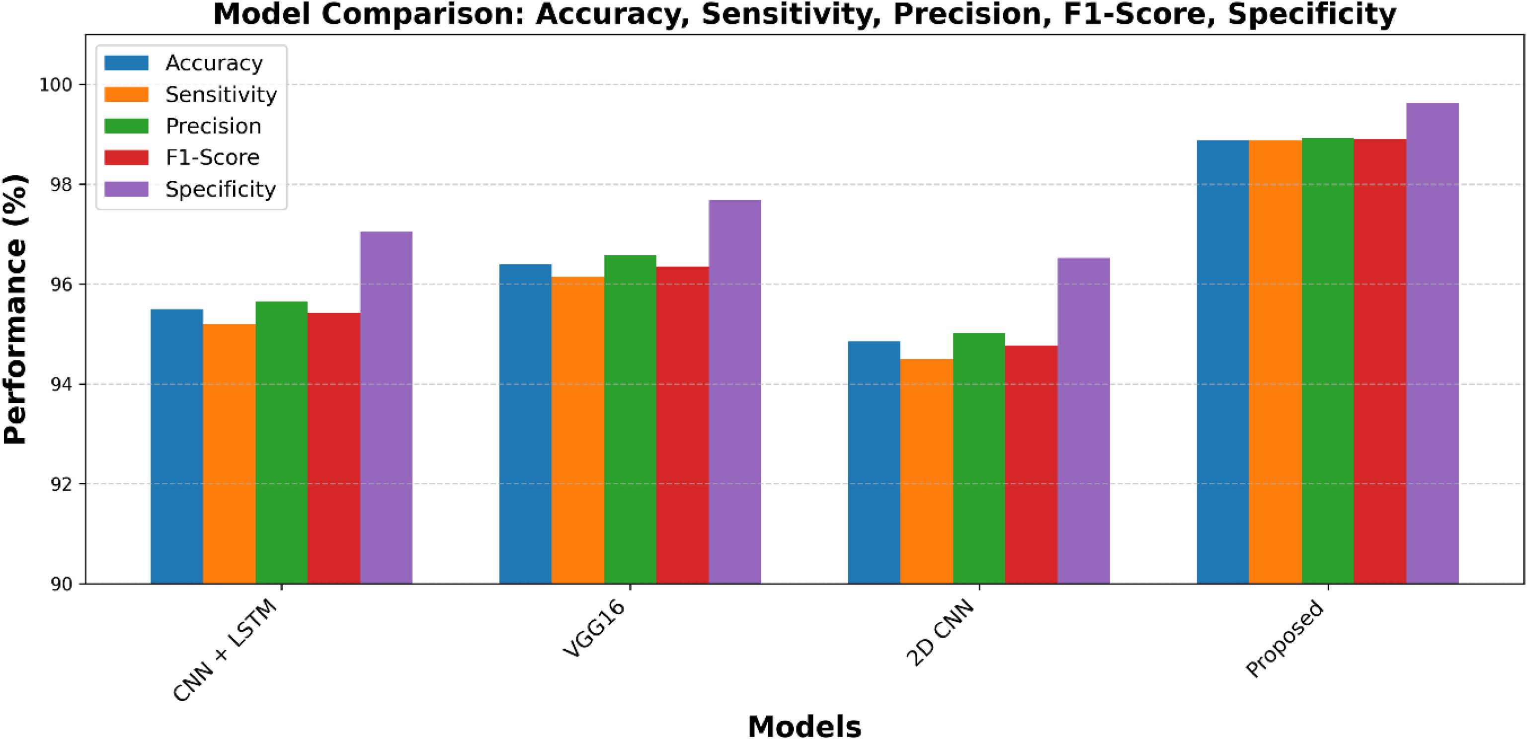
Comparative performance analysis of baseline models and proposed method.

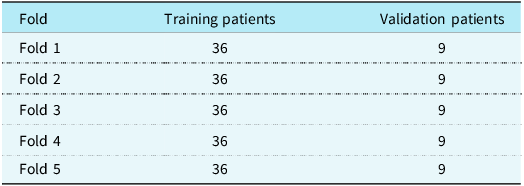
Figure 2 demonstrates the performance comparison of the proposed and other existing approaches across various evaluation metrics. The proposed model attained high accuracy (98.89%), F1-score (98.90%), and specificity (99.63%), showing superior discriminative capability for cardiac MRI classification. This enhancement confirms the effectiveness of hybrid convolutional neural network-transformer feature fusion and enhanced preprocessing in learning complex cardiac representations. External multi-centre validation wasn’t performed due to the limited publicly available annotated cardiac MRI datasets. Table 3 represents the patients distribution across five cross-validation folds
Figure 2 presents the comparative performance analysis of the proposed CardioFusion-XAI framework against convolutional neural network + LSTM, VGG16, and conventional 2D convolutional neural network models. The proposed framework achieved the highest performance across all evaluation metrics, attaining an accuracy of 98.89%, sensitivity of 98.89%, precision of 98.93%, F1-score of 98.90%, and specificity of 99.63%. Among the baseline methods, VGG16 demonstrated the strongest performance with an accuracy of 96.4% and specificity of 97.7%, whereas the conventional 2D convolutional neural network achieved the lowest overall performance. The superior results of CardioFusion-XAI can be attributed to the integration of WaveCLAHE preprocessing, complementary convolutional neural network-based feature extraction using EfficientNet-B0 and ResNet-50, and Vision Transformer-based feature fusion, which effectively capture both local anatomical patterns and global contextual information. These findings confirm the effectiveness of the proposed hybrid architecture for accurate and robust cardiac MRI classification. Table 4 presents the Comparison Table for proposed work.
Discussion
The proposed CardioFusion-XAI framework attained superior cardiac MRI classification performance via the integration of WaveCLAHE preprocessing, hybrid convolutional neural network-based feature extraction, vision transformers-based contextual fusion, and regularised classification. The proposed model attained 98.89% accuracy and 98.90% F1-score compared to other existing approaches. The preprocessing stage enhances image quality, local contrast, and the hybrid feature extraction strategy effectively captures both the local textures and global anatomical dependencies. The proposed model attained high performance but lacks several challenges. Although the proposed framework achieved promising performance, its evaluation was limited to the Sunnybrook Cardiac Dataset, containing four cardiac condition categories. Therefore, the findings should be interpreted as evidence of effectiveness for four-class cardiac MRI classification rather than comprehensive cardiovascular disease diagnosis. Additional validation on larger multi-centre datasets encompassing a wider range of cardiac pathologies is required before considering broader clinical applicability. The Sunnybrook dataset lacks several subjects, and doesn’t perform external multi-centre validation. Additionally, the proposed framework only evaluates adult cardiac MRI datasets but does not include paediatric or congenital cardiac conditions. Future research focuses on larger multi-centre datasets, multimodal learning, and privacy-preserving collaborative training strategies.
Although Grad-CAM and t-SNE provided useful visual insights into feature learning and model attention, these methods offer only partial explanations of the proposed framework. Specifically, Grad-CAM primarily reflects convolutional neural network-derived activations and does not fully capture the contribution of transformer-based contextual fusion. Similarly, t-SNE visualises feature-space organisation but does not establish causal or clinically interpretable reasoning. Therefore, the explainability results should be viewed as supportive qualitative analyses rather than complete explanations of model decision-making.
Despite being evaluated on an adult cardiac MRI dataset, the proposed framework addresses methodological challenges that are also relevant to paediatric cardiac imaging, including robust feature extraction, multi-scale representation learning, and explainable decision support. These capabilities may be valuable for future AI-assisted analysis of paediatric and congenital heart disease imaging, where diagnostic accuracy and interpretability are particularly important. The comparative experiments indicate that the performance improvement is not solely attributable to increased model complexity. Individual convolutional neural network and transformer backbones achieved lower classification accuracy than the proposed hybrid framework, while the removal of preprocessing or feature-fusion components resulted in measurable performance degradation. These findings support the contribution of each module within the CardioFusion-XAI architecture.
Data and code availability
The Sunnybrook Cardiac Dataset used in this study is publicly available. The source code, trained model weights, and patient-level data split files will be made publicly available upon acceptance of the manuscript to facilitate reproducibility and future research.
Conclusion
This study proposed CardioFusion-XAI, an explainable hybrid deep learning framework for four-class cardiac MRI classification using the Sunnybrook Cardiac Dataset. The framework integrates WaveCLAHE preprocessing, convolutional neural network-based feature extraction, Vision Transformer-based feature fusion, and explainable AI techniques to improve classification performance and interpretability. However, the proposed framework was evaluated only on an adult cardiac MRI dataset and was not validated on paediatric or congenital heart disease populations. Therefore, its applicability to young patients remains unconfirmed and requires further investigation using clinically relevant paediatric cardiac datasets.
Experimental results demonstrated strong classification capability, achieving an accuracy of 98.89% and an F1-score of 98.90%. The integration of multi-scale feature learning and transformer-based contextual modelling contributed to improved discrimination among heart failure without infarction, heart failure, hypertrophy, and normal cardiac categories. However, these findings were obtained from a relatively small single-centre dataset, and external validation was not available. Therefore, the reported performance should be interpreted as promising rather than conclusive, and further evaluation on larger independent cohorts is necessary before broader clinical adoption can be considered.
Nevertheless, the study was evaluated on a single dataset containing four cardiac condition classes and a limited number of subjects. Therefore, the findings should not be interpreted as evidence of a comprehensive cardiovascular disease diagnosis or immediate clinical deployment. Future work will focus on validation using larger multi-centre datasets, additional cardiac pathologies, multimodal imaging data, and privacy-preserving collaborative learning strategies.
Patient distribution across five cross-validation folds

Table 3. Long description
A table comparing the performance of different model variants for cardiac MRI classification. The table has 7 rows and 6 columns. The columns are labeled Model variant, Accuracy, Precision, Recall, F1 score, and AUC. The rows are labeled as follows: Without vision transformers, Without ResNet-50 branch, Without EfficientNet-B0 branch, Without Hybrid preprocessing (WaveCLAHE Net), Without CLAHE, Without wavelet, and Full CardioFusion-XAI. The values in the table are as follows: Row 1: Without vision transformers, Accuracy 0.9554, Precision 0.9561, Recall 0.9554, F1 score 0.9555, AUC 0.9691. Row 2: Without ResNet-50 branch, Accuracy 0.9812, Precision 0.9820, Recall 0.9812, F1 score 0.9810, AUC 0.9918. Row 3: Without EfficientNet-B0 branch, Accuracy 0.9835, Precision 0.9842, Recall 0.9833, F1 score 0.9837, AUC 0.9929. Row 4: Without Hybrid preprocessing (WaveCLAHE Net), Accuracy 0.9678, Precision 0.9685, Recall 0.9674, F1 score 0.9675, AUC 0.9787. Row 5: Without CLAHE, Accuracy 0.9845, Precision 0.9851, Recall 0.9849, F1 score 0.9852, AUC 0.9934. Row 6: Without wavelet, Accuracy 0.9817, Precision 0.9822, Recall 0.9825, F1 score 0.9821, AUC 0.9918. Row 7: Full CardioFusion-XAI, Accuracy 0.9889, Precision 0.9893, Recall 0.9889, F1 score 0.9890, AUC 0.9998.
Comparison table for proposed work

Table 4. Long description
A table comparing the performance metrics of different models. The table has five rows and six columns. The columns are labeled Model, Accuracy (percent), Sensitivity (percent), Precision (percent), F1-Score (percent), and Specificity (percent). The rows are labeled with different models: Convolutional neural network + LSTM, VGG16, 2D convolutional neural network, and Proposed CardioFusion-XAI. Row 1: Convolutional neural network + LSTM, Accuracy 95.5, Sensitivity 95.2, Precision 95.6, F1-Score 95.4, Specificity 97.0. Row 2: VGG16, Accuracy 96.4, Sensitivity 96.1, Precision 96.6, F1-Score 96.4, Specificity 97.7. Row 3: 2D convolutional neural network, Accuracy 94.9, Sensitivity 94.5, Precision 95.0, F1-Score 94.8, Specificity 96.5. Row 4: Proposed CardioFusion-XAI, Accuracy 98.89, Sensitivity 98.89, Precision 98.93, F1-Score 98.90, Specificity 99.63.
Supplementary material
The supplementary material for this article is available at https://doi.org/10.1017/S1047951126123671.