Introduction
Worldwide, people suffer every day as a result of injuries or diseases, and in many cases, the only possible cure is to completely replace some of the organs or tissue. Transplants can come from a living donor, from an immunologically compatible deceased donor, or, in some cases, from the patient himself. Obtaining a compatible donor is usually a complicated and prolonged process. Furthermore, in cases in which an autograft is possible, it can be painful and can compromise the normal performance of other healthy organs or tissues [Reference O’Brien1].
Tissue engineering aims to replace affected tissues and organs using structures known as scaffolds, which mimic the characteristics of the extracellular matrix of those to be replaced. Healthy cells obtained from the patient can be subsequently cultured in the scaffold using an appropriate medium to guarantee their in vitro proliferation, acceptance, and normal tissue growth at implantation time [Reference Migliaresi and Motta2]. Scaffolds can be biodegradable and completely replaceable by new tissue, such as polylactic acid (PLA), which degrades to form lactic acid [Reference Gregor, Filová, Novák, Kronek, Chlup, Buzgo, Blahnová, Lukášová, Bartoš, Nečas and Hošek3, Reference Elsayed, Zocca, Schmidt, Günster, Colombo and Bernardo4, Reference Bruyas, Lou, Stahl, Gardner, Maloney, Goodman and Yang5]. However, there are also biocompatible ceramic scaffolds [Reference Elsayed, Zocca, Schmidt, Günster, Colombo and Bernardo4, Reference Bruyas, Lou, Stahl, Gardner, Maloney, Goodman and Yang5, Reference Ma, Feng, Chang and Wu6], some of which do not degrade and others which are bioinert. This is the case with alumina scaffolds, which, despite not interacting with the surrounding tissues, are nevertheless studied, due to their excellent mechanical properties [Reference Moon, Choi, Koh and Kim8, Reference Al-Ketan, Abu Al-Rub and Rowshan9, Reference Montazerian, Davoodi, Asadi-Eydivand, Kadkhodapour and Solati-Hashjin10, Reference Maskery, Sturm, Aremu, Panesar, Williams, Tuck, Wildman, Ashcroft and Hague11].
Scaffolds for tissue engineering should have a porosity percentage range, mechanical properties, and a pore size similar to the tissue they will replace, in order to facilitate fluid exchange and angiogenesis, and thus promote cell growth [Reference Gregor, Filová, Novák, Kronek, Chlup, Buzgo, Blahnová, Lukášová, Bartoš, Nečas and Hošek3]. Scaffolds based on triply periodic minimal surfaces (TPMS) guarantee, through their design, an interconnected porosity and control of porosity percentage and pore size, due to their intrinsic definition, where a periodic signal constructs three-dimensional lattice structures by tessellating a unit cell [Reference Al-Ketan, Abu Al-Rub and Rowshan9, Reference Montazerian, Davoodi, Asadi-Eydivand, Kadkhodapour and Solati-Hashjin10, Reference Maskery, Sturm, Aremu, Panesar, Williams, Tuck, Wildman, Ashcroft and Hague11]. These structures, along with the proliferation of additive manufacturing techniques, allow obtaining highly complex pieces. They have also facilitated the design and manufacture of new and better scaffolds for tissue engineering [Reference Afshar, Anaraki, Montazerian and Kadkhodapour12].
Additive manufacturing, or 3D printing, is used to obtain three-dimensional pieces based on a digital file. They can be designed based on computer-aided drawing (CAD) software, obtained through the use of a 3D scan, or derived from computerized axial tomography or magnetic resonance. These files are represented in a format known as STL to be processed by the computer-aided manufacturing (CAM) software, which will generate instructions to be followed by the 3D printer. The printer manufactures the desired piece layer by layer, adding material in different ways, depending on the type of technology used. Among the most common types of additive manufacturing technologies are fused deposition modeling (FDM), liquid deposition modeling (LDM), and stereolithography (STL) [Reference Gross, Erkal, Lockwood, Chen and Spence13, Reference Campbell, Bourell and Gibson14]. These techniques have been widely used for polymers, but 3D ceramics printing is an emerging field [Reference Mamatha, Biswas, Ramavath, Das and Johnson15].
It has been demonstrated that the geometric characteristics of scaffolds such as surface curvature, pore shape, and pore size exert a great influence on the scaffolds’ performance for bone regeneration, where concave surfaces and pore sizes between 300 and 1200 µm are the most recommended parameters [Reference Montazerian, Davoodi, Asadi-Eydivand, Kadkhodapour and Solati-Hashjin10, Reference Zadpoor16, Reference Bidan, Kommareddy, Rumpler, Kollmannsberger, Fratzl and Dunlop17, Reference Bidan, Kommareddy, Rumpler, Kollmannsberger, Bréchet, Fratzl and Dunlop18]. These characteristics can be achieved with scaffolds based on TPMS, due to their intrinsic properties and their having a zero mean curvature, which resembles the average curvature of trabecular bone [Reference Bobbert, Lietaert, Eftekhari, Pouran, Ahmadi, Weinans and Zadpoor19].
In the present investigation, the effect of the geometry and the type of material used in scaffold manufacturing on cell proliferation was evaluated. For this, two types of scaffolds were designed: The first consisted of a parallelepiped with square profile through-hole pores, which is widely used in 3D cell cultures [Reference Gregor, Filová, Novák, Kronek, Chlup, Buzgo, Blahnová, Lukášová, Bartoš, Nečas and Hošek3, Reference Elsayed, Zocca, Schmidt, Günster, Colombo and Bernardo4, Reference Bruyas, Lou, Stahl, Gardner, Maloney, Goodman and Yang5, Reference Montazerian, Davoodi, Asadi-Eydivand, Kadkhodapour and Solati-Hashjin10]. The second was based on a surface belonging to the TPMS family, known as Schwarz D or Diamond. The two geometries were compared using scaffolds made of the same material, in this case PLA. Two different materials, alumina and PLA, were also tested, maintaining the same geometry, in this case the Schwarz D minimal surface, in order to compare the performance of a common inert ceramic bone substitute [Reference Lauria, Kramer, Schröder, Kant, Hausmann, Böke, Leube, Telle and Fischer7] with a widely used biocompatible polymer when a TPMS is used to model the internal architecture. These scaffolds’ complex internal architectures cannot be obtained by traditional manufacturing techniques; they only can be achieved through the use of additive manufacturing approaches. For the printing process, a commercial PLA filament and a novel alumina printable paste were used to obtain the desired scaffolds. Even though alumina is a widely used ceramic material, there are few published reports about its use in additive manufacturing, especially in extrusion-based techniques [Reference Mamatha, Biswas, Ramavath, Das and Johnson15], so we developed an alumina paste to be used in LDM processes that was suitable for the fabrication of highly complex geometries such as TPMS, which exhibited higher performance related to cell proliferation than the common rectangular-shaped scaffold. Cell proliferation kinetics were studied through DNA quantification on the third and seventh days of culturing in scaffolds of different geometries and materials using the CHO-K1 cell line (Chinese hamster ovary fibroblasts).
Results
Obtained scaffolds
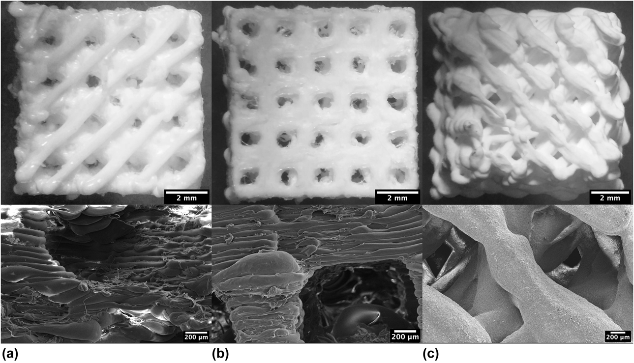
The scaffolds obtained at the end of the printing process for the PLA scaffolds and at the end of the sintering process in the case of alumina scaffolds can be seen in Figs. 1(a)–1(c). Upon completing the printing of PLA scaffolds, it is necessary to manually remove the underlying raft.
Obtained scaffolds at the end of the printing process. The top row shows stereo microscopic images, while the bottom row shows SEM images. (a) Schwarz D-based scaffold fabricated with PLA, (b) rectangular scaffold fabricated with PLA, and (c) Schwarz D-based scaffold fabricated with alumina.

A bumpy surface, typical in 3D printed parts, can be observed in all the scaffolds. Nevertheless, this does not modify the macroscopic structure; it only impacts the microscopic structure (bottom of Fig. 1), which could represent a higher concentration of cells in the space between two contiguous bumps, independently of the material or design but directly related to the printing parameters. However, this was not evident here; conversely, the cells seemed to be indiscriminately distributed into grooves and bumps. Although the actual rectangular scaffold does not have 90° corners as designed, it has sharp corners, vertical flat walls, and holes that go through the scaffold in a single direction [Fig. 1(b)]; the Schwarz D scaffold, on the other hand, exhibits truncated porosity and curved walls [Figs. 1(a) and 1(b)]. The overall dimensions of the designed PLA scaffold were maintained, and the alumina scaffolds were oversized, considering the ceramic paste contraction and anisotropy of the additive manufacturing parts (14.8% shrinkage over the x and y axes and 7.1% over the z axis).
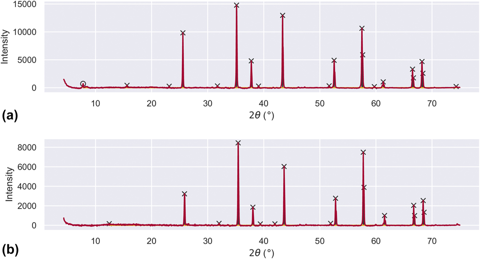
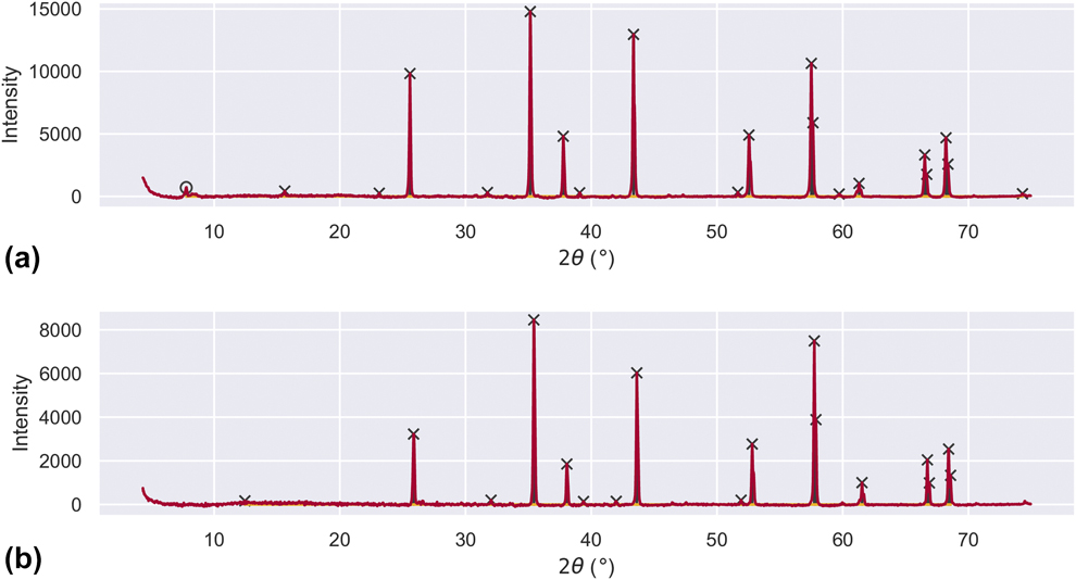
Figure 2(a) shows the X-ray diffractogram of the alumina paste prior to heat treatment. It can be seen that the predominant phase is corundum, with a rhombohedral crystallographic system with 2θ peaks located at 28.5°, 35.1°, 37.8°, 43.4°, and 57.6°. These peaks are identified by an X mark in the figure. A peak at 2θ = 7.8°, corresponding to palygorskite or attapulgite, can also be seen, which is identified by an O mark in the figure. Figure 2(b) shows the X-ray diffractogram after heat treatment at 1480 °C for four hours, with a heating curve designed for alumina sintering. Alumina is still present in the sample in its corundum crystallographic form, and the peak located at 2θ = 7.8°, corresponding to the attapulgite phase, is appreciably diminished. This can be attributed to the fact that the palygorskite phase loses its crystalline structure above 800 °C [Reference Manzanilla20]. This is evidence that the addition of palygorskite as a rheological agent, used as a binder material for paste fabrication, did not modify the desired phase of the alumina once the thermal treatment was performed.
Alumina paste X-ray diffractogram: (a) before thermal treatment and (b) after thermal treatment. The O corresponds to palygorskite and the X corresponds to corundum.
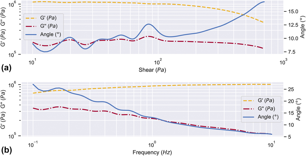
The rheological behavior of alumina in its printable paste form was characterized by performing an amplitude sweep test, as shown in Fig. 3(a). The test was carried out considering the shear rate the paste is subjected to in the printing process. The graph allows observing the elastic modulus, viscous modulus, and phase angle against strain. It was determined that the paste has a viscoelastic linear region until it reaches a strain of 158.5 Pa.
Rheological characterization: (a) alumina paste amplitude sweep and (b) alumina paste frequency sweep.
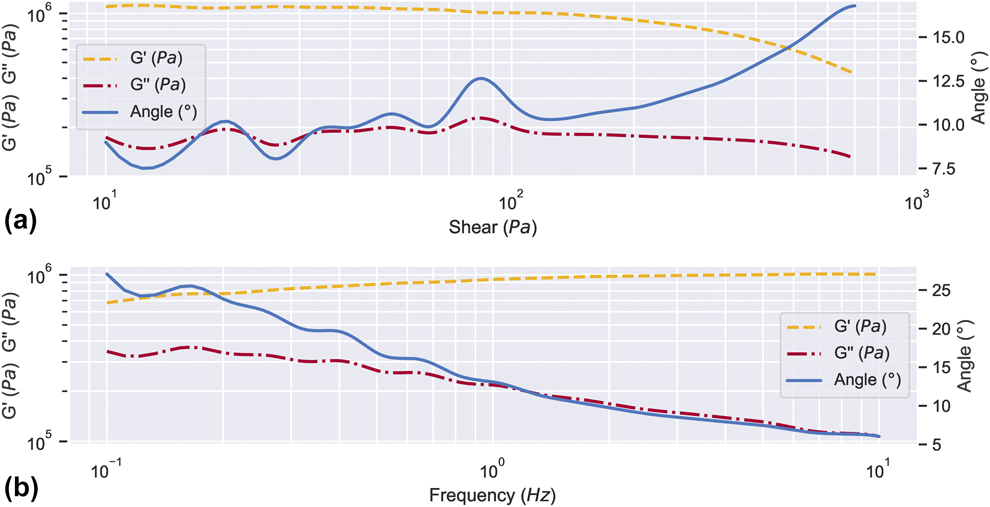
A rheological frequency sweep test, shown in Fig. 3(b), was carried out considering the shear rate of the linear viscoelastic region. The phase angle decreases as the frequency increases, and in addition, it is always lower than 45°, indicating that as the frequency increases, the paste develops a viscoelastic solid behavior. Since the elastic modulus (G′) is greater than the viscous modulus (G″), the behavior of the paste is considered to be that of a solid, aiding the self-support of the piece in green.
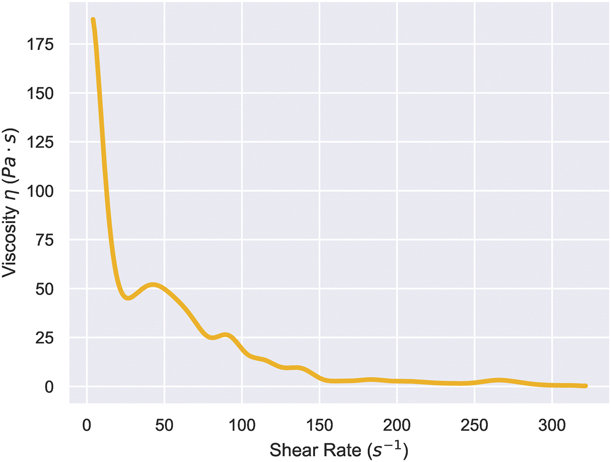
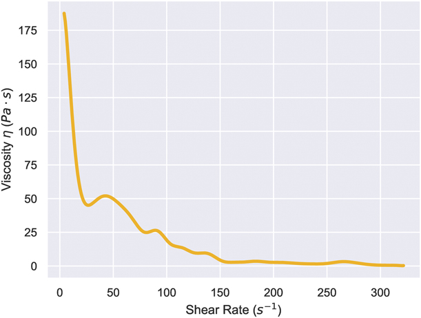
The flow diagram shown in Fig. 4 shows a high viscosity when low shear rates are applied and a rapid decrease in material viscosity as the shear rate increases, confirming that the material exhibits good performance for extrusion processes, allowing it to flow when a force is applied through the printing system but maintaining the shape once that force is removed.
Alumina paste flow diagram.
Cell culturing
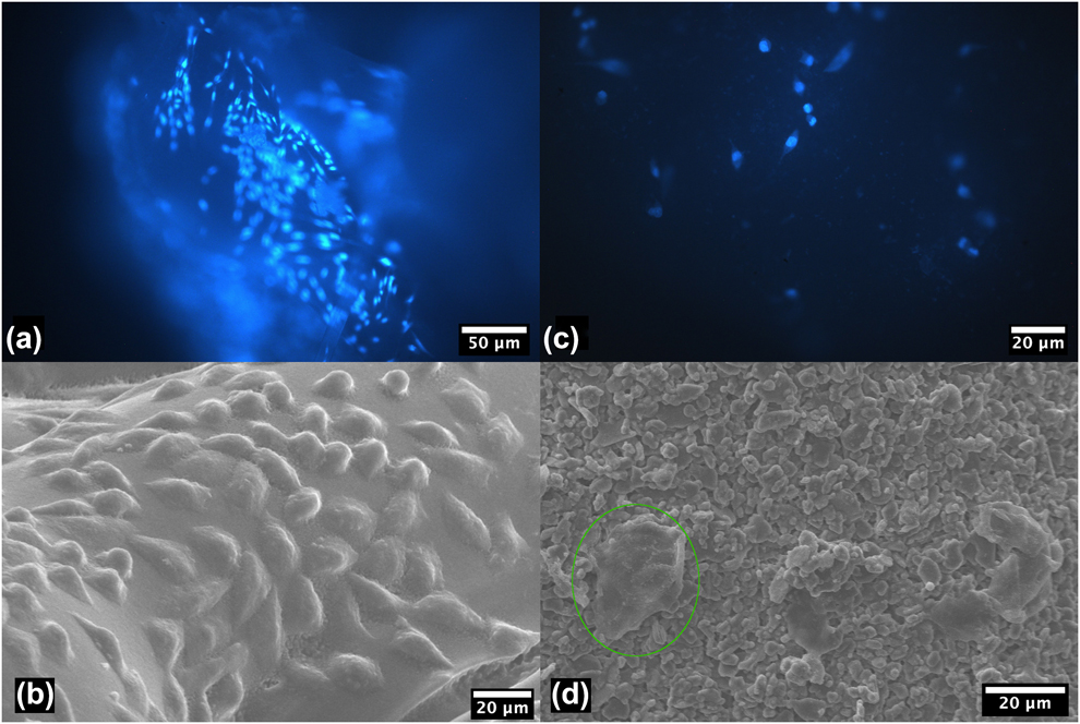
Fluorescence microscopy and scanning electron microscopy (SEM) images of external surface of the PLA scaffolds are shown in Fig. 5, in which the effect of the geometry on cell proliferation was assessed. A higher density of cells can be observed in Figs. 5(a) and 5(b), corresponding to Schwarz D geometry, than in Figs. 5(c) and 5(d), corresponding to the rectangular geometry. In addition, in the latter, the absence of cells in the flat walls is evident all around the scaffold but concentrated in the curved protuberances, as indicated in Fig. 5(d).
PLA scaffolds: (a) Schwarz D-based scaffold fluorescence image, (b) Schwarz D-based scaffold SEM image, (c) rectangular scaffold fluorescence image, and d) rectangular scaffold SEM image.
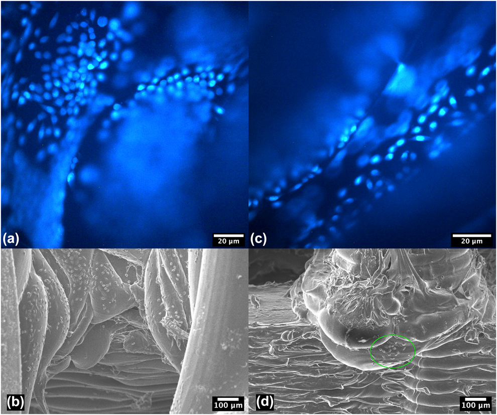
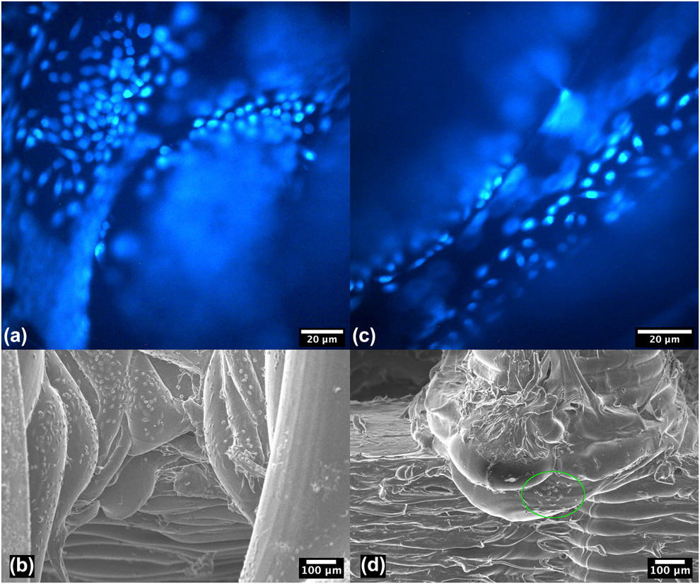
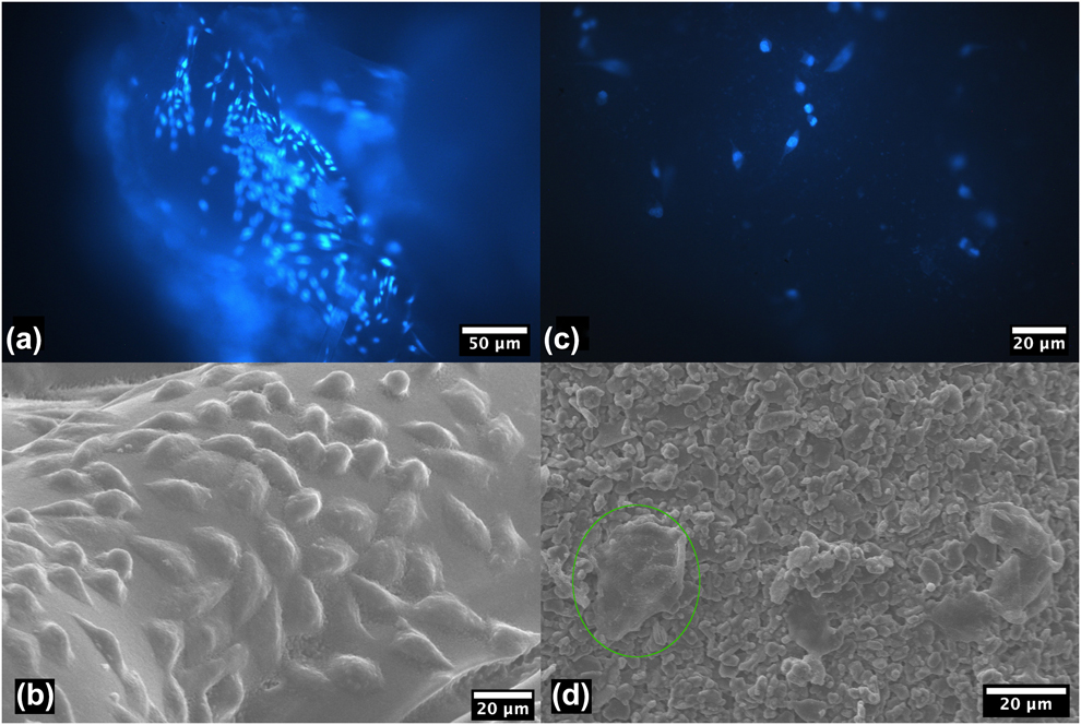
Figure 6 shows the fluorescence and SEM images of scaffolds based on Schwarz D surfaces, in which the effect of the material on cell proliferation was assessed. Comparing Figs. 6(a) and 6(c), a greater density of cells can be observed in the former, which corresponds to PLA scaffolds based on the Schwarz D surface. In Fig. 6(c), corresponding to the alumina scaffold based on the same geometry, a significant decrease in the number of cells can be seen; furthermore, a change in their morphology was observed, as indicated in Fig. 6(d).
Schwarz D-based scaffolds: (a) PLA scaffold fluorescence image, (b) PLA scaffold SEM image, (c) alumina scaffold fluorescence image, and (d) alumina scaffold SEM image.
Quantification
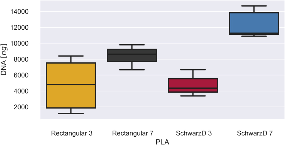
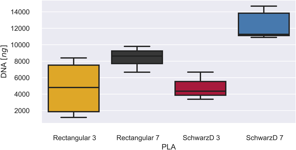
The results of quantification of the DNA present in the PLA scaffolds, measured by NanoDrop spectrophotometry (ThermoFisher Scientific, Waltham, Massachusetts), are shown in Fig. 7. On the left side of this figure, measurements of the scaffolds based on the Schwarz D surface are shown, while on the right side measurements based on rectangular geometry are shown. A statistically significant difference was found, with a P value < 0.05 between the amount of DNA present at 3 and 7 days for both geometries, with the amount of DNA present at 7 days of culturing being greater, confirming that cells thrived and proliferated in both models. In addition, there was a statistically significant difference, with a P value < 0.05, between the amount of DNA in scaffolds based on the Schwarz D surface and that in scaffolds based on rectangular geometry after 7 days of culturing, the amount of DNA being greater in the former. No statistically significant difference between the models was found after 3 days of culturing.
DNA quantification of PLA scaffolds.
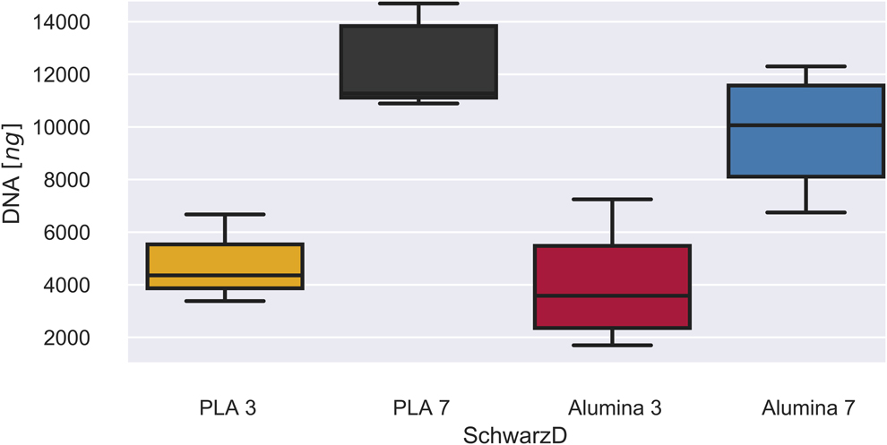
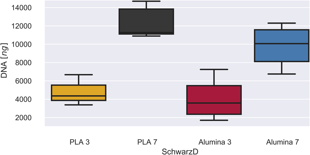
The quantification data of the DNA present in scaffolds based on the Schwarz D surface, measured via spectrophotometry in NanoDrop, are shown in Fig. 8, on the left side those manufactured with PLA and on the right side those manufactured with alumina. In this case, a statistically significant difference was also found in the amount of DNA present at 3 and 7 days of culturing in both cases, with a P value < 0.05, with the amount of DNA being greater after 7 days of culturing. However, in this case, no statistically significant difference was found between the amount of DNA present in PLA and alumina scaffolds after 7 days of culturing. Also, as in the previous case, no statistically significant difference was found in the amount of DNA after 3 days of culturing.
DNA quantification of Schwarz D-based scaffolds.
Conclusions
Scaffolds with the proposed geometries and with a pore size of less than 1000 μm were obtained, in both PLA and alumina. The alumina paste preserves its main crystallographic phase (corundum) and its shape after heat treatment. Rheological tests carried out on the paste demonstrated an ideal behavior for extrusion processes, and it does not lose its shape when deposited. On analyzing the DNA quantification, it was shown that the geometry plays a fundamental role in cell proliferation. Given that there is a statistically significant difference between the amount of DNA present in scaffolds based on the Schwarz D surface and scaffolds based on a rectangular geometry, the amount of DNA present in the former confirms that curved surfaces favor the cell proliferation rate of the CHO-K1 cell line. On the other hand, no statistically significant difference was found between PLA and alumina scaffolds when the geometry used was the Schwarz D surface, which indicates that alumina scaffolds could achieve performance comparable to PLA scaffolds. Although neither of the two materials exhibited evident cytotoxicity, since there was a statistically significant difference between the amount of DNA at 3 and 7 days of culturing, a change in cell morphology on the surface of the alumina scaffolds was observed. This could be due to the irregular surface of the material after heat treatment; however, changes could occur at the metabolic level. It is recommended that tests related to cell metabolism as well as changes in gene expression and genomic stability be performed, in order to guarantee that the cells exhibit disease-free proliferation when cultured on these materials. Future investigations could assess the behavior of mesenchymal stem cells for bone tissue regeneration when differentiating into osteoblasts, using TMPS-based scaffolds, since they have been shown to exhibit better performance of the cell proliferation rate.
Methodology
Scaffold design
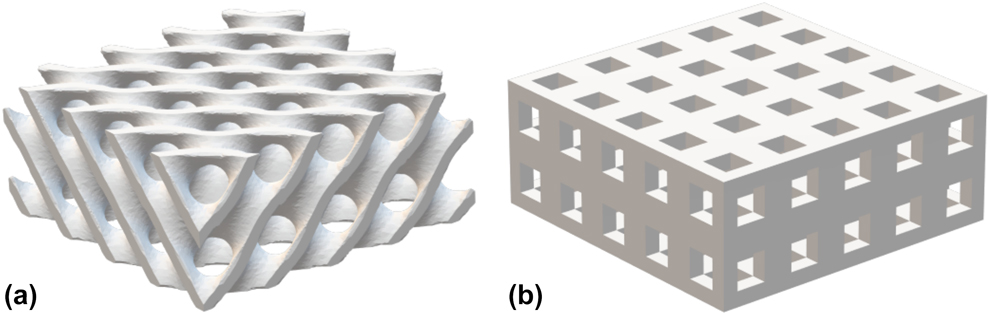
To evaluate the effect of geometry on cell proliferation and adhesion, two different geometric shapes were evaluated: The first consisted of a square-shaped parallelepiped (rectangular scaffold) of 9 mm sides by 3.6 mm height, with through-holes with a square profile of 0.9 mm, as shown in Fig. 9(b). The second belonged to a family of surfaces known as TPMS, characterized by an interconnected porosity and high porosity [Reference Fantini, Curto and De Crescenzio21]. A solid construct of these surfaces can be approximated using an implicit representation through trigonometric combinations of periodic sine and cosine functions [Reference Maskery, Sturm, Aremu, Panesar, Williams, Tuck, Wildman, Ashcroft and Hague11]. The details of the design process can be found in a previous investigation [Reference Restrepo, Ocampo, Ramírez, Paucar and García22]. In order to compare the structures properly, 5 unit cells in the x and y axes and 2 unit cells in the z axis were used, and the overall dimensions were the same for both geometries (9 × 9 × 3.6 mm). For this investigation, the surface named Schwarz D (diamond) was selected [Fig. 9(a)], since it has a high compressive strength without sacrificing its Young’s modulus, as shown previously [Reference Restrepo, Ocampo, Ramírez, Paucar and García22].
Proposed models: (a) Schwarz D and (b) rectangular.

Production of alumina paste
For the fabrication of ceramic scaffolds, a printable alumina paste was developed. Scaffolds based on the Schwarz D surface were constructed with this paste in order to compare their use in three-dimensional cell cultures with their PLA counterparts. Scaffolds made with this paste were oversized, considering a 14.8% shrinkage along the x and y axes and a 7.1% shrinkage along the z axis. The alumina paste had a composition of 95% wt/wt alumina (Nabalox, Nabaltec, Schwandorf, Germany, D50 of 1.96 μm) and 5% wt/wt attapulgite (MIN-U-GEL 400, Active Minerals International (Sparks, Maryland), LLC, D50 of 4.20 μm) as binder material. The dry solids were homogenized and then mixed with 28% wt/wt distilled water in order to adjust their rheological behavior and make them suitable for extruding. The optimal concentrations were determined by trial and error. DRX analysis was performed in order to verify that the initial phase of the alumina, corundum, did not undergo modification when the attapulgite was added.
Fabrication of scaffolds
The scaffolds were constructed using a Wasp 20 × 40 delta 3D printer. Rectangular and Schwarz D surface-based scaffolds were printed with natural commercial PLA, using the printer’s polymer extruder, which has a 0.4-mm nozzle. To generate the G code, which is the set of instructions that the machine follows, Cura 3.3.1 software (Ultimaker, Utrech, The Netherlands) was used, models were imported, and the printing parameters were configured, in this case a layer height of 0.06 mm, a printing speed of 60 mm/s, a printing temperature of 210 °C, and a bed temperature of 60 °C. In order to easily remove the scaffolds from the printing surface, a 10-mm raft was used. This raft must be removed manually at the end of the printing process.
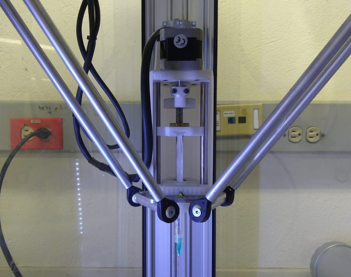
To construct the alumina scaffolds, an advanced ceramics LDM extruder adapted to a Wasp 20 × 40 3D printer, designed and manufactured in the Ceramics and Vitreous Laboratory of Universidad Nacional de Colombia at Medellin, was used (Fig. S1). The material was placed in 5 mL syringes, avoiding bubbles. The syringes were placed in the extrusion head, where the pistons are displaced by a lead screw mechanism, with the aim of extruding the paste through a G23 flat needle with an internal diameter of approximately 0.3 mm. The G code was created with Cura 15.04.2 software (Ultimaker, Utrech, The Netherlands), where a layer height of 0.4 mm and a printing speed of 15 mm/s were set, and the nozzle temperature and bed temperature were disabled in the software (room temperature).
Ceramic pieces were printed on a glass substrate for easy manipulation, following a drying procedure at room temperature for 24 h and then in a Binder drying chamber for another 24 h at a temperature of 100 °C. Finally, they were sintered, following a heating curve (Fig. S2) designed for alumina sintering with a maximum plateau at 1480 °C for four hours in a Sentro Tech Corp SA 1700X furnace.
To characterize the alumina paste, an X-ray diffractogram and rheological tests were performed. The X-ray diffractogram was performed with a copper source, a current of 40 mA, and a voltage of 40 kV, at room temperature and with a continuous scan type. The sweep was done between 0° < 2θ < 80°, with a step of 0.1″. For the rheological characterization, an amplitude sweep between 101 and 103 Pa, frequency sweep between 10-1 and 101 Hz, and flow diagram between 0 and 350 s−1 were performed in a Kinexus Pro+ rotational rheometer with the plate–plate configuration, considering the printing conditions.
Cell culturing
The scaffolds were immersed in absolute alcohol for 12 h and irradiated with UV for 15 min for sterilization. They were then washed with PBS and placed in 24-well culture plates. For carrying out the in vitro tests, cell line CHO-K1 was used under standard culture conditions (5% CO2, 98% RH, and 37 °C). The culture medium used was RPMI1640 (Caisson, Smithfield, Utah) supplemented with 1% L-glutamine, 1% antibiotic, and 5% fetal bovine serum (FBS).
Each scaffold was immersed in 1 mL of supplemented medium, where 100,000 cells were seeded per scaffold, obtaining the deposition of cells on their surface. Two replicates of each experiment were performed, and each scaffold was evaluated in triplicate. The amount of DNA was measured via NanoDrop spectrophotometry after three and seven days of incubation. Each scaffold was discarded once the measurement was performed.
Cell adhesion was verified via fluorescence microscopy (Nikon Eclipse 80i, Nikon, Minato, Tokyo, Japan) and scanning electron microscopy (Zeiss Evo, Zeiss, Oberkochen, Germany), using Hoechst staining dye over the scaffolds’ external surface for nucleus identification. No quantification can be done with these images, due to the difficulty of observing the interior of the scaffolds.
DNA extraction and purification
To determine the final amount of DNA present in the scaffolds, an extraction procedure was performed after three and seven days of culturing, and then it was quantified via spectrophotometry using NanoDrop. Each scaffold was removed from the well, washed with PBS, and placed in a 15-mL centrifuge tube, where a 3-mL volume was filled with PBS. The cells were lysed using ultrasonic probe equipment (Cole-Parmer 500, Cole-Parmer, Vernon Hills, Illinois), for which the equipment’s microprobe was used at 40% power (200 W) at 20 kHz for three seconds. The scaffolds were removed from each tube and washed with two drops of PBS in order to remove any traces of DNA present inside them.
The total volume of each sample was recorded, and 200 μL of each was taken. They were placed in 1.5 mL tubes, an equivalent volume of chloroform was added, and the mixture was gently shaken. The tubes were centrifuged at 10,000 rpm for 5 min at room temperature in order to separate the organic and aqueous phases. The supernatant was transferred to a new tube, and then 200 μL of isopropyl alcohol previously incubated at −20 °C was added and centrifuged at 12,000 rpm for 15 min at 4 °C. The isopropanol was discarded, and the precipitant was washed with 500 μL of 70% ethanol and centrifuged at 12,000 rpm for 5 min at room temperature. The supernatant was discarded, and the pellets were allowed to dry in air at room temperature for 15–20 min. Finally, the DNA was rehydrated with ultra-pure water and stored at −20 °C for further quantification via spectrophotometry [Reference Baena, Valle, José, Moreno, Gómez Alegría, Esther and Camargo23].
Quantification
DNA quantification was performed via spectrophotometry, using a NanoDrop 2000 (Thermo Fisher Scientific, Waltham, Massachusetts). Three measurements of DNA concentration were made per sample, taking 1 µL of the previously stored homogenized cell suspension. From these data, and considering the initial volume, the total amount of DNA was calculated in nanograms.
Statistical analysis
Normality tests were performed for all statistical groups, followed by a 2T test for those that exhibited normal distribution, or Mood’s medians test for the opposite cases, in order to evaluate the statistical differences between the groups of interest. The confidence intervals are presented graphically. Each scaffold was evaluated in triplicate, and two replicates were performed.
Supplementary material
To view supplementary material for this article, please visit https://doi.org/10.1557/jmr.2019.323.
Acknowledgments
We especially appreciate the collaboration of the Colombian company Sumicol for providing the Nabalox alumina and attapulgite for the fabrication of the alumina paste.