Introduction
Thylacocephala is a peculiar group of arthropods within Crustacea (Lange et al., Reference Lange, Hof, Schram and Steeman2001) and even possibly within Eucrustacea (Haug et al., Reference Haug, Briggs, Mikulic, Kluessendorf and Haug2014). In general, they have a pair of triangular or trapezoidal valves that nearly enclose the body inside and often carry remarkable anterior and posterior spines. From the optic notch near the anterior end of the carapace, a pair of compound eyes projects forward, sometimes arising from elongate stalks. In some Mesozoic forms such as Dollocaris, the eyes could be huge with visual hypertrophy (Vannier et al., Reference Vannier, Schoenemann, Gillot, Charbonnier and Clarkson2016). Many species have three pairs of spiny raptorial appendages, either big such as Clausocaris lithographica (Oppenheim, Reference Oppenheim1888) or small such as Thylacares brandonensis Haug et al., Reference Haug, Briggs, Mikulic, Kluessendorf and Haug2014, extending from the carapace ventrally (Haug et al., Reference Haug, Briggs, Mikulic, Kluessendorf and Haug2014). During the past decade, as various new techniques such as X-ray tomography were applied to study this group, previously unknown details of soft parts, such as eye structure, have been revealed with high accuracy (Haug et al., Reference Haug, Briggs, Mikulic, Kluessendorf and Haug2014; Vannier et al., Reference Vannier, Schoenemann, Gillot, Charbonnier and Clarkson2016). However, most of the species in this group are known exclusively from their carapace, which preserves more easily than the rest of the body. As a result, the taxonomy of Thylacocephala is based mainly on the morphology and ornamentation of the carapace.
Thylacocephala has a long stratigraphic record and was especially highly diversified in the Mesozoic. Their fossil record dates back to the Silurian (Haug et al., Reference Haug, Briggs, Mikulic, Kluessendorf and Haug2014) and possibly even earlier to the Cambrian (Vannier et al., Reference Vannier, Chen, Huang, Charbonnier and Wang2006), and extends to as recently as the Late Cretaceous (Dames, Reference Dames1886; Schram et al., Reference Schram, Hof and Steeman1999; Charbonnier et al., Reference Charbonnier, Teruzzi, Audo, Lasseron, Haug and Haug2017). Multiple localities have been reported with records spanning from Europe, Central and East Asia, Madagascar, and Australia (see Schram, Reference Schram2014 and Braig et al., Reference Braig, Haug, Schädel and Haug2019 for a review). Recently, as abundant materials were reported from Japan, South China, Italy, and North America, the Triassic appears to have been the period with the highest taxonomic richness of thylacocephalans (Table 1; Ehiro et al., Reference Ehiro, Sasaki, Kano, Nemoto and Kato2015, Reference Ehiro, Sasaki, Kano and Nagase2019; Ji et al., Reference Ji, Tintori, Jiang and Motani2017; Teruzzi and Muscio, Reference Teruzzi and Muscio2018; Charbonnier et al., Reference Charbonnier and Brayard2019).
List of the Triassic thylacocephalans with their geographic distribution.

Ji et al. (Reference Ji, Tintori, Jiang and Motani2017) reported for the first time the occurrence of thylacocephalans in the Early Triassic of South China. Their locality, the Chaohu area of Anhui Province, has been proved to have one of the best paleontological records of the early recovery of marine vertebrate fauna after the largest biological crisis at ca. 252 Ma, known as the end-Permian mass extinction (EPME) (Jiang et al., Reference Jiang, Motani, Tintori, Rieppel, Chen, Huang, Zhang, Sun and Ji2014, Reference Jiang, Motani, Huang, Tintori, Hu, Rieppel, Fraser, Ji, Kelley, Fu and Zhang2016; Ji et al., Reference Ji, Zhang, Jiang, Bucher, Motani and Tintori2015; Motani et al., Reference Motani, Jiang, Chen, Tintori, Rieppel, Ji and Huang2015a, Reference Motani, Jiang, Tintori, Rieppel, Chen and Youb, Reference Motani, Jiang, Tintori, Rieppel, Chen and Youc; Zhou et al., Reference Zhou, Fu, Zhang, Ni and Ji2015, Reference Zhou, Jiang, Motani, Tintori, Ji, Sun, Ni and Lu2017). Along with vertebrates, a few invertebrates, including ammonoids, bivalves, and coleoids, were also discovered, among which the most common group appears to have been the thylacocephalan crustaceans (Ji et al., Reference Ji, Tintori, Jiang and Motani2017; this paper). During the past decade, bed-by-bed collecting revealed new material of this group with precise occurrence information, forming a sound basis for further study on its biostratigraphy and biochronology (Motani et al., Reference Motani, Jiang, Tintori, Ji and Huang2017). The thylacocephalans from different beds of the Majiashan Quarry vary remarkably in taxonomic composition, as inferred from both the size and ornamentation of carapaces. The purpose of this paper is to report a hitherto unrecognized thylacocephalan assemblage from a stratigraphic horizon that differs from what was reported by Ji et al. (Reference Ji, Tintori, Jiang and Motani2017)—Bed 605 of the Majiashan Quarry—and to discuss its implications on the taxonomy and paleoecology of Microcarididae.
Geological setting
Materials for this study were collected from the Majiashan Quarry located to the east of Chaohu Lake (Anhui Province, South China Block) (Fig. 1). Here, a nearly complete sedimentary succession is exposed, ranging from the upper part of the Yinkeng Formation (Early Triassic) to the Dongmaanshan Formation (early Middle Triassic). For the Early Triassic, three formations can be recognized in ascending order: the Yinkeng, Helongshan, and Nanlinghu formations. The upper Yinkeng Formation consists mainly of thin-bedded, yellowish-green to dark-brown mudstone with intercalated calcareous mudstone and micro-nodular limestone. The Helongshan Formation is marked by two thick limestone layers at the bottom and is composed largely of rhythmic alternations of thin-bedded dark-gray mudstone and limestone. This unit is only ~17 m thick and well exposed in this area, facilitating the stratigraphic correlation of outcrops. Near the top of the formation are a few layers of fossiliferous limestone nodules containing three-dimensionally preserved fish, bivalves, and coprolites (Zhou et al., Reference Zhou, Fu, Zhang, Ni and Ji2015). Similar fossiliferous nodules have been recorded in many Early Triassic localities around the world (e.g., Jurong of Jiangsu Province, Madagascar, Spitsbergen, and Greenland), and they may be approximately coeval (Tintori et al., Reference Tintori, Hitij, Jiang, Lombardo and Sun2014a). However, the horizon at the top of the Helongshan Formation is probably the youngest among all other pre-Spathian ones (Tintori et al., Reference Tintori, Hitij, Jiang, Lombardo and Sun2014a), being located near the Smithian/Spathian boundary. On the bedding surface instead of inside nodules, fishes, shrimps, bivalves, as well as rare thylacocephalans and ammonoids were recorded during our fieldwork. The Nanlinghu Formation is ~160 m thick, and its three members are recognized on the basis of lithology. The lower member is 48.6 m thick and consists mainly of yellow to gray, thin- to medium-bedded mudstone and thin- to thick-bedded limestone intercalated with micro-nodular limestone. The mid-member is 46.8 m thick and consists mainly of gray, medium- to thick-bedded micro-nodular limestone and calcareous mudstone. The rocks of the lower part may appear reddish in color upon weathering. Many layers are rich in bioclusters and bioturbation, containing abundant bivalves, ammonoids, and thylacocephalans. The upper member is 64.5 m thick and comprises gray, thin- to medium-bedded limestone intercalated with yellowish calcareous marls. In this member, abundant fossils, including ammonoids, bivalves, thylacocephalans, fishes, and marine reptiles, have been recorded (Chen, Reference Chen1985; Tong et al., Reference Tong, Zakharov and Wu2004; Zhao et al., Reference Zhao, Orchard, Tong, Sun, Zuo, Zhang and Yun2007; Sun et al., Reference Sun, Tintori, Jiang and Motani2013; Jiang et al., Reference Jiang, Motani, Tintori, Rieppel, Chen, Huang, Zhang, Sun and Ji2014; Motani et al., Reference Motani, Jiang, Tintori, Rieppel and Chen2014, Reference Motani, Jiang, Chen, Tintori, Rieppel, Ji and Huang2015a, Reference Motani, Jiang, Tintori, Rieppel, Chen and Youb, Reference Motani, Jiang, Tintori, Rieppel, Chen and Youc; Tintori et al., Reference Tintori, Huang, Jiang, Sun, Motani and Chen2014b; Ji et al., Reference Ji, Zhang, Jiang, Bucher, Motani and Tintori2015). Fossil preservation varies depending on the lithotypes in the Nanlinghu Formation. In general, the fossils are rare in lime-rich beds and more abundant in marly-mudstone layers, where preservation is also usually better than in limestones. Facies analysis suggests that the Majiashan Quarry was deposited on a deepwater carbonate slope during the end-Smithian to Spathian, exhibiting a strong meter-scale sedimentary cyclicity (Fu et al., Reference Fu, Jiang, Montanẽz, Meyer, Motani and Tintori2016).
(1) Map of the section location (red stars). (2) Occurrence of the newly described thylacocephalan material from the Majiashan section (red dot).

Materials and methods
The studied specimens were collected during intensive fieldwork in the quarries of Majiashan from 2010 to 2016. In May 2010, some specimens were collected from a single thin horizon at the southern rim of the quarry before a stratigraphic investigation. This horizon has since been correlated to Bed 605 on the basis of a thorough stratigraphic study (Fig. 1). The majority of the specimens were collected from the bottom of the lower level in a new quarry in May 2013. Unfortunately, this outcrop has subsequently been covered by soil and is unavailable for further research. The bed yielding these materials is again correlated to Bed 605 (Fig. 1). The age of the bed is ca. 248.8 Ma on the basis of astronomical cycles of carbon cycling (Fu et al., Reference Fu, Jiang, Montanẽz, Meyer, Motani and Tintori2016). The bedding planes in the region are vertical and mudstones are heavily weathered, making it difficult to retrieve the fossils completely. Nevertheless, sufficient numbers of thylacocephalan, coleoid, and possible malacostracan remains have been recorded to warrant a paleontological investigation.
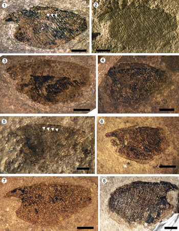
Observations were made using a binocular microscope Olympus SZX10 with photographic device and Ultra-depth three-dimensional screen visual microscopy system KEYENCE VHX-6000. To enhance the visibility, the specimens were soaked in ethylic alcohol under a microscope (Figs. 2–4).
Microcaris rectilineatus n. sp. from Bed 605 of the Nanlinghu Formation, Chaohu, South China. (1, 2) Holotype NIGPAS 171265: (1) taken under alcohol (arrows point to a series of tubercles/short ridges as lateral carina); (2) taken under normal light. (3) Paratype NIGPAS 171266. (4) Paratype NIGPAS 171268. (5) Paratype NIGPAS 171267 (arrows point to a series of tubercles/short ridges as lateral carina). (6) Paratype NIGPAS 171269. (7) Paratype NIGPAS 171270. (8) Paratype NIGPAS 171271. Scale bars = 2 mm.

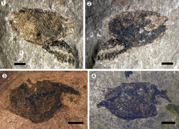
Microcaris rectilineatus n. sp. from Bed 605 of the Nanling Formation, Chaohu, South China: (1, 2) NIGPAS 171272, with posterior appendages; (3) NIGPAS 171273; (4) NIGPAS 171274. Scale bars = 2 mm.

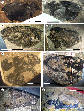
Four kinds of thylacocephalans with large carapaces. (1, 2) NIGPAS 171275, holotype of Diplacanthocaris chaohuensis n. gen. n. sp. with a large, smooth carapace, very short anterior and posterior spines, and small optic notch. (3) NIGPAS 171276, Ankitokazocaris sp. with a large, nearly triangular and smooth carapace and a large optic notch; similar to A. chaohuensis from Bed 680. (4) NIGPAS 172397, Ankitokazocaris sp. (5, 6) NIGPAS 171277, Microcaris sp. (7) NIGPAS 171278, Microcaris sp. with a large, nearly triangular-shaped carapace, a large optic notch, and long and nearly straight striations extending from the dorsal margin and slightly curved toward the ventral margin. The carapace length could be over 50 mm on the basis of (7). (8) NIGPAS 171279, Kitakamicaris sp. from Bed 680. Scale bars = 10 mm.

To compare the shape of thylacocephalan carapaces, a total of 32 species, including one new genus and two new species (compared with Braig et al., Reference Braig, Haug, Schädel and Haug2019) were selected for the outline-based geometric morphometric analysis (see Appendix for details). All taxa selected were based on a complete carapace in lateral view, respectively. As most species were based on a limited number of specimens and the sample size often did not warrant a quantitative analysis within each species, we here chose one specimen of each species, mostly the holotype unless it was not available or was incomplete. A total of 200 points were placed along the outline of each specimen's entire body using tpsDig version 2.32 (Rohlf, Reference Rohlf2020). After outlining, each tps curve was converted into landmark points with XY coordinates using tpsUtil and used as the raw data for subsequent analyses. The thin-plate splines (TPS) procedure was used to analyze and display shape variations among different species. A generalized Procrustes analysis (GPA) was then performed to superimpose landmark configurations using least-square estimates for translation and rotation parameters (Adams et al., Reference Adams, Rohlf and Slice2004), eliminating any variation due to differences in translation, orientation, and size. Elliptical Fourier shapes (EFS) using the algorithm in Paleontological Statistics (PAST) software version 4.03 (Hammer et al., Reference Hammer, Harper and Ryan2001) was then used as a standard method for carrying out outline analysis (Ferson et al., Reference Ferson, Rohlf and Koehn1985). Data after GPA were used for a principal component analysis (PCA) to explore patterns of variation in the shape of the thylacocephalans.
Repository and institutional abbreviation
Specimens in this study are deposited in the Nanjing Institute of Geology and Palaeontology, Chinese Academy of Sciences (NIGPAS).
Systematic paleontology
Thylacocepha Pinna et al., Reference Pinna, Arduini, Pesarini and Teruzzi1982
Concavicarida Schram, Reference Schram2014
Microcarididae Schram, Reference Schram2014
Included genera
Microcaris Pinna, Reference Pinna1974, Ferrecaris Calzada and Mañé, Reference Calzada and Mañé1993, Kitakamicaris Ehiro and Kato in Ehiro et al., Reference Ehiro, Sasaki, Kano, Nemoto and Kato2015, and Miyagicaris Ehiro et al., Reference Ehiro, Sasaki, Kano and Nagase2019.
Diagnosis
Small to medium size, with an anteriorly pointed rostrum and vertical parallel ridges or terraces on the whole carapace. Incomplete intercalated ridges from both dorsal and ventral margins. Carapace outline trapezoidal with short truncate posterior edge.
Remarks (amended after Schram, Reference Schram2014)
This family was erected by Schram (Reference Schram2014) for Microcaris Pinna, Reference Pinna1974, Atropicaris Arduini and Brasca, Reference Arduini and Brasca1984, Ferrecaris Calzada and Mañé, Reference Calzada and Mañé1993, and Thylacocephalus Lange et al., Reference Lange, Hof, Schram and Steeman2001. The diagnostic characters he defined for this family include small to modest size when compared with the largest species, Ostenocaris cypriformis (Arduini et al., Reference Arduini, Pinna and Teruzzi1980), thin, pointed, and anteriorly directed rostrum, and carapace surface with rugations or terraces. Kitakamicaris has recently been added to the family. Charbonnier et al. (Reference Charbonnier, Teruzzi, Audo, Lasseron, Haug and Haug2017, p. 5) pointed out the feeble bases of Schram's subdivision but kept using it without redefining this family. Moreover, Charbonnier et al. (Reference Charbonnier, Teruzzi, Audo, Lasseron, Haug and Haug2017) made the situation more confusing at least for a few genera/families. Regarding Microcarididae, Charbonnier et al. (Reference Charbonnier, Teruzzi, Audo, Lasseron, Haug and Haug2017, p. 14) assigned Keelicaris, Microcaris, Atropicaris, Rugocaris, Ferrecaris, Thylacocephalus, and Kitakamicaris to the family only on the basis of the presence of ornamentation. However, Charbonnier et al. (Reference Charbonnier, Teruzzi, Audo, Lasseron, Haug and Haug2017, p. 12) assigned Thylacocephalus to Dollocarididae while Charbonnier et al. (Reference Charbonnier, Teruzzi, Audo, Lasseron, Haug and Haug2017, table 1) again assigned it to Microcarididae while Keelicaris was assigned to Protozoeidae. Charbonnier et al. (Reference Charbonnier and Brayard2019) removed Keelicaris and Rugocaris from Microcarididae and included only Atropicaris, Ferrecaris, Microcaris, Kitakamicaris, and Parisicaris. Schram (Reference Schram2014) already pointed out that Thylacocephalus was assigned to Microcaridida only on the basis of a similar, but not identical, ornamentation while its body outline is very different from other Triassic genera, especially in the presence of a long posterior spine rather than the anterior rostrum. Following Charbonnier et al. (Reference Charbonnier, Teruzzi, Audo, Lasseron, Haug and Haug2017, p. 12), we exclude Thylacocephalus from this family because of its unique ornamentation (the presence of posterior and ocular spines and several vertical rows of pits) and carapace outline. In addition, we follow Charbonnier et al. (Reference Charbonnier, Teruzzi, Audo, Lasseron, Haug and Haug2017, table 1) in considering Keelicaris as a Protozoeidae instead of a Microcarididaea (as in Charbonnier et al., Reference Charbonnier, Teruzzi, Audo, Lasseron, Haug and Haug2017, p. 14). The ornamentation and shape of the carapace of Rugocaris (Tintori et al., Reference Tintori, Bigi, Crugnola and Danini1986) is totally different from those of Microcaris, so we leave it as incerte sedis for the family.
Tintori et al. (Reference Tintori, Bigi, Crugnola and Danini1986) suggested that Microcaris and Atropicaris could be considered synonyms. Arduini (Reference Arduini1988) rejected this hypothesis along with the fact that thylacocephalans have a bivalved carapace. Pinna (Reference Pinna1974, p. 30–31) clearly stated that Microcaris had two valves, and this has later been confirmed in many other species (Schram, Reference Schram2014). Regarding Microcaris and Atropicaris, some morphological differences considered by Arduini (Reference Arduini1988) actually resulted from preservation, as the type species of the two genera were from different localities and lithostratigraphic units: Microcaris from limestone in Val Seriana of Bergamo and Atropicaris from mudstone in Valle Imagna of Bergamo. Moreover, because the preservation was poor, microcarids from Norian rocks of northeast Italy (Dalla Vecchia and Muscio, Reference Dalla Vecchia and Muscio1990) and central Italy (Dalla Vecchia, Reference Dalla Vecchia1993) were difficult to separate and were assigned mostly to Microcaris. Thus, given the remarkable intraspecific variability on the body outline of thylacocephalan species (Arduini, Reference Arduini1988; Ehiro et al., Reference Ehiro, Sasaki, Kano, Nemoto and Kato2015; Ji et al., Reference Ji, Tintori, Jiang and Motani2017), we consider Atropicaris a subjective junior synonym of Microcaris. Very recently, Teruzzi and Muscio (Reference Teruzzi and Muscio2018) confirmed the synonymy between the two genera. In addition, the ages of Microcaris minuta Pinna, Reference Pinna1974 and Atropicaris rostrata Arduini and Brasca, Reference Arduini and Brasca1984 type materials are very close, both around the Alaunic/Sevatian (middle/late Norian, Rigo et al., Reference Rigo, Galli and Jadoul2009) boundary, and only about 50 stratigraphical meters apart at maximum. Thus, pending a detailed revision of all available Norian material, we could have only a single genus, and probably just a single species (namely Microcaris minuta) of microcarid in the Italian Norian units.
Glaessner (Reference Glaessner1931) described a crustacean assemblage from the lower Carnian of Lunz (Austria), erected the species Austriocaris striata Glaessner, Reference Glaessner1931, and ascribed it to Phyllocarida. Pinna (Reference Pinna1974) noted similarities between A. striata and his new material of Microcaris minuta, which was considered to be Phyllocarida too. Arduini and Brasca (Reference Arduini and Brasca1984) assigned A. striata to their new genus Atropicaris, which was followed by Schram (Reference Schram2014). As we consider Atropicaris a synonym of Microcaris, A. striata is also assigned to Microcaris herein, thus extending the duration of this genus to the early Carnian. Thylacocephalans from the late Carnian have recently been recorded from mass mortality surfaces from Slovenia (Hitij et al., Reference Hitij, Gašparič, Žalohar, Jurkovšek and Kolar-Jurkovšek2019, figs. 34, 35), clearly showing characters close to Microcaris.
Microcaris were reported from the late Anisian (Middle Triassic) of the Monte San Giorgio area (Affer and Teruzzi, Reference Affer and Teruzzi1999) and Carnic Alps (Teruzzi and Muscio, Reference Teruzzi and Muscio2018). In both cases, the preservation is quite poor, and Affer and Teruzzi (Reference Affer and Teruzzi1999) inferred the fragmentation of the specimens to be from predation because thylacocephalan remains were often discovered inside coprolites. A few specimens described as Microcaris minuta were reported from probably the middle Anisian of Slovenia (Križnar and Hitij, Reference Križnar and Hitij2010).
Kitakamicaris is clearly very close to Microcaris in its body outline and ornamentation. The size of K. utatsuensis Ehiro and Kato in Ehiro et al., Reference Ehiro, Sasaki, Kano, Nemoto and Kato2015 is small among thylacocephalans and falls well within a possible intrageneric variability in Microcaris (Vannier et al., Reference Vannier, Chen, Huang, Charbonnier and Wang2006). Microcaris rostrata (Arduini and Brasca, Reference Arduini and Brasca1984) n. comb. ranges from 18 to 27 mm long (Arduini and Brasca, Reference Arduini and Brasca1984) while M. minuta could reach up to 28 mm (Pinna, Reference Pinna1976); thus, there are limited differences between this size range and that from the published material of Kitakamicaris, from 17 to 35 mm (Ehiro et al., Reference Ehiro, Sasaki, Kano, Nemoto and Kato2015). One distinctive difference between the two genera is in the rostrum. Ehiro et al. (Reference Ehiro, Sasaki, Kano, Nemoto and Kato2015) described a central and two lateral carinae in Kitakamicaris. However, these carinae are just parts of narrow keels (marginal rim in Vannier et al., Reference Vannier, Chen, Huang, Charbonnier and Wang2006) that underline the whole valve margin. Taphonomic processes may be responsible for enough displacement to superimpose the two valves on each other, thus making one of the rims appear to be a lateral carina. Another difference is in the number of ridges on the carapace. In K. utatsuensis there are 45–55 ridges, being similar to M. rostrata, while the type species M. minuta has about 40 ridges. Consequently, in Microcaris the ridges are mostly parallel and unbranched, and some appear intercalated and incomplete in the center. Ehiro et al. (Reference Ehiro, Sasaki, Kano, Nemoto and Kato2015) described the presence of sharp low ridges in between the central vertical ridges and named this structure the dorsal carina (‘dc’ in their fig. 8.6a, b) in the dorsolateral region of a few specimens but did not label it in their figure 8.5. However, what is labelled as ‘dc’ in their figure 8.1 is simply the dorsal rim of one of the superimposed valves. Thus, it appears that Ehiro et al. (Reference Ehiro, Sasaki, Kano, Nemoto and Kato2015) named two different structures in the same way, as is clear in their figure 4. In some unpublished specimens from the Norian of Italy, a similar structure, a sparse series of small tubercles, is present (A. Tintori, personal observation). However, it is not always present in specimens of Microcaris, as in Kitakamicaris (Ehiro et al., Reference Ehiro, Sasaki, Kano, Nemoto and Kato2015). As this ‘lateral carina’ (we prefer to use the terminology of Charbonnier et al., Reference Charbonnier and Brayard2019 instead of ‘dorsal carina’ of Ehiro et al., Reference Ehiro, Sasaki, Kano, Nemoto and Kato2015) is not always present in either species, its taxonomic implication is not yet clear. The structure may be related to sexual dimorphism or sexual maturity, but further studies are necessary to test these interpretations.
More recently, Parisicaris triassica Charbonnier in Charbonnier et al., Reference Charbonnier and Brayard2019 was erected from the early Spathian of North America (Charbonnier et al., Reference Charbonnier and Brayard2019). Preservation of these specimens is mainly fragmentary, and only the holotype appears ‘complete.’ The most diagnostic feature of this species is a large ventral notch. However, as the marginal rim of the ventral notch is absent, the observed notch could have resulted from incomplete preservation because there is no trace of this structure in the paratype. Differences from other species of Microcaris are limited and related mostly to the presence of a presumed ventral notch ‘shown’ in the holotype and the larger size (40 mm) of the holotype. However, this material shows a similar, but shorter, ‘carina’ in the dorsolateral region of the valves (‘lateral carina’ in Charbonnier et al., Reference Charbonnier and Brayard2019). As in Kitakamicaris, this ‘carina’ is not a continuous ridge but is made of short ridges between the central vertical ridges. Therefore, there is insufficient reason to sustain a new genus, and here we consider Parisicaris triassica as belonging to Microcaris, being close to Kitakamicaris utatsuensis.
Genus Microcaris Pinna, Reference Pinna1974
Type species
Microcaris minuta Pinna, Reference Pinna1974.
Other species
Microcaris rostrata (Arduini and Brasca, Reference Arduini and Brasca1984) n. comb., Microcaris striata (Glaessner, Reference Glaessner1931) n. comb., Microcaris triassica (Charbonnier in Charbonnier et al., Reference Charbonnier and Brayard2019) n. comb., Microcaris rectilineatus n. sp.
Diagnosis
Microcarids with relatively small carapaces, less than 40 mm long, and with a narrowly pointed rostrum and well-developed vertical (transverse) ornamentation. Some ribs not completely developed, just in dorsal or ventral regions of the carapace, and irregularly intercalated to complete ones. Almost no ridge branching. Optical edge almost straight or nearly round, with large and unstalked compound eyes (when preserved). Posterior edge straight to gently concave. A series of tubercles/short ridges as lateral carina in some specimens.
Occurrence
Microcaris covers almost the complete stratigraphic range of the Triassic: M. triassica and M. rectilineatus n. sp. from the Spathian (Olenekian, late Early Triassic), possible Microcaris minuta from the middle Anisian of Slovenia, Microcaris sp. from the late Anisian (Middle Triassic), and M. minuta and M. rostrata from the middle/late Norian (Late Triassic) (Pinna, Reference Pinna1974; Arduini, Reference Arduini1988; Affer and Teruzzi, Reference Affer and Teruzzi1999; Križnar and Hitij, Reference Križnar and Hitij2010; Teruzzi and Muscio, Reference Teruzzi and Muscio2018; Charbonnier et al., Reference Charbonnier and Brayard2019). So far, Late Triassic species have been found in several localities in central and northern Italy and in Austria, while Early Triassic species are found in North America (M. triassica) and central eastern China (M. rectilineatus). Specimens from the Middle Triassic are found in different localities from the Southern Calcareous Alps in northern Italy. Thus, the genus appears to span the complete Tethys and is also distributed along the western coast of Panthalassa.
Remarks
Neither Pinna (Reference Pinna1974) nor Arduini and Brasca (Reference Arduini and Brasca1984) gave a diagnosis for their new genera Microcaris and Atropicaris, but only separately described the holotype and paratypes. Schram (Reference Schram2014) did not specify diagnostic characters for each genus either when erecting the new family Microcarididae. One of the co-authors (A. Tintori) personally checked the type series of the two species and the unreported Norian specimens in the Bergamo Museum Natural History, Paleontological Museum of the Milano University, Milano Museum Natural History, and Friuli Museum of Natural History in Udine collections. Thus, owing to their ornamentation and body outline, we consider the majority of specimens collected from Bed 605 from the new Majiashan Quarry as belonging to Microcaris.
Type material
Holotype, NIGPAS 171265. Paratypes, NIGPAS 171266, 171267, 171268, 171269, 171270, 171271, 171272, 171273, 171274.
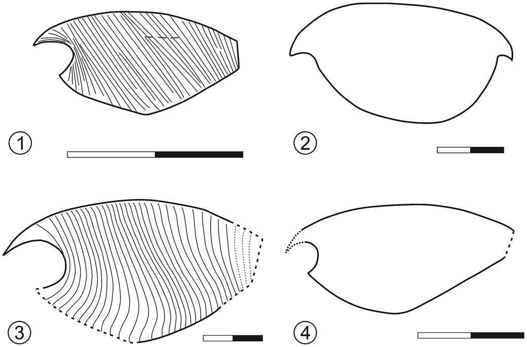
Sketch reconstructions of the carapaces of four species from Bed 605: (1) Microcaris rectilineatus n. sp.; (2) Diplacanthocaris chaohuensis n. gen. n. sp.; (3) Microcaris sp.; (4) Ankitokazocaris sp. Scale bars = 10 mm.

Diagnosis
Carapace small, round, and triangular shaped, length nearly twice that of depth. Ventral margin angulate in the middle, and dorsal margin evenly convex. Anterior spine present and rather long. Optic notch round and nearly symmetrical. Posterior margin vertical and straight. Carapace covered with 30–35 well-developed long and nearly straight ribs, running anterodorsally to posteroventrally.
Occurrence
Most of the referred specimens came from Bed 605 of the upper Majiashan Quarry in Chaohu District, Anhui Province, PR China; the rest were not collected directly from the bed but from fallen slabs around it. The age is Spathian (Olenekian, Early Triassic).
Description
The carapace is trapezoidal or round triangular shaped. The anteroposterior length of the carapace is within 10–13 mm. The dorsal margin is slightly and evenly convex, and the ventral margin is angulate in the middle, forming an angle of 130–135°. The anterior optic notch is semicircular and nearly symmetrical and forms an acute or nearly right angle with the ventral margin. The posterior margin is short and straight, forming a 120° angle with the dorsal and ventral margins, respectively. The anterior spine is not remarkably long and points anteroventrally (Fig. 3.3). The carapace length is about twice the carapace depth. The entire carapace is developed with thin and densely spaced ribs running from the dorsal to the ventral margin. The number of ribs cannot be accurately counted but is mostly around 30 and up to 35. The ribs are nearly straight and parallel, being almost perpendicular to the posterior part of the ventral margin. Secondary ribs are developed occasionally, being intercalated between the primary ribs from both the dorsal and ventral margins. Near the anteroventral margin, the ribs are curved anteriorly. The posterior appendages are short and have been pushed outside the carapace (Fig. 3.1, 3.2). However, it is impossible to count the exact number of appendages due to the preservation.
Etymology
From Latin rectus (straight) and lineatus (with lines), in reference to the straight lines (ridges) on the carapace.
Remarks
M. rectilineatus n. sp. is different from M. minuta in the anteroventral marginal process and rib orientation. The ribs are nearly parallel and strongly inclined posteriorly in M. rectilineatus while they are nearly vertical and slightly and evenly curved posteriorly near the dorsal margin in M. minuta. M. rectilineatus is different from M. rostrata in its smaller size, deep optic notch, longer anterior spine, and strongly inclined and straight ribbing. It is distinct from M. striata on the basis of the posterior margin and anterior optic notch. It is different from M. triassica due to its smaller size and straighter, parallel ribbing.
Microcaris sp.
Figures 4.5–4.7, 5.3
Description
Two specimens (NIGPAS 171277, 171278) with ribs were found along with Microcaris rectilineatus n. sp. from Bed 605. They are estimated to have a carapace length of 50 mm and 40 mm, respectively, which is much bigger than M. rectilineatus. The number of ribs is estimated to be around 30. The ribs are mostly parallel and straight, without bifurcation, and slightly curve toward the ventral margin, which is different from M. rectilineatus. The anterior notch is partly preserved in NIGPAS 171277 (Fig. 4.5, 4.6) and appears semicircular. The ventral margin is angulate in the middle, forming an angle of ~140°.
Materials
NIGPAS 171277, 171278.
Remarks
The two specimens resemble M. rectilineatus in the number and morphology of ribs on the carapace. The ribs appear less dense than those of M. rectilineatus mostly because of a widened space between the adjacent ribs, which in turn is correlated with the increase in carapace size. The subdivision of our specimens in two well-separated size groups (Figs. 2, 3 versus Fig. 4.5–4.7) raises a question about their taxonomic placement. Microcaridid species show a wide size range (see the preceding) but are usually within a continuous size variation when a number of specimens are collected (Pinna, Reference Pinna1974, Reference Pinna1976; Dalla Vecchia and Muscio, Reference Dalla Vecchia and Muscio1990; Dalla Vecchia, Reference Dalla Vecchia1993; Ehiro et al., Reference Ehiro, Sasaki, Kano, Nemoto and Kato2015, fig. 7). However, in all previous cases, the specimens did not come from a single layer, but mostly from a single fossiliferous site with no further stratigraphic indications (Ehiro et al., Reference Ehiro, Sasaki, Kano, Nemoto and Kato2015) or from different localities of a wide geographical and stratigraphic range (Pinna, Reference Pinna1974, Reference Pinna1976; Tintori et al., Reference Tintori, Bigi, Crugnola and Danini1986; Dalla Vecchia and Muscio, Reference Dalla Vecchia and Muscio1990; Dalla Vecchia, Reference Dalla Vecchia1993), so that the size variability could be related to differences in paleoenvironment and age or preservational and collection biases. As we collected our material from a rock layer just a few centimeters thick, it is reasonable to assume that they belong to the same group, as both large and small individuals were part of the same biocoenosis. It is possible that they were buried together due to unknown taphonomic processes because most of the small individuals are complete while the large individuals are mostly fragments. However, since the two specimens are not complete and are still missing many diagnostic features, we assign them as Microcaris sp. in this study before more material is discovered.
Genus Kitakamicaris Ehiro and Kato in Ehiro et al., Reference Ehiro, Sasaki, Kano, Nemoto and Kato2015
Type species
Kitakamicaris utatsuensis Ehiro and Kato in Ehiro et al., Reference Ehiro, Sasaki, Kano, Nemoto and Kato2015 (IGPS collection catalog number 111448) from the middle part of the upper Olenekian Osawa Formation to the north of Cape Tatezaki, Utatsu, Minamisanriku Town, Miyagi Prefecture, Japan, by original designation.
Kitakamicaris sp.
Figure 4.8
Description
One specimen from Bed 680 is found to have a trapezoidal carapace with vertical ribs. The left carapace is exposed and mostly complete, missing the anterior spine and the ventral and posterior margins. The length of the carapace is estimated to be 35–40 mm. The anterior optic notch, similar to that of Ankitokazocaris, is clearly present. The angle formed by the anterior and ventral margin is 75°. The entire carapace bears vertical and nearly parallel ribs. The number of primary ribs is close to 50, but the precise number is not available because the posterior margin is incomplete. The ribs are mostly curved anteriorly near the dorsal margin and slightly anteriorly near the ventral margin, therefore forming a sigmoidal shape. At the anteroventral part of the carapace, the ribs are curved remarkably and nearly parallel to the anterior margin of the carapace. Some ribs start from the dorsal margin and stop before reaching the medial line of the carapace, and some ribs branch off, especially at the anteroventral part of the carapace.
Materials
NIGPAS 171279.
Remarks
The ribs of this specimen strongly resemble those of Kitakamicaris described by Ehiro et al. (Reference Ehiro, Sasaki, Kano, Nemoto and Kato2015). Since the anterior spine and ventral margin are missing, we leave the taxonomic assignment as Kitakamicaris sp. before more material is found. It can be easily distinguished from the Microcaris rectilineatus n. sp. of this same assemblage due to its larger size, nonparallel rib orientation, and larger number of ribs.
Order and Family uncertain
Genus Diplacanthocaris new genus
Type species
Diplacanthocaris chaohuensis n. gen. n. sp.
Diagnosis
As for the type species by monotypy.
Occurrence
Bed 605 of Nanlinghu Formation (Olenekian, Lower Triassic) at the upper Majiashan Quarry (Chaohu, Anui Province, PR China).
Etymology
From old Greek, diplacanthus, referring to the two spines (anterior and posterior) on the carapace, and caris, a general name for crustaceans.
Remarks
This genus has a unique sinuous carapace shape with short anterior and posterior spines. The general shape of the carapace is largely different from the majority of other groups (see the following analysis on the carapace shape for details) and thus warrants the erection of a new genus.
Diplacanthocaris chaohuensis new genus new species
Figures 4.1, 4.2, 5.2
Holotype
NIGPAS 171275.
Diagnosis
Carapace medium sized, smooth without ornamentation. Anterior spine very short, anterior notch with a nearly right angle. Posterior spine short. Ventral margin evenly and strongly convex. Dorsal margin slightly convex.
Occurrence
Same as the genus.
Description
The carapace is smooth without any kind of ornamentation. The maximal length is 33 mm, and the maximal depth is 17 mm. The dorsal margin of the carapace is slightly and gently curved dorsally. The anterior process is small, forming a nearly right angle with the anteroventral margin. The ventral margin is strongly convex, without any angulation in the middle, and nearly symmetrical anteroposteriorly. A posterior spine is also present, although short. No traces of internal organs are available.
Etymology
Named after the city of Chaohu near the type locality.
Remarks
So far, only one specimen is known, with no trace of soft tissue; therefore, the species is erected on the basis of its carapace only. This genus has a unique carapace shape in which it differs from all other Triassic thylacocephalans with smooth carapaces, such as Ankitokazocaris from the same assemblage. The short anterior and posterior spines and the general shape of the carapace resemble those of Convexicaris mazonensis Schram, Reference Schram1990 from the late Carboniferous—their carapaces are both smooth without strong ornamentation. Diplacanthocaris is distinct from the latter in that the dorsal margin is much less convex and the angulate part appears more posteriorly located along the ventral margin. The carapace size of Diplacanthocaris n. gen. is around 33 mm, slightly larger than that of C. mazonensis (11.2–23.3 mm). Diplacanthocaris has longer anterior and posterior spines compared with the latter. Therefore, we here erect a new taxon for Diplacanthocaris n. gen. for now.
Family Concavicaridae Schram, Reference Schram2014
Genus Ankitokazocaris Arduini, Reference Arduini1990
Type species
Ankitokazocaris acutirostris Arduini, Reference Arduini1990 (n. cat. MSNM/i 10836) from Ambatolokobè, near Ankitokazo, Ambilobè Region, Madagascar, by original designation.
Ankitokazocaris sp.
Figures 4.3, 4.4, 5.4
Description
Two specimens from Bed 605 show a round triangular-shaped carapace, strongly resembling that of Ankitokazocaris chaohuensis Ji et al., Reference Ji, Tintori, Jiang and Motani2017 (e.g., NIGPAS 171276, 172397). The carapace is completely smooth. The optic notch is clearly present, but the anterior spine is not preserved in the available specimens. The ventral margin is angulate in the middle, forming an angle of nearly 140°.
Materials
NIGPAS 171276, 172397.
Remarks
The species A. chaohuensis is very common in Beds 677 and 680 in the same quarry, but specimens from Bed 605 are too fragmentary to have a sound, specific attribution. Although we confidently think that they could be conspecific to A. chaohuensis, we prefer to leave the new material as Ankitokazocaris sp.
Very recently, a new genus, Ligulacaris, has been erected from the early Spathian Paris Biota (Idaho, USA.) by Charbonnier (Charbonnier et al., Reference Charbonnier and Brayard2019), with the type species L. parisiana Charbonnier in Charbonnier et al., Reference Charbonnier and Brayard2019. The new taxon is based on just two specimens, neither of which shows a complete outline. The general shape can be estimated to be well in the range of variability shown by Ankitokazocaris (see for example Ji et al., Reference Ji, Tintori, Jiang and Motani2017, fig. 3), and the supposed posteroventral notch is not visible in the holotype (Charbonnier et al., Reference Charbonnier and Brayard2019, fig. 4A, B), and is only traced with a broken line in the ‘interpretative line drawing’ (Charbonnier et al., Reference Charbonnier and Brayard2019, fig. 4C). New unpublished material confirms that the posteroventral margin is without any notch (A. Tintori, personal observation). In our opinion, these specimens could be easily considered conspecific to A. chaohuensis, or at least to a similar species belonging to Ankitokazocaris.
When describing the type species, A. acutirostris, Arduini (Reference Arduini1990) did not cite a lateral carina on the type specimens. Probably owing to preservation, the surfaces of the specimens appear wrinkled so that it is difficult to observe small details (A. Tintori, personal observation). This kind of carina (similar in position to that present in Microcaris and Kitakamicaris) appears to be present in at least a few specimens of A. bandoi Ehiro and Kato in Ehiro et al., Reference Ehiro, Sasaki, Kano, Nemoto and Kato2015 (ridge and furrow in Ehiro et al., Reference Ehiro, Sasaki, Kano, Nemoto and Kato2015, fig. 6) as well as in L. parisiana (A. Tintori, personal observation). No similar structure has been detected in the specimens on which the erection of A. chaohuensis was based.
Morphometrical analysis results
The result of the outline-based morphometric analysis is shown in Figure 6. The first and second principal components (PCs) account for the majority of shape variations and are adopted to compare the shapes of the species. PC 1 is mainly influenced by the relative length of the carapace (including the anterior and posterior spines), and the second dimension (PC 2) is highly weighed by shape changes due to the curvature of the dorsal and ventral margin. This result is somewhat different from that of Braig et al. (Reference Braig, Haug, Schädel and Haug2019), which is based on 40 specimens belonging to 27 species. The difference likely resulted from the different taxon selection since the current analysis is based on 32 specimens of 32 species. In addition, we expanded the data set to incorporate the recently discovered taxa to obtain a more comprehensive result.
Result of the principal component analysis from the elliptic Fourier analysis on the carapace shape of 32 thylacocephalan species. Principal components 1 (PC 1) and 2 (PC 2) represent 58.9% and 22.8% of the total variation, respectively. The variation of carapace shapes associated with negative and positive ends of PC 1 and PC 2 are illustrated in gray. Numbers 1–32 correspond to the species listed in the Appendix. Diplacanthocaris chaohuensis n. gen. n. sp. is marked with a gray dashed line. Microcaris and Atropicaris are marked with gray solid lines.

Dollocaris ingens van Straelen, Reference van Straelen1923, D. michelorum Polz, Reference Polz2001, and Mayrocaris bucculata Polz, Reference Polz1994 are regarded as having similar body shapes, and they are closely grouped in the morphospace as indicated by plots of PC 1 and PC 2 (Fig. 6). However, four species of Concavicaris are quite far apart from each other on the basis of the different values of PC 1, suggesting this genus includes variable carapace shapes due to its long evolutionary history throughout most of the late Paleozoic. It is noteworthy that Diplacanthocaris chaohuensis n. gen. n. sp. displays a relatively distinct carapace shape compared with the other thylacocephalans of the same period; therefore, it is reasonable to erect it as a new genus. It is close to Convexicaris mazonensis from the late Carboniferous (Schram, Reference Schram1990) due to possessing a similar length/height proportion and varies slightly on the dorsal margin and posterior spine. Microcaris minuta, M. rostrata, and M. rectilineatus n. sp. have generally similar carapace shapes but vary slightly in the length/depth ratio. This is not surprising since the size ranges of these species do not completely overlap.
Discussion and conclusions
Our knowledge of Early Triassic thylacocephalans has largely been expanded on both taxonomy and paleogeographic distribution due to recent discoveries from South China (Ji et al., Reference Ji, Tintori, Jiang and Motani2017), Japan (Ehiro et al., Reference Ehiro, Sasaki, Kano, Nemoto and Kato2015, Reference Ehiro, Sasaki, Kano and Nagase2019), and North America (Charbonnier et al., Reference Charbonnier and Brayard2019) (Table 1). Thylacocephalans are thought to have reached one of the highest diversity levels in the Early Triassic, despite a few questionable taxonomic assignments. They have been reported from many localities, including Madagascar, Japan, South China, Italy, Australia, and the United States, spreading over Tethys and Panthalassa. Therefore, their geographic dispersal was fast and efficient during this period.
In the Chaohu fauna, vertebrates such as Chaohusaurus and fishes are widely distributed throughout the sections and are occasionally found in concentrations possibly formed by mass mortality events (Fu et al., Reference Fu, Jiang, Montanẽz, Meyer, Motani and Tintori2016). On the contrary, thylacocephalans appear to be concentrated in only a few layers. Their stratigraphic distribution most likely resulted from their depositional environments. Unlike vertebrates with big bones, the preservation of the organic exoskeleton of arthropods highly depends on special taphonomic processes such as anoxia or rapid burial by sediments. On the basis of the present new materials, there were at least two assemblages during the late Spathian (Early Triassic) in the Chaohu fauna, from Beds 605 and 677/680, respectively. Both assemblages contain at least two taxa but are dominated by one genus (species). The assemblage from Beds 677/680 is dominated by Ankitokazocaris chaohuensis, which has a relatively large and smooth carapace (Ji et al., Reference Ji, Tintori, Jiang and Motani2017). Thylacocephalans with ornamented carapaces such as Kitakamicaris sp. are very rare. Fishes and other invertebrates (coleoids, malacostracan shrimps, and bivalves) are also scarce in these beds. Because A. chaohuensis was extremely abundant in several lamina of Beds 677 and 680, this species must have been very common and dominated the community during the time necessary for the deposition of these fossiliferous layers, spanning at least a few thousand years (Fu et al. Reference Fu, Jiang, Montanẽz, Meyer, Motani and Tintori2016).
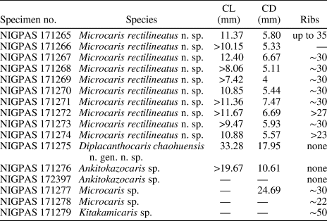
The assemblage from Bed 605 is dominated by Microcaris rectilineatus n. sp., which is relatively small sized but has well-developed ornamentation on the carapace. Other genera are relatively scarce. Among all thylacocephalans in the assemblage, two groups can be clearly observed, being characterized by ornamented versus smooth carapaces, respectively. Most of the specimens have ornamented carapaces, while only a few have smooth carapaces (Figs. 2–4). Among the ornamented ones, two groups can be further separated by their size, although they appear to share a similar carapace shape (round triangular) and ornamentation pattern (nearly straight and vertical ridges). Between the two groups, we compared differences on the number of ridges and the distance between the two neighboring ridges. It is clear that all large specimens are similar in size, approximately 35–40 mm in length, and the small specimens are all around 10 mm except an exceptional specimen of 15 mm long (Table 2). We cannot rule out the possibility that taphonomic processes might have caused the fragmentation of most of the large specimens. However, this could also be a result of a biological process (such as predation) because thylacocephalan fragments are sometimes discovered in coprolites (Affer and Teruzzi, Reference Affer and Teruzzi1999). A recent study suggests that thylacocephalans could have been an important food source for large predators such as chondrichthyans and placoderms during the Late Devonian (Jobbins et al., Reference Jobbins, Haug and Klug2020). Therefore, their huge population could also have served as the food source of fishes and ichthyosaurs during the Early Triassic.
Measurements of the thylacocephalan specimens from Bed 605 of the Majiashan Quarry. CL = length of carapace; CD = depth of carapace; Ribs = number of ribs on carapace.

Microcaris has been considered a bottom dweller because its terraced ornamentation appears to be related to an endobenthic way of life (Briggs and Rolfe, Reference Briggs, Rolfe, Briggs and Lane1983; Tintori et al., Reference Tintori, Bigi, Crugnola and Danini1986). This is in contrast to a more recent interpretation that regards smooth thylacocephalans as nektonic organisms living near the water surface (Jobbins et al., Reference Jobbins, Haug and Klug2020). However, considering that these narrow organisms could generate very low lift and had rather small swimming appendages (Jobbins et al., Reference Jobbins, Haug and Klug2020), it is probable that thylacocephalans could not swim well enough to live far from the sea bottom. Tintori et al. (Reference Tintori, Bigi, Crugnola and Danini1986) pointed out that the depositional environment of thylacocephalans was different from their living environment (at that time, thylacocephalans were considered mainly epibenthic) as they were usually preserved in well-laminated, anoxic rocks. Both thylacocephalan assemblages from the Majiashan section are preserved in laminated rocks containing abundant framboidal pyrite, suggesting that the waters near the bottom were mostly anoxic (Brett and Baird, Reference Brett and Baird1986), thus unfavorable for a benthic lifestyle. As a matter of fact, we do not know exactly whether those thylacocephalans lived near the water surface of a well-oxygenated environment or closer to the anoxic region with a limited oxygen supply. However, the assemblages of Bed 677/680 and 605 both appear poorly diversified, as thylacocephalans are the dominant fossils and other macrofossils are very rare. This might suggest that the thylacocephalans lived in a low-oxygen environment, where many other organisms could not survive. This might also be the reason thylacocephalans recovered quickly and became the dominant taxa shortly after the Permian–Triassic mass extinction.
Among the thylacocephalans, ornamented taxa, such as in the Microcaridae, appear to be mostly restricted to the Triassic. In most of the thylacocephalan faunas where microcarids are present (Ehiro et al., Reference Ehiro, Sasaki, Kano, Nemoto and Kato2015; Hitij et al., Reference Hitij, Gašparič, Žalohar, Jurkovšek and Kolar-Jurkovšek2019; A. Tintori and F. Confortini, personal observation; this paper), the ornamented taxa are often dominant or even represent the only thylacocephalan of the respective fauna, especially in the Upper Triassic. While in the deeper lagoon environments where microcarids were absent, only ‘smooth’ specimens could be collected. Such cases can be found in the lower Carnian of Raibl/Cave del Predil or in the lower Ladinian sections of the Northern Grigna (Tintori, Reference Tintori2013). Regarding the Beds 677/680 assemblage, the thylacocephalans are considered to have lived in shallow water over a fine calcareous sand bottom (Tintori, Reference Tintori2013). This environment is quite different from those in the assemblages containing microcarids—they are typically found in muddy bottoms, from Early Triassic to Middle Triassic Norian. During the Mesozoic, thylacocephalans appear to be recorded mainly in shallow waters with muddy bottoms or in the calcareous bottoms of shallow intracarbonate platform basins. The only exception is the Lower Jurassic Ostenocaris, which is from the Moltrasio Limestone (Tintori et al., Reference Tintori, Bigi, Crugnola and Danini1986) and the Middle Jurassic assemblage from La Voulte (Charbonnier et al., Reference Charbonnier, Vannier, Hantzpergue and Gaillard2010), representing deep-water environments. From the Late Jurassic to the time of their extinction, thylacocephalans are recorded mainly from whitish and pure limestones in Germany, Lebanon, and Mexico, and they are always deposited in rather shallow lagoons surrounded by carbonate platforms.
Acknowledgments
This research is supported by the strategic priority research program of the Chinese Academy of Sciences (grant no. XDB26000000), the National Natural Science Foundation of China (41972003, 41920104001, 41402014), the Youth Innovation Promotion Association of the CAS (to C.J.), and the State Key Laboratory of Palaeobiology and Stratigraphy (Nanjing Institute of Geology and Palaeontology, CAS) (no. 20182107). We deeply thank the journal editors, J. Kastigar and B. Hunda, for the thorough corrections. We thank T. Hegna and reviewers J. Haug and J. Vannier for their constructive suggestions, which greatly improved the manuscript. We thank A. Brayard (Dijon, France), M. Valle (Bergamo, Italy), D. Milesi (S. Pellegrino, Italy), L. Simonetto (Udine, Italy), and A. Alessandrello (Milano, Italy) for kindly showing us specimens in their care. We are also deeply indebted to M. Malzanni (Bergamo, Italy) for information related to fossiliferous sites in the Bergamo area. Z. Chen (NIGPAS, China) helped with photographing.
Appendix 1. Source table of reconstruction drawings and images for the elliptic analysis.