Non-technical Summary
The squat lobster family Munididae is one of the most diverse groups of decapods, widely distributed in the world's oceans. However, its fossil records in the North Pacific have been poorly understood. In this study, three fossil species, including a newly discovered species, of squat lobsters from the Miocene era (18–13 million years ago) in Japan are described and characterized.
Introduction
The squat lobster family Munididae Ahyong et al., Reference Ahyong, Baba, Macpherson and Poore2010 has a worldwide distribution in the present oceans. Munididae is one of the most diverse families within the anomuran decapods and includes over 500 species in 21 genera (Machordom et al., Reference Machordom, Ahyong, Andreakis, Baba, Buckley, García-Giménez, McCallum, Rodríguez-Flores and Macpherson2022). The oldest record of the family dates back to the Upper Jurassic (Tithonian) of Austria (Robins et al., Reference Robins, Feldmann and Schweitzer2012; Schweitzer et al., Reference Schweitzer, Feldmann, Karasawa, Klompmaker and Robins.2023). The fossil records of Munididae are known from the Upper Cretaceous of Morocco, Paleogene of Norway, Denmark, Italy, Hungary, United States, and Argentina, and Neogene of Slovakia, Italy, United States, Argentina, and Japan (Schweitzer et al., Reference Schweitzer, Feldmann, Karasawa, Klompmaker and Robins.2023).
The hitherto known munidid fossils of Japan are represented by two species assigned to Munida Leach, Reference Leach and Cuvier1821 [1820] from the Miocene deposits. Munida nishioi Karasawa, Reference Karasawa1993 occurs from the various localities of central and southwestern Honshu (Karasawa, Reference Karasawa1993; Kato, Reference Kato1996; Ando et al., Reference Ando, Kato and Saito2019). Munida ogaensis (Hatai and Kotaka, Reference Hatai and Kotaka1970), originally described as the problematic fossil Kazuoia ogaensis Hatai and Kotaka, Reference Hatai and Kotaka1970, is known only from the type locality of the Oga Peninsula, northeastern Honshu (Hatai and Kotaka, Reference Hatai and Kotaka1970; Ando et al., Reference Ando, Kato and Saito2019). In addition, an undetermined species assigned to Munida was reported from the Itahana and Nawashiroda formations, central Honshu (Kato, Reference Kato2001; Ando et al., Reference Ando, Kato and Saito2019).
The purpose of this paper is to redescribe Munida nishioi Karasawa, Reference Karasawa1993 and Kazuoia ogaensis Hatai and Kotaka, Reference Hatai and Kotaka1970 and to reevaluate the generic status for both species on the basis of careful observations of the type and additional specimens. Furthermore, a new species of Trapezionida Macpherson and Baba in Machordom et al., Reference Machordom, Ahyong, Andreakis, Baba, Buckley, García-Giménez, McCallum, Rodríguez-Flores and Macpherson2022 is described from the lower Miocene Yonekawa Formation of central Japan.
Materials and methods
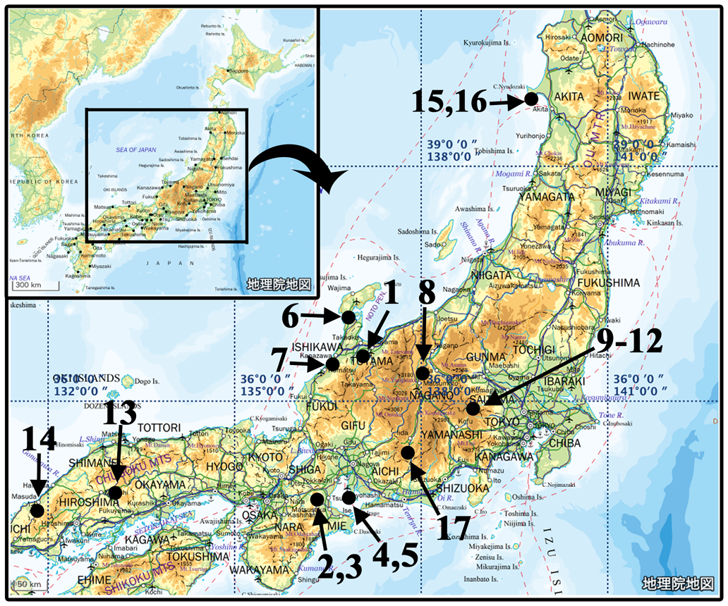
We examined specimens collected from eight localities (Fig. 1.1–1.5, 1.15–1.17), deposited in the Natural History Museum and Institute, Chiba, Mizunami Fossil Museum, and Tohoku University Museum. Some specimens were prepared using Murray Engineering Micro-Jacks. When the specimens were represented by the external molds, latex casts were made using GC Exaflex Putty Standard Pack. The original specimens and latex casts were photographed with digital cameras, Olympus E-PL5 with Olympus OM-system Zuiko Auto Macro 50 mm lens (Olympus, Tokyo, Japan) and Nikon D7200 with Micro Nikkor 60 mm. Some images coated with ammonium chloride or magnesium oxide were also prepared. The images were toned for contrast and brightness using Adobe Photoshop CS14 for Macintosh.
Occurrences of the Miocene Munididae of Japan. (1–5) Localities of the examined specimens of Grimothea nishioi: (1) Murasugi (type locality); (2) Ashizaka; (3) Iedokoro; (4) Hashizume; (5) Iwaya. (6–14) Other localities of G. nishioi: (6) Kanmachi; (7) Tatsumi; (8) Ooguchi; (9) Oofuchi; (10) Obashira; (11) Onohara; (12) Terao; (13) Kamifuno; (14) Okuda. (15, 16) Notake, localities of G. ogaensis. (17) Chiyo, type locality of Trapezionida chiyoensis. The map is derived from the Geospatial Information Authority of Japan (http://geolib.gsi.go.jp/).

The terminology used largely follows Baba et al. (Reference Baba, Macpherson, Lin and Chan2009, Reference Baba, Ahyong, Macpherson, Poore, Ahyong and Taylor2011). The measurements of specimens are of the maximum carapace width (CW) and the carapace length excluding the rostrum (CL). Abbreviations used are P1–P5 (pereiopods 1–5).
Repositories and institutional abbreviations
Types and figured and compared specimens examined in this study are deposited in the following institutions: The Natural History Museum and Institute, Chiba (CBM-PI), Chiba, Japan; Mizunami Fossil Museum, Gifu, Japan (MFM); Tohoku University Museum (IGPS), Sendai, Japan.
Systematic paleontology
Family Munididae Ahyong et al., Reference Ahyong, Baba, Macpherson and Poore2010
Genus Grimothea Leach, Reference Leach and Cuvier1821 [1820]
Type species
Galathea gregaria Fabricius, Reference Fabricius1793 by monotypy.
Diagnosis
Carapace with distinct setiferous transverse ridges, setae non-iridescent; moderately convex; pair of epigastric spines directly behind ocular peduncles usually flanked by spines; anterolateral spine distinct; five to seven spines on branchial margin; posterior margin unarmed; pterygostomian flap unarmed and sometimes visible in dorsal view. Rostrum spiniform, laterally compressed and with dorsal spines in some species, flanked by supraocular spines; shallow longitudinal grooves between bases of rostral and supraocular spines. Epistome with ridge arising from marginal ridge of mouth, laterally leading to level of antennal gland aperture. Telson with posterior plates wide, each plate with length/breadth ratio of 0.6–0.7. Thoracic sternum longer than wide or slightly wider than long; maximum width at sternite 7; posterior margin of sternite 3 contiguous to slightly narrower than anterior margin of sternite 4. Eyes freely movable, cornea dilated, eyelashes present. Antennular article 1 moderately long and slender, nearly twice longer than wide, with two distal and two lateral spines; distal half as long as or slightly longer than wide. Antennal article 1 fused with epistome, with distomesial spine; article 2 usually with small distomesial and distolateral spines. Maxilliped 3 slender, ischium elongate. P1 fingers as long as or slightly longer than palm, usually unarmed. P2–4 spinose, relatively slender; flexor margin of dactyli with row of seta-like spines, each arising from very low, small process; P4 moderately long and slender, mero-carpal articulation not reaching anterior margin of carapace. Gonopods 1 and 2 present (Macpherson and Baba in Machordom et al., Reference Machordom, Ahyong, Andreakis, Baba, Buckley, García-Giménez, McCallum, Rodríguez-Flores and Macpherson2022, p. 956–957).
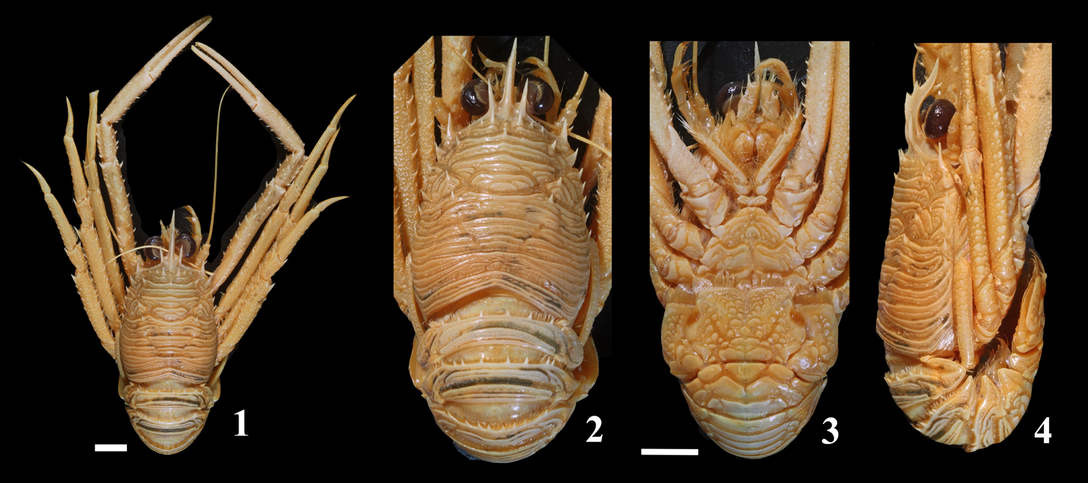
Compared material (Fig. 2)
Grimothea princeps (Benedict, Reference Benedict1902), MFM192120, CW/CL = 30.8/39.6 mm, Suruga Bay, Japan.
Grimothea princeps (Bennedict, Reference Benedict1902) from the Suruga Bay of Japan, extant, compared specimen, MFM192120. (1, 2) Dorsal view. (3) Ventral view. (4) Lateral view. Scale bars = 10 mm.

Remarks
During the major revisional work on the systematics of Munididae, Machordom et al. (Reference Machordom, Ahyong, Andreakis, Baba, Buckley, García-Giménez, McCallum, Rodríguez-Flores and Macpherson2022) recognized Grimothea Leach, Reference Leach and Cuvier1821 [1820] as a valid genus. At that time, they included 14 extant species previously assigned to three munidid genera, Cervimunida Benedict, Reference Benedict1902, Munida Leach, Reference Leach and Cuvier1821 [1820], and Pleuroncodes Stimpson, Reference Stimpson1860, in this genus. After that, Tiwari et al. (Reference Tiwari, Padate and Cubelio2023) described the new species, Grimothea krishaha Tiwari et al., Reference Tiwari, Padate and Cubelio2023, from India; therefore, Grimothea consists of 15 extant species.
Among species of Grimothea, G. princeps (Benedict, Reference Benedict1902) and G. johni (Porter, Reference Porter1903) were formerly placed in Cervimunida. This genus is well defined by having the dorsal and ventral spines of the rostrum, which are not present within the other munidid species (Baba, Reference Baba1988; Baba et al., Reference Baba, Macpherson, Lin and Chan2009; Macpherson and Baba, Reference Macpherson, Baba, Poore, Ahyong and Taylor2011). However, Machordom et al. (Reference Machordom, Ahyong, Andreakis, Baba, Buckley, García-Giménez, McCallum, Rodríguez-Flores and Macpherson2022) recognized the Grimothea clade containing Cervimunida, Pleuroncodes, and two species previously assigned to Munida within Munididae, using molecular and morphological-based phylogenetic analyses. They concluded that Cervimunida and Pleuroncodes were the junior synonyms of Grimothea.
Two Japanese Miocene species, Munida nishioi and Kazuoia ogaensis, are redescribed and transferred to Grimothea in the following. Both species have dorsal and ventral spines of the rostrum spine, which are unique characters of G. princeps and G. johni, but both species are readily distinguished from G. johni in that the pterygostomian region is not visible dorsally and the dorsal spines of the carapace are well developed.
Grimothea nishioi has close affinities with G. princeps in that the carapace is dorsally armed with six epigastric, two parahepatic, two postcervical, and two anterior branchial spines, the lateral margin of the carapace bears seven spines, the rostrum is laterally compressed and arched, and the pleonal somites 2–4 are armed with spines on the anterior ridge. However, G. nishioi differs from G. princeps by having the supraocular spines nearly parallel to the rostral spine rather than divergent and the pereiopod 1 merus with five spines on the dorsal margin. Furthermore, the anterior ridge on the pleonal somite 2 is armed with 10 spines in G. nishioi whereas this ridge is armed with 8 in G. princeps, and the anterior ridge on pleonal somite 4 is armed with 5 spines on G. nishioi and with 8 in G. princeps.
Hatai and Kotaka (Reference Hatai and Kotaka1970) originally described Kazuoia ogaensis, a new genus and a new species of the problematical fossil, from examination of only one specimen (IGPS no. 86726) from the middle Miocene Nishikurosawa Formation. Hatai and Kotaka (Reference Hatai and Kotaka1970) described it as the thorax and pleon of an isopod and referred it to the isopod family Cymothoidae Leach, 1814. Although Karasawa and Kato (Reference Karasawa, Kato, Ikeya, Hirano and Ogasawara2001) assigned the species to Munida, they did not give any description and discussion for it. When the body orientation of the holotype is routed 180 degrees, it is truly identical to the carapace of Munididae. Furthermore, examination of the holotype and additional specimen shows that Kazuoia ogaensis should be included in Grimothea. The species is similar to Grimothea nishioi, but a narrow posterior margin of the carapace readily distinguishes this species from G. nishioi. In addition, the rostrum of G. ogaensis is longer than that of G. nishioi.
Kazuoia Hatai and Kotaka, Reference Hatai and Kotaka1970 is a monotypic genus. Kazuoia becomes a junior synonym of Grimothea because Kazuoia ogaensis is removed to Grimothea Leach, Reference Leach and Cuvier1821 [1820].
Miller (Reference Miller1951) noted the occurrence of Pleuroncodes sp. from the upper Miocene Capistrano Formation of California, but he did not describe and illustrate it. Robins (Reference Robins2013) also regarded it as an undetermined species of Pleuroncodes on the basis of examination of the specimen (USNM 373110) deposited in the United States National Museum of Natural History, Smithsonian Institution. Pleuroncodes is now a junior synonym of Grimothea; therefore, this species is moved to Grimothea.
Grimothea nishioi (Karasawa, Reference Karasawa1993) new combination
Figures 3–5
- Reference Karasawa1993
Munida nishioi Karasawa, Reference Karasawa1993, p. 38, pl. 5, figs. 5, 6, 9.
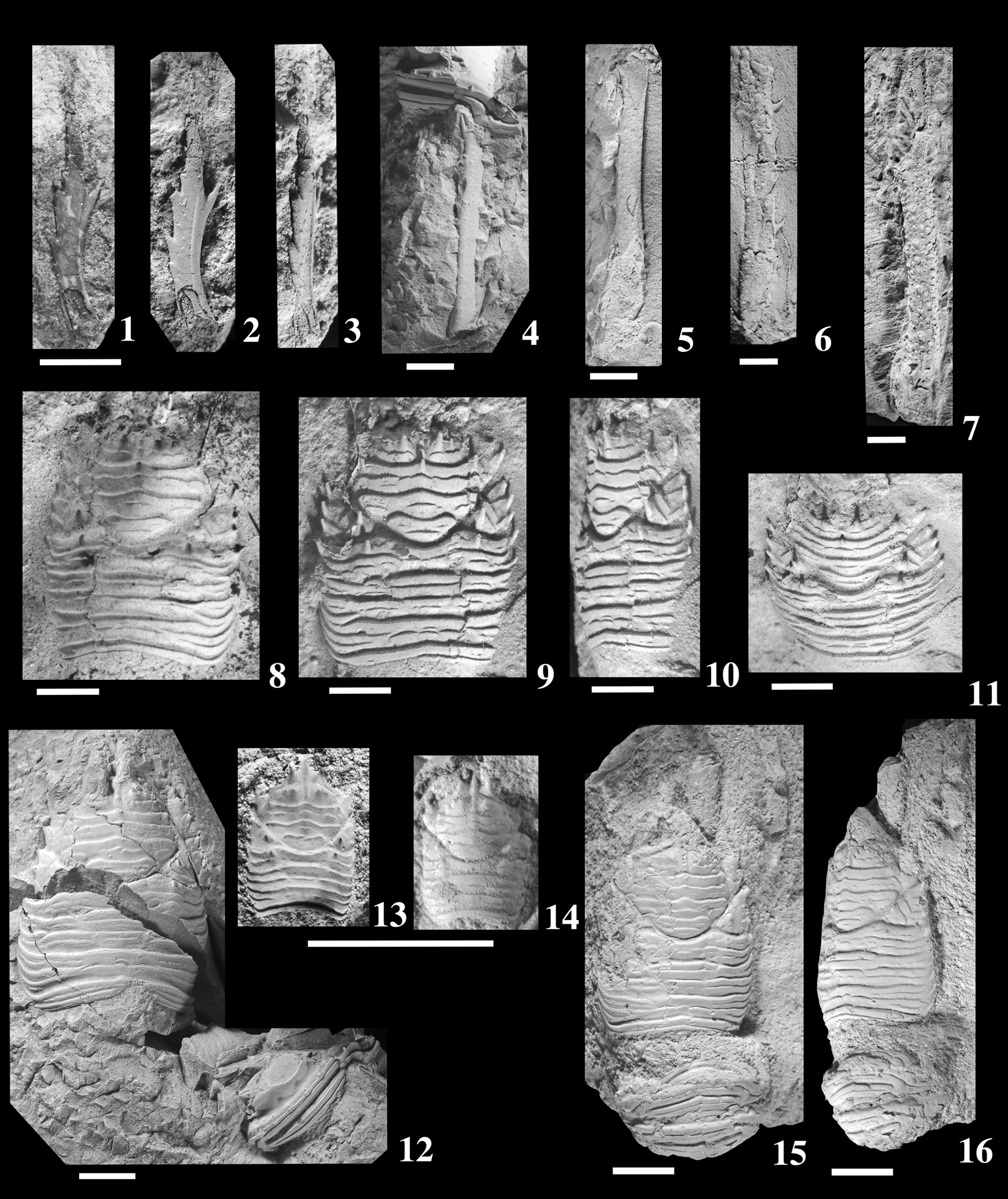
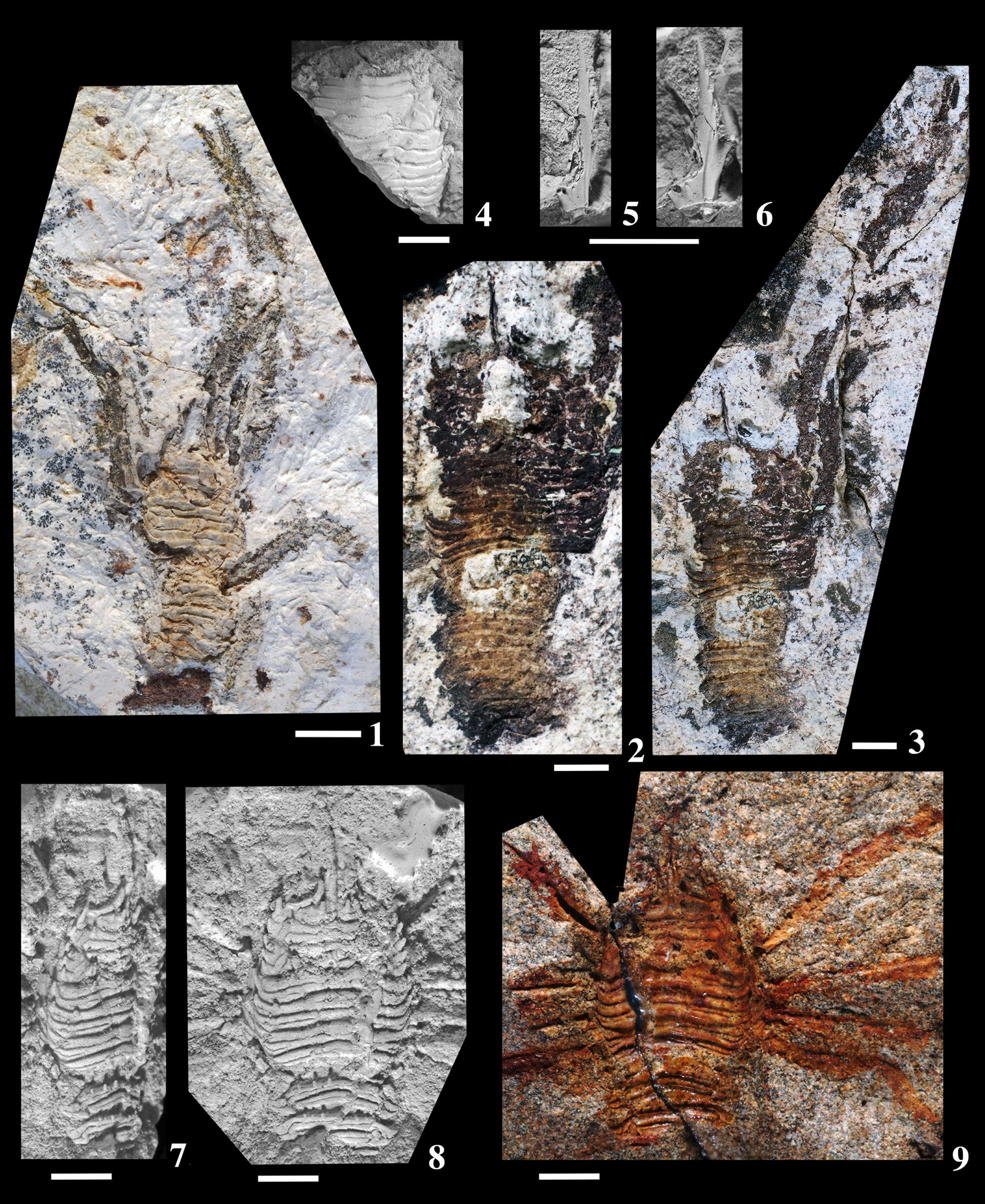
Grimothea nishioi (Karasawa, Reference Karasawa1993) new combination from the Higashibessho Formation, upper lower Miocene, Murasugi (Fig. 1.1). (1–3) Rostrum, MFM84140: (1, 2) lateral view; (3) dorsal view. (4, 5) Meri of pereiopods, MFM84142: (4) lateral view; (5) mesial view. (6) Dorsal view of merus of pereiopod 1, MFM83418. (7) Dorsal view of propodus and dactylus of pereiopod 1, MFM83419. (8) Inner view of external mold of carapace, MFM83054, holotype. (9–11) Latex cast of MFM83054: (9) dorsal view; (10) lateral view; (11) anterior view. (12) Dorsal view of carapace and pleonal somite 4, MFM84142. (13) Inner view of external mold carapace, MFM84141. (14) Dorsal view of latex cast of MFM83141. (15, 16) Carapace, pleon, and pereiopods, MFM84139: (15) dorsal view; (16) ventral view. (2–16) Images coated with ammonium chloride. Scale bars = 5 mm.

Grimothea nishioi (Karasawa, Reference Karasawa1993) new combination from the Morozaki Group, lower Miocene, Iwaya (Fig. 1.4). (1) Inner view of carapace, pleon, and pereiopods, MFM83998. (2–4) Latex cast of MFM83998: (2, 3) lateral views of carapace and pleon; (4) dorsal view of carapace, pleon, and pereiopods. (2–4) Images coated with ammonium chloride. Scale bars = 5 mm.

Grimothea nishioi (Karasawa, Reference Karasawa1993) new combination. (1) Dorsal view of carapace, pleon, and pereiopods, MFM83999, Morozaki Group, lower Miocene, Iwaya (Fig. 1.5). (2, 3) Inner view of carapace, pleon, and pereiopods, MFM83400, Morozaki Group, lower Miocene, Hashizume (Fig. 1.4): (2) closeup image of carapace and pleon; (3) whole image. (4) Dorsal view of carapace, MFM39143, Ichishi Group, lower Miocene, Ashizaka (Fig. 1.2). (5, 6) Rostrum of MFM39143: (5) dorsal view; (6) lateral view. (7, 8) Latex cast of MFM39144, Ichishi Group, lower Miocene, Iedokoro (Fig. 1.3), carapace and pleon: (7) lateral view; (8) dorsal view. (9) Inner view of external mold of carapace, pleon, and pereiopods, MFM39144, Ichishi Group, lower Miocene, Iedokoro (Fig. 1.3). (4–6, 7, 8) Images coated with ammonium chloride. Scale bars = 5 mm.

Holotype
External mold of carapace (MFM83054) from the upper lower Miocene Higashibessho Formation, Toyama, Japan (Karasawa, Reference Karasawa1993, pl. 5, fig. 5).
Emended diagnosis
Carapace subrectangular, longer than wide. Rostrum spiniform, slender, long, about 0.4 times as long as carapace, gently arched anteriorly, depressed laterally, with rimmed lateral margins; dorsal carina with five spines; one ventral spine just below third dorsal spine. Frontal margins slightly concave, oblique laterally, rimmed; supraocular spines slender, long, nearly parallel. Lateral margins feebly convex with seven spines. Posterior margin slightly wider than frontal margin, convex forward, with smooth rim. Dorsal surface moderately convex longitudinally, gently convex transversely, covered with main transverse terraced ridges and secondary transverse ridges; anterior margins of ridges fringed with dense, minute denticles. Epigastric regions with transverse row of three spines, spine posterior to base of supraocular spine strongest, transverse ridges just behind spines; two small spines present behind carina of rostrum. Short median protogastric spine present. Cervical and postcervical grooves distinct. Parahepatic, branchial dorsal, and postcervical spines present on each side of carapace. Pterygostomian regions not visible dorsally. Terga of pleonal somites 2–5 ornamented with two transverse ridges, each ridge interrupted by transverse stria; anterior ridges with 10, eight, and six spines on somites 2, 3, and 4, respectively. P1 slender, long, three times carapace length excluding rostrum; surfaces with variably sized tubercules and spines; merus with five spines on mesial margin. P2–4 slender, long, with variably sized tubercles and spines on surfaces.
Occurrences (Fig. 1)
The present species is known from the early–middle Miocene deposits of the following 14 localities in Japan.
Loc. 1.––Murasugi (type locality), Yatsuo, Toyama City, Toyama Prefecture (36°34′29″N, 137°09′13″E); mudstone of the Higashibessho Formation, Yatsuo Group; late early Miocene, NPD3A–NPD3B Zone of Yanagisawa and Akiba's (Reference Yanagisawa and Akiba1998) scale of diatoms by Yanagisawa (Reference Yanagisawa1999) (Karasawa, Reference Karasawa1993 as Munida nishioi).
Loc. 2.––Ashizaka, Misato, Tsu City, Mie Prefecture (34°43′05″N, 136°23′30″E); mudstone of the Oi Formation, Ichishi Group; early Miocene, NPD2B Zone of Yanagisawa and Akiba's (Reference Yanagisawa and Akiba1998) scale of diatoms by Oshida et al. (Reference Oshida, Hayashi, Yanagisawa, Kurihara and Hoshi2018) (Karasawa, Reference Karasawa1993 as Munida nishioi; this work).
Loc. 3.––Iedokoro, Misato, Tsu City, Mie Prefecture (34°43′38″N, 136°23′59″E); mudstone of the Oi Formation, Ichishi Group; early Miocene, NPD2B Zone of Yanagisawa and Akiba's (Reference Yanagisawa and Akiba1998) scale of diatoms by Oshida et al. (Reference Oshida, Hayashi, Yanagisawa, Kurihara and Hoshi2018) (Karasawa, Reference Karasawa1993 as Munida nishioi; this work).
Loc. 4.––Hashizume, Yamami, Minamichita-cho, Aichi Prefecture (34°43′43″N, 136°52'39E); tuffaceous sandstone of the Yamami Formation, Morozaki Group; early Miocene, NPD2B Zone of Yanagisawa and Akiba's (Reference Yanagisawa and Akiba1998) scale of diatoms by Ito et al. (Reference Ito, Irizuki and Iwai1999) (Takeda et al., Reference Takeda, Mizuno and Yamaoka1986 as Munida sp.; Karasawa et al., Reference Karasawa, Mizuno, Hachiya and Ando2017 as Munididae; this work).
Loc. 5.––Iwaya, Minamichita-cho, Aichi Prefecture (34°43′34″N, 136°54′52″E); tuffaceous sandstone of the Yamami Formation, Morozaki Group; early Miocene, NPD2B Zone of Yanagisawa and Akiba's (Reference Yanagisawa and Akiba1998) scale of diatoms by Ito et al. (Reference Ito, Irizuki and Iwai1999) (Takeda et al., Reference Takeda, Mizuno and Yamaoka1986 as Munida sp.; Karasawa et al., Reference Karasawa, Mizuno, Hachiya and Ando2017 as Munididae; this work).
Loc. 6.––Kanmachi, Nakajima, Nanao City, Ishikawa Prefecture (37°07′18″N, 136°50′29″E); mudstone of the Hamada Formation; late early Miocene by Kaseno (Reference Kaseno1990) (Imaizumi, Reference Imaizumi1969 as Eumunida sp.).
Loc. 7.––Tatsumi, Kanazawa City, Ishikawa Prefecture (36°30′21″N, 136°42′02″E); mudstone of the Asagaya Formation; late early Miocene by Kaseno (Reference Kaseno1990) (Karasawa, Reference Karasawa1993 as Munida nishioi).
Loc. 8.––Ooguchi, Toyoshina-machi, Azumino City, Nagano Prefecture (36°18′10″N, 137°57′32″E); sandstone of the Aoki Formation; middle Miocene by Karasawa et al. (Reference Karasawa, Narita, Sakumoto, Koike and Mizuno1999) and Itoigawa and Yanagisawa (Reference Itoigawa and Yanagisawa2002) (Karasawa et al., Reference Karasawa, Narita, Sakumoto, Koike and Mizuno1999 as Munida nishioi).
Loc. 9.––Ofuchi, Minano-cho, Saitama Prefecture (36°04′40″N, 139°05′35″E); sandstone of the Ushikubitoge Formation; late early Miocene by Takahashi (Reference Takahashi2008) (Obata, Reference Obata1995 as Munida nishioi; Kato, Reference Kato1996 as Munida sp. cf. M. nishioi).
Loc. 10.––Obashira, Chichibu City, Saitama Prefecture (36°03′60″N, 139°05′07″E); mudstone of the Ushikubitoge Formation; late early Miocene by Takahashi (Reference Takahashi2008) (Obata, Reference Obata1995 as Munida nishioi; Kato, Reference Kato1996 as Munida sp. cf. M. nishioi).
Loc. 11.––Onohara, Chichibu City, Saitama Prefecture (36°02′23″N, 139°05′35″E); mudstone of the Nagura Formation; early middle Miocene by Takahashi (Reference Takahashi2008) (Kato, Reference Kato1996 as Munida sp. cf. M. nishioi).
Loc. 12.––Terao, Chichibu City, Saitama Prefecture (35°57′29″N, 139°00′39″E); mudstone of the Nagura Formation; early middle Miocene by Takahashi (Reference Takahashi2008) (Kato, Reference Kato1996 as Munida sp. cf. M. nishioi).
Loc. 13.––Kamifuno, Funo-cho, Miyoshi City, Hiroshima Prefecture (34°54′26″N, 132°46′41″E); sandstone of the Lower Sandstone Member of the Bihoku Group; late early Miocene, NPD3A–NPD3B Zones of Yanagisawa and Akiba's (Reference Yanagisawa and Akiba1998) scale of diatoms by Watanabe et al. (Reference Watanabe, Miyake, Nozaki, Yamamoto, Takemura and Nishimura1999) (Sakumoto, Reference Sakumoto1997 as Munida nishioi).
Loc. 14.––Okuda, Masuda City, Shimane Prefecture (34°38′51″N, 131°49′04″E); Sandstone of the Toyoda Formation of the Masuda Group; late early Miocene by Goto et al. (Reference Goto, Irizuki, Hayashi, Tanaka, Matsuyama and Iwatani2013) (Sakumoto, Reference Sakumoto1997 as Munida nishioi).
Emended description
Carapace excluding rostrum, subrectangular, 1.2 times longer than wide, widest at mid-length. Rostrum spiniform, slender, long, 0.4 times as long as carapace, gently arched anteriorly, depressed laterally, flanked by supraocular spines, with rimmed lateral margins; dorsal carina well developed with five dorsal spines directed anteriorly; anterior third spine largest, followed by anterior fourth, anterior second, last, and first spines; one ventral spine directed anteriorly, just below second dorsal spine; lateral surface with a row of small tubercles along dorsal spines and three short lateral spines directed anteriorly on forepart of base of ventral spine; shallow longitudinal grooves between bases of rostral and supraocular spines. Frontal margins slightly concave, rather oblique, rimmed; supraocular spines slender, long, 0.4 times as long as rostrum, nearly parallel to rostrum, directed slightly upward. Lateral margins feebly convex; anterolateral spines slender, long, reaching half-length of supraorbital spine, directed anterolaterally; hepatic margin with one short spine directed anterolaterally in front of deep cervical notch; anterior branchial margins with three short, anterolaterally directed spines; posterior branchial margin bearing two short spines directed anterolaterally behind shallow postcervical notch, posterior one smallest. Posterior margin slightly wider than frontal margins, convex forward, with smooth rim. Dorsal surface moderately convex longitudinally, gently convex transversely, covered with main, transverse terraced ridges gently sloping posteriorly and steeply sloping anteriorly; secondary transverse ridges continuous or discontinuous between main ridges; anterior margins of main and secondary ridges fringed with dense, minute denticles. Epigastric regions with transverse row of three spines, spine posterior to base of supraocular spine strongest, transverse ridges just behind spines; two small spines longitudinally arranged behind carina of rostrum. Short median protogastric spine present on anterior ridge of protogastric region. Cervical and postcervical grooves distinct. Short parahepatic, branchial dorsal, and postcervical spines present on each side of carapace. Pterygostomian regions not visible dorsally.
Tergites of pleonal somites 2–5 with two transverse ridges, each ridge interrupted by transverse stria. Anterior ridges with 10, eight, and six spines on somites 2, 3, and 4, respectively.
P1–4 poorly preserved. P1 slender, long, three times as long as carapace length excluding rostrum; surfaces covered with variably sized tubercules and spines inclined distally; merus about 1.3 times carapace length, with five large spines on mesial margin and short spines on lateral margin: carpus short; propodus long; both fingers shorter than palm. Details of P2 unknown, but merus 0.85 times carapace length. P3 and P4 long, slender; meri, carpi, and propodi covered with irregular-sized tubercles. P3 merus about 0.8 times carapace length, extensor and flexor margins with spines directed distally; carpus about 0.3 length of merus with three spines inclined distally on extensor margin. P4 merus 0.7 times carapace length, extensor and flexor margins with short spines inclined distally; mero–carpal articulation not reaching anterior margin of carapace; carpus about 0.3 length of merus; propodus about 2.2 length of carpus; dactylus half-length of propodus.
Examined and illustrated materials
MFM39143, carapace and pereiopods, from Loc. 2; MFM39144, external mold of carapace, pleon and pereiopods, and latex cast from Loc. 3; MFM83054 (holotype), external mold of carapace and latex cast from Loc. 1; MFM83148, pereiopod 1 merus from Loc. 1; MFM83149, pereiopod 1 propodus and dactylus from Loc. 1; MFM83998, external mold of carapace, pleon and pereiopods, and latex cast from Loc. 5; MFM83999, carapace, pleon, and pereiopods from Loc. 5; MFM84000, external mold of carapace, pleon, and pereiopods from Loc. 4; MFM84139, carapace, pleon, and pereiopods from Loc. 1; MFM84140, carapace and pereiopods from Loc. 1; MFM84141, external mold and latex cast from Loc. 1; MFM84142, carapace, pleon, and pereiopod meri from Loc. 1.
Measurements (mm)
MFM83054 (holotype), 11.9 × 14.3; MFM39144, 11.7 × 13.4; MFM83998, 13.9 × 17.1; MFM84139, 12.1 × 16.8; MFM84000, 14.4 × 16.7; MFM84141, 3.4 × 4.3; MFM84142, 17.2 × 21.2.
Remarks
The holotype is an incomplete carapace lacking the rostrum and supraocular spines. Therefore, the emended diagnosis and description are given for the species, based on examination of additional specimens allowing a more detailed description.
Grimothea ogaensis (Hatai and Kotaka, Reference Hatai and Kotaka1970) new combination
Figure 6
- Reference Hatai and Kotaka1970
Kazuoia ogaensis Hatai and Kotaka, p. 2, figs. 1, 2.
- Reference Karasawa, Kato, Ikeya, Hirano and Ogasawara2001
Munida ogaensis; Karasawa and Kato, p. 428.
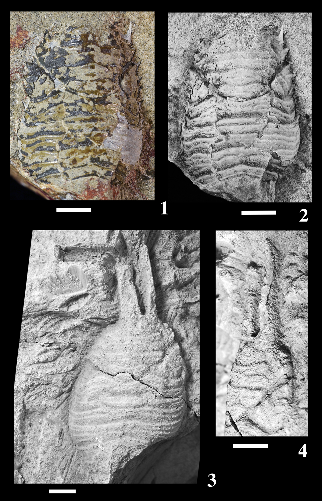
Grimothea ogaensis (Hatai and Kotaka, Reference Hatai and Kotaka1970) new combination from the Nishikurosawa Formation, middle Miocene. (1, 2) Dorsal view of carapace, IGPS 86726, holotype, Notake (Fig. 1.15). (3, 4) Carapace and fragments of pereiopods, CBM-PI 2752, Notake (Fig. 1.16): (3) dorsal view; (4) lateral view. (2–4) Images coated with ammonium chloride. Scale bars = 5 mm.

Holotype
Carapace (IGPS no. 86726) from the middle Miocene Nishikurosawa Formation, Akita, Japan (Hatai and Kotaka, Reference Hatai and Kotaka1970, figs. 1, 2).
Diagnosis
Carapace subrectangular, longer than wide. Rostrum spiniform, slender, long, half-length of carapace, gently arched anteriorly, upturned, depressed laterally; dorsal carina with dorsal spines; one ventral spine just below second dorsal spine; small lateral spine in front of base of ventral spine. Frontal margins oblique laterally. Lateral margins widened posteriorly with seven spines. Posterior margin as long as frontal margin. Dorsal surface moderately convex longitudinally, gently convex transversely, covered with main transverse terraced ridges and secondary transverse ridges. Cervical and postcervical grooves distinct. Parahepatic, branchial dorsal, and postcervical spines present. Pterygostomian regions not visible dorsally.
Occurrences (Fig. 1)
This species is known only from the middle Miocene Nishikurosawa Formation exposed at the type locality and its adjacent locality.
Loc. 15.––Notake, Daijima (type locality), Funagawaminato, Oga City, Akita Prefecture (detailed information not known, approximately 39°51′52″N, 139°48′08″E); mudstone of the Nishikurosawa Formation; middle Miocene NPD4Bb Zone of Yanagisawa and Akiba's (Reference Yanagisawa and Akiba1998) scale of diatoms by Kano et al. (Reference Kano, Ohguchi, Yanagisawa, Awata, Kobayashi, Sato, Hayashi, Kitazato, Ogasawara and Komazawa2011).
Loc. 16.––Notake, Daijima, Funagawaminato, Oga City, Akita Prefecture (39°51′52″N, 139°48′08″E); mudstone of the Nishikurosawa Formation; middle Miocene NPD4Bb Zone of Yanagisawa and Akiba's (Reference Yanagisawa and Akiba1998) scale of diatoms by Kano et al. (Reference Kano, Ohguchi, Yanagisawa, Awata, Kobayashi, Sato, Hayashi, Kitazato, Ogasawara and Komazawa2011).
Original description of holotype
The fossil is elongate-quadrate in shape and narrowed slightly at both anterior and posterior extremities, which are both bluntly rounded, separated in two distinct parts of the thorax or anterior approximately two-thirds and the abdomen or posterior approximately one-third by a distinct groove of anteriorly directed rather narrowly convex shape; exposed dorsal surface roundly convex and with about 16 narrow appendages, each of posterior six appendages terminating laterally into posteriorly directed sharp and curved spines, posteriormost spine strongest, longest, and measuring about 2 mm in length, others shorter, narrower, and more sharply pointed than largest one just mentioned. The appendages on anterior (thorax) part of specimen narrow, each measuring about 1 mm in width, axially curved backward on anterior part, curvature becoming indistinct posteriorly, appendages laterally weakly sigmoidal on anterior part, terminating into posteriorly directed sharp but short spines; on posterior abdomen part of specimen each of six appendages measuring about 1.5 to 1 mm in width, not curved backward along axial part, laterally showing sigmoidal structure, terminating into well-developed spines, among which posteriormost is strongest, largest, and outstanding. Abdomen and thorax extremities not preserved. Entire specimen as preserved measures 2 cm in length, 5 mm in height or convexity, largest and posteriormost spine about 2 mm in length, all others shorter and slenderer (Hatai and Kotaka, Reference Hatai and Kotaka1970, p. 2).
Revised description of holotype
Carapace subrectangular, about 1.3 times longer than wide, widest at posterior one-fifth, moderately convex longitudinally, slightly convex transversely. Cuticles of carapace partly preserved; therefore, details of dorsal surface unknown. Rostrum and supraorbital spines not preserved. Frontal margin oblique laterally, 0.65 times maximum width. Lateral margins widened posteriorly, anterior half nearly straight, posterior half gently convex; anterolateral spines slender, long, directed anterolaterally; hepatic margin with one short spine directed anterolaterally in front of deep cervical notch; anterior branchial margins with three short, anterolaterally directed spines; posterior branchial margin bearing two short spines directed anterolaterally behind shallow postcervical notch. Posterior margin as long as frontal margin, convex forward, with smooth rim. Dorsal surface covered with main, transverse terraced ridges gently sloping posteriorly and steeply sloping anteriorly; secondary transverse ridges between main ridges; anterior margins of main and secondary ridges fringed with dense, minute denticles. Spines on epigastric regions not observed. Cervical and postcervical grooves distinct. Bases of parahepatic, branchial dorsal, and postcervical spines present. Pterygostomian regions not visible dorsally.
Description of additional specimen
Carapace deformed, lacking anterior region. Rostrum slightly transported from its natural position, spiniform, slender, long, about half-length of carapace, gently upturned over proximal third, downturned over middle third, upturned distal third, depressed laterally; dorsal carina with four dorsal spines, anterior second spine largest; one ventral spine just below second dorsal spine; small lateral spine in front of base of ventral spine. Dorsal surface covered with main transverse terraced ridges; secondary transverse ridges present between main ridges; cervical and postcervical grooves distinct; bases of parahepatic and branchial dorsal spines present. Fragments of pereiopods preserved with tuberculated surfaces.
Examined and illustrated materials
IGPS 86726 (holotype), carapace, from Loc. 15; CBM-PI 2752, carapace and pereiopods, from Loc. 16.
Measurements (mm)
IGPS no. 86726 (holotype), 22.3 × 16.0; CBM-PI 2752, 25.2 × 21.3.
Remarks
The revised description is given for the holotype as it is regarded as the carapace of a munidid decapod, not the thorax and pleon of an isopod. An additional specimen remaining, the rostrum, is also described from Loc. 16 near the type locality. Hatai and Kotaka (Reference Hatai and Kotaka1970) gave no diagnosis for this species; therefore, a diagnosis is herein provided.
Genus Trapezionida Macpherson and Baba in Machordom et al., Reference Machordom, Ahyong, Andreakis, Baba, Buckley, García-Giménez, McCallum, Rodríguez-Flores and Macpherson2022
Type species
Munida acantha Macpherson, Reference Macpherson and Crosnier1994 by original designation.
Diagnosis
Carapace with lateral margin straight or slightly convex; dorsal surface with distinct transverse setigerous ridges, setae non-iridescent; pair of epigastric spines directly behind ocular peduncles flanked by spines; protogastric spines absent; anterolateral spine distinct; branchial lateral margin with three to five spines; posterior margin unarmed; pterygostomian flap unarmed and invisible in dorsal view. Rostrum spiniform, flanked by supraocular spines; shallow longitudinal grooves between bases of rostral and supraocular spines. Epistome with ridge arising from marginal ridge of mouth, laterally leading to level of antennal gland aperture. Abdominal somites with transverse ridges. Telson with posterior plates wide, each plate as long as wide. Thoracic sternum wider than long; maximum width at sternite 7; sternite 3 is three to four times wider than long; sternite 4 trapezoidal, anterior margin transverse, widely contiguous or subparallel to posterior margin of sternite 3 for at least three-quarters of the width; lateral parts of posterior sternites with or without carinae. Eyes freely movable, cornea dilated; eyelashes present. Antennular article 1 moderately long and slender, approximately twice longer than wide, with two distal and two lateral spines; distomesial spine more than half-length of distolateral spine. Antennal peduncle sometimes reduced; article 1 not fused with epistome, with distomesial spine well developed, usually reaching or exceeding end of article 2. Maxilliped 3 slender, ischium elongate. P1 fingers shorter than palm. P2–4 spinose, relatively slender; flexor margin of dactyli with row of seta-like spines, each arising from very low, small process; P4 merus moderately long, approximately two-thirds length of P2 merus, not reaching anterior margin of carapace. Gonopods 1 and 2 present (Macpherson and Baba in Machordom et al., Reference Machordom, Ahyong, Andreakis, Baba, Buckley, García-Giménez, McCallum, Rodríguez-Flores and Macpherson2022, p. 960–961).
Remarks
Machordom et al. (Reference Machordom, Ahyong, Andreakis, Baba, Buckley, García-Giménez, McCallum, Rodríguez-Flores and Macpherson2022) revised the classification of Munididae and classified Munida containing 301 extant species into 14 genera. In their work, Munida was redefined with only six extant species from the Northeast Pacific. Among munidid genera, Trapezionida is the most diverse group, including over 155 species, distributed in the Indo-West Pacific (Machordom et al., Reference Machordom, Ahyong, Andreakis, Baba, Buckley, García-Giménez, McCallum, Rodríguez-Flores and Macpherson2022). After that, additional species of Trapezionida are newly described (Tiwari et al., Reference Tiwari, Padate, Cubelio and Osawa2022; Reference Tiwari, Padate and Cubelio2023; Macpherson et al., Reference Macpherson, Rodríguez-Flores and Machordom2024), and Trapezionida comprises 164 extant species.
A new species described in the following has the definitive characters of Trapezionida: the carapace has an unarmed posterior margin; the thoracic sternite 4 is trapezoidal, and the anterior margin is contiguous to the posterior margin of sternite 3 for about 0.75 width of sternite 3; P4 merus is moderately long, about 0.7 the length of P2 merus, and has a mero-carpal articulation not reaching the anterior margin of carapace; therefore, this species is assigned to Trapezionida.
Trapezionida chiyoensis new species
Figure 7
- Reference Imaizumi1971
Eumunida ? sp.; Imaizumi, p. 173 with an unnumbered figure.
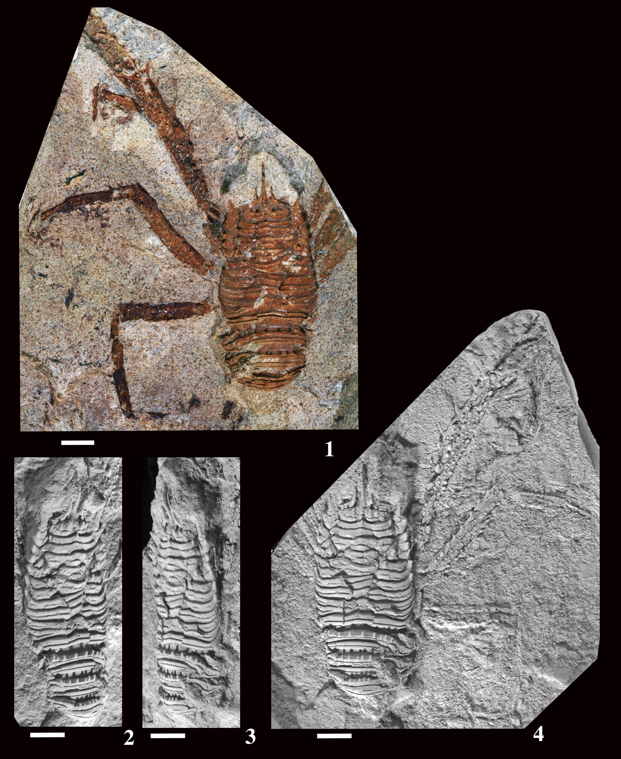
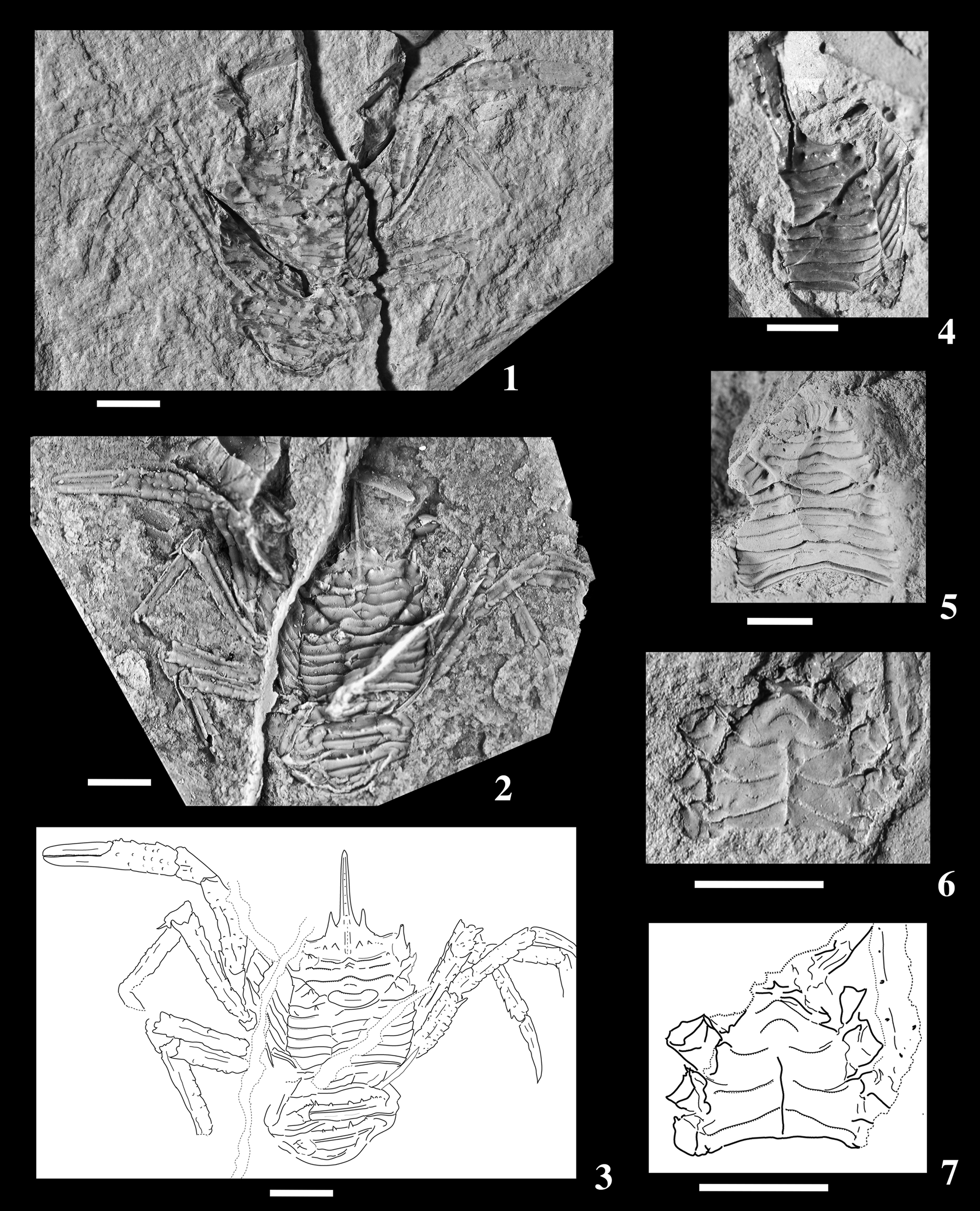
Trapezionida chiyoensis new species from the Yonekawa Formation, lower Miocene, Chiyo (Fig. 1.17). (1) Inner view of external mold of carapace, pleon, and pereiopods, IGPS 109984, holotype. (2) Dorsal view of latex cast of IGPS 109984. (3) Line drawing of latex cast of IGPS 109984. (4) Inner view of external mold of carapace, IGPS 109985, paratype. (5) Inner view of external mold of carapace figured by Imaizumi (Reference Imaizumi1971), IGPS 112851, paratype. (6) Ventral view of external mold of thoracic sternum, IGPS 109986, paratype. (7) Line drawing of IGPS 10998. (1, 2) Images coated with magnesium oxide. Scale bars = 2 mm.

Types
Holotype, IGPS 109984, external mold of carapace, pleon, and pereiopods; paratype, IGPS 109985, external mold of carapace; paratype, IGPS 109986, external mold of thoracic sternum; paratype, IGPS 112851, external mold of carapace; all from the lower Miocene Yonekawa Formation, Chiyo, Nagano, Japan.
Diagnosis
Carapace excluding rostrum, subrectangular, slightly longer than wide, widest at posterior one-fifth. Rostrum spiniform, slender, long, about 0.6 times as long as carapace, slightly directed upward, gently arched anteriorly, with rimmed lateral margins; dorsal carina well developed, dentated dorsally, extending to protogastric region. Frontal margins gently concave, rather oblique, rimmed. Lateral margins nearly straight, widened posteriorly, with seven spines. Posterior margin convex forward, rimmed. Dorsal surface gently convex longitudinally and transversely, covered with main, transverse terraced ridges; secondary transverse ridges present only on gastric regions, weak, discontinuous; anterior margins of main and secondary ridges fringed with dense, minute denticles. Epigastric regions with row of spines; inner spine posterior to base of supraocular spine largest, followed by spine on midpoint of epigastric region and spine on lateral corner, other spines subtle; transverse ridge behind row of epigastric spines obscure; small median spine behind rostral carina. Median spine present on anteriormost ridge of protogastric region. Cervical and postcervical grooves distinct. Parahepatic, branchial dorsal, and postcervical spines present. Pterygostomian regions not visible dorsally. Tergites of pleonal somites 2 with five pairs of spines on anterior ridge; somite 3 with two pairs of spines on anterior ridge. P1–4 long, slender; lateral surfaces of merus, carpus, and palm covered with sparsely arranged tubercles and spines inclined distally. P1 two times as long as carapace length excluding rostrum; merus 0.8 times carapace length; propodus with median row of seven spines on lateral surface; both fingers slightly shorter than palm with straight opposable margins. Meri, carpi, and propodi of P2–4 armed with spines on extensor and flexor margins; spines of flexor margins larger than those of extensor margins. P2 merus as long as P1 merus, with seven spines on both margins. P3 merus 0.8 times as long as P2 merus, with five spines on both margins. Merus of P4 0.7 times as long as that of P2, with five spines on both margins. Thoracic sternum trapezoidal, as long as wide, widest at sternite 7; sternite 3 3.5 times as wide as long; sternite 4 trapezoidal, 0.45 times wider than long; anterior margin of sternite 4 transverse, contiguous to sternite 3 for 0.75 width of sternite 3.
Occurrences (Fig. 1)
The present species are only known from the type locality.
Loc. 17.––Cliff in Chiyo, Yonekawa, Iida City, Nagano Prefecture (35°25′10″N, 137°51′49″E); mudstone of the Yonekawa Formation; early Miocene (NPD2B Zone of Yanagisawa and Akiba's (Reference Yanagisawa and Akiba1998) scale of diatoms by Ito et al., Reference Ito, Irizuki and Iwai1999) (Imaizumi, Reference Imaizumi1971 as Eumunida ? sp.).
Description
Carapace excluding rostrum, subrectangular, slightly longer than wide, widest at posterior one-fifth. Rostrum spiniform, slender, long, 0.6 times as long as carapace excluding rostrum, slightly directed upward, gently arched anteriorly, flanked by supraocular spines, with rimmed lateral margins; dorsal carina well developed, dentated dorsally, extending to protogastric region; shallow longitudinal grooves between bases of rostral and supraocular spines. Frontal margins gently concave, slightly oblique laterally, rimmed, 0.8 times maximum width; bases of supraocular spines preserved. Lateral margins nearly straight, widened posteriorly, with rounded posterolateral corners; anterolateral spines slender, long, directed slightly anterolaterally; hepatic margin with one short spine directed anterolaterally in front of deep cervical notch; anterior branchial margins with three short spines directed anterolaterally; posterior branchial margin bearing two short spines directed anterolaterally behind shallow postcervical notch. Posterior margin about as long as frontal margin, convex forward, with smooth rim. Dorsal surface gently convex longitudinally and transversely, covered with main, transverse terraced ridges gently sloping posteriorly and steeply sloping anteriorly; secondary transverse ridges present only on gastric regions, weak, discontinuous; anterior margins of main and secondary ridges fringed with dense, minute denticles. Anterior part of epigastric regions with row of spines; inner spine posterior to base of supraocular spine largest, followed by spine on midpoint of epigastric region and spine on lateral corner, other spines subtle; transverse ridge behind row of epigastric spines obscure; small median spine behind rostral carina. Short median spine present on anteriormost ridge of protogastric region. Cervical and postcervical grooves distinct. Short parahepatic, branchial dorsal, and postcervical spines present on each side of carapace. Pterygostomian regions not visible dorsally.
Tergites of pleonal somites 2 and 3 with two transverse ridges, each ridge interrupted by transverse stria. Somite 2 bearing five pairs of spines on anterior ridge; median and submedian pairs of spines evenly spaced; third, fourth, and fifth pairs on lateral side close to each other. Anterior ridge of somite 3 with two pairs of spines evenly spaced.
P1–4 long, slender; lateral surfaces of merus, carpus, and palm covered with sparsely arranged tubercles and spines inclined distally. Left P1 two times as long as carapace length excluding rostrum; merus 0.8 times carapace length; carpus 0.4 length of merus; propodus three times as long as carpus, with a median row of seven spines directed distally on lateral surface; both fingers slightly shorter than palm, bearing longitudinal groove on lateral surface, with straight opposable margins. Meri, carpi, and propodi of P2–4 armed with spines on extensor and flexor margins; spines of flexor margins larger than those of extensor margins. Merus of P2 as long as that of P1, bearing seven spines on both margins, distal spine of flexor margin strong; carpus 0.3 length of merus, with relatively long distal spine on extensor margin; propodus 0.7 length of merus; dactylus 0.8 length of merus. Merus of P3 0.8 times as long as that of P2, armed with five spines on both margins, distal spine of flexor margin large; carpus 0.4 length of merus; propodus 0.85 length of merus; dactylus 0.6 length of merus. Merus of P4 0.7 times as long as that of P2, bearing five spines on both margins, distal spine of flexor margin strong; carpus 0.4 length of merus; propodus as long as merus; dactylus 0.7 length of merus. P5 merus preserved, half-length of P2 merus, much slender than other meri.
Thoracic sternum trapezoidal, as long as wide, widest at sternite 7. Sternite 3 3.5 times as wide as long. Sternite 4 trapezoidal, 0.45 times wider than long, with convex groove medially; anterior margin transverse, contiguous to sternite 3 for 0.75 width of sternite 3; sternites 5–7 with median groove; sternal sutures 4/5 and 5/6 incomplete, suture 6/7 nearly complete.
Etymology
The specific name is derived from “Chiyo” in Yonekawa, the place name of the type locality.
Examined and illustrated materials
IGPS 109984 (holotype), external mold of carapace, pleon and pereiopods, and latex cast; IGPS 109985 (paratype), external mold of carapace; IGPS 109986 (paratype), internal mold of thoracic sternum; IGPS 112851 (paratype, figured in Imaizumi, Reference Imaizumi1971), external mold of carapace.
ZooBank
urn:lsid:zoobank.org:act:B3099E47-8DF8-47BB-98C2-30887901290C
Measurements (mm)
IGPS 109984 (holotype), 4.0 × 4.3.
Remarks
The present species is the first fossil record for Trapezionida. Karasawa (Reference Karasawa1993) identified Eumunida ? sp. reported from the Yonekawa Formation of Chiyo by Imaizumi (Reference Imaizumi1971) (Fig. 7.5) with Munida nishioi (as Grimothea nishioi in this work) by having dorsal spines of the carapace. Examination of the additional specimens collected from the same locality by R. Imaizumi and stored in the Tohoku University Museum shows that these specimens differ from those of Grimothea nishioi because the carapace has the rostrum with a dentate median carina and the epigastric region with a row of three distinct and subtle spines, and the pereiopod 1 is about two times as long as the carapace length excluding the rostrum. The anterior ridge of the pleonal somite 3 is armed with four spines, but that in G. nishioi has eight spines.
Acknowledgments
This study is dedicated to our best colleague, the late R.M. Feldmann (Kent State University), whose advice, support, and kindness helped us in different times. We are indebted to J. Nemoto (Tohoku University Museum) for his assistance in reviewing the holotype of Kazuoia ogaensis, T. Ichihara (College of Nagoya Bunri University), T. Kaede (Mizunami), K. Nishio (Tokai Fossil Society), T. Sato (Akita University), and A. Ujihara (formerly Nagoya University) for donating additional specimens for our study, and T. Goda (Konan), M. Ichiwara (Mobara), and T. Kaede (Mizunami) for preparing some specimens. We appreciate the reviews of A.A. Klompmaker (guest editor of this volume, University of Alabama), C.M. Robins (University of Alabama), and an anonymous reviewer, whose comments greatly improved the manuscript.
Declaration of competing interests
The authors declare none.