Repair of common arterial trunk using an extracardiac right ventricular to pulmonary arterial conduit is the preferred method in most cardiac surgical centres. Reoperation is a fact of life for survivors of common arterial trunk and related cardiac lesions who have undergone such repairs. Long-term survivors may require periodic conduit revisions, with a potentially escalating technical difficulty and risk. Herein we present an analysis of the currently available choices for extracardiac conduits, and outline what we consider to be a safe strategy for conduit revision. The discussion also applies to related anatomic situations for which ventricular to pulmonary arterial conduits are employed, such as pulmonary atresia with ventricular septal defect, discordant transposition with ventricular septal defect and left ventricular outflow tract obstruction, etc. The general principles and strategy outlined herein have been employed by my own surgical teams in three institutions, and my personal experience includes over 200 conduit revisions with one mortality.
The scope of the problem (conduit failure)
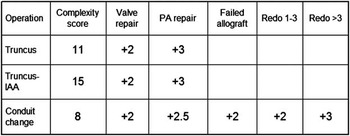
There are a number of potential indications for reoperation in common arterial trunk, including persistent anatomic lesions, truncal valve dysfunction, pulmonary artery stenosis, pulmonary insufficiency following valveless repair, endocarditis, and others. However, the most common indication by far is dysfunction – insufficiency, stenosis, or both – of a right ventricular to pulmonary arterial conduit placed at the time of primary operation. Although most reoperations for conduit replacement are straightforward, occasionally they can become more challenging. In Figure 1, the Aristotle complexity score for common arterial trunk repair and reoperations is presented. One can see that, by consensus of experienced surgeons, there is an escalating complexity for successive operations, especially when additional concurrent procedures are required.
Aristotle complexity score for circulatory arrest time (CAT) operations and reoperations. Complexity score for reoperation increases with successive procedures, failed homograft conduit type, and when concurrent corrective procedures are required. Theoretically, the complexity score for reoperation can exceed that of the primary CAT repair.

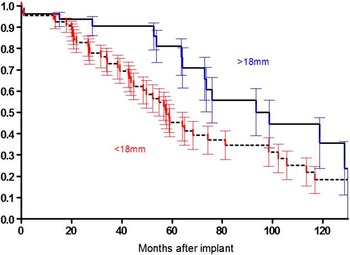
It is therefore fundamental to attempt to limit the number of conduit revisions required over a patient's lifetime. If a right ventricular to pulmonary arterial conduit is used at the initial repair of common arterial trunk, the most important factor in achieving this aim will be conduit longevity, a composite goal definable as freedom from reoperation combined with freedom from conduit dysfunction. In a Melbourne report going back to 1991, we noted that for 215 primary right ventricular to pulmonary arterial conduit implants performed for common arterial trunk and related anomalies the early mortality risk was 3.7% (confidence limits 1–7%), with a survival probability beyond 52 months of 95% (confidence limits 91–99%; Fig 2).Reference Sano, Karl and Mee 1 These favourable survival statistics were compromised by the high probability of need for reoperation for conduit replacement, and the results were especially disappointing for conduits <18 mm in diameter, irrespective of conduit type (xenograft versus allograft). In the cited series, the median interval to conduit replacement was 56.7 months (Fig 1). Similar results have been noted and reported in the literature by virtually every large paediatric cardiac surgical centre over the past three decades, with only limited improvement as new conduits were introduced – and in some cases withdrawn.
Actuarial freedom from conduit replacement for 215 primary right ventriclular to pulmonary arterial conduit implants for varying anatomic indications, at median age 23 months (0–325). Overall early mortality risk was 3.7% and survival probability beyond 52 months was 94.8%. The median interval to conduit replacement was 56.7 months (12.9–151). Results were similar for allografts and xenografts, but significantly worse for conduits <18 mm in diameter 9 = 0.024, hazard ratio = 1.86).Reference Sano, Karl and Mee 1 These findings have been reproduced in many other studies.

Are results better in the current decade? A recent report from a well-experienced team in Sankt Augustin relates specifically to common arterial trunk primary repairs using either allografts or xenograft conduits and defines our current expectations well.Reference Sinzobahamvya, Boscheinen and Blaschczok 2 In this study, 10-year freedom from any first, second, and third conduit reintervention was 17.9%, 46.1%, and 81.9%, respectively, whereas freedom from the first conduit replacement was 87.5%, 64.1%, and 39.5% at 1, 3, and 5 years, respectively. On the basis of this study, 2.6 interventions per survivor per 10 years of follow-up could be anticipated.
Why do conduits fail? Multiple recognised factors culled from a meta-analysis of the surgical literature suggest that a number of variables influence the time interval from conduit implant to failure.Reference Askovich, Hawkins and Sower 3 – Reference Mokhles, Rizopoulos and Andrinopoulou 5 Although experience is somewhat variable in different units, most surgical teams would consider some or all of the following to be important:
-
1. High right ventricular pressure (pulmonary vascular disease, branch pulmonary arterial stenosis, pulmonary arterial arborisation abnormalities)
-
2. Conduit–patient size mismatch (Z-score outside “ideal” range of +1 to +3)
-
3. Patient age <1 year (or smaller diameter conduit)
-
4. Conduit type and preservation technique (allograft versus xenograft)
-
5. Extra-anatomic course of conduit (as compared with Ross operation)
-
6. Directly retrosternal conduit position
-
7. ABO mismatch (allografts).
Valved conduits suitable for the pulmonary circulation
Most surgical teams view the allograft pulmonary valve as the gold standard for comparison of all new valves suitable for use in right ventricular to pulmonary arterial extracardiac conduits, and certainly there is a long and successful history in cardiac surgery.Reference Askovich, Hawkins and Sower 3 – Reference O'Brien, Harrocks and Stafford 6 There have been historical claims for the superior longevity of ABO-matched cryopreserved pulmonary allografts over other conduit valve types, but not all implanting institutions would support the argument.Reference Homann, Haehnel and Mendler 7 Setting conduit longevity aside for the moment, one important consideration in the employment of allograft valves is availability, which in Australia is especially poor for conduits under 18–20 mm in diameter. The situation is most problematic for neonates with common arterial trunk who are unable to wait indefinitely for availability of a valve, an issue that seems unlikely to be resolved in the near future. Therefore, we have explored the option of reduction plasty of larger allografts to form bicuspid valves suitable for neonates. Our own experience suggests that results are superimposable on those of intact trileaflet allografts, as reported previously by Michler et alReference Michler, Chen and Quaegebeur 8 and Breamer et al.Reference Bramer, Mokhles, Takkenberg and Bogers 9
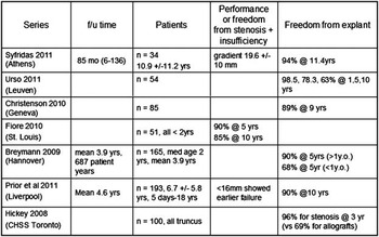
The conduit that perhaps has received the most intense scrutiny is the Contegra (Medtronic) xenograft.Reference Sfyridis, Avramidis, Kirvassilis, Zavaropoulos, Papagiannis and Sarris 10 – Reference Hickey, McCrindle and Blackstone 15 The introduction of the Contegra conduit in Europe and subsequently the United States of America in the 1990s generated a great deal of interest, as it was seen as a possible solution to some of the problems inherent in allograft valves. Contegra is a glutaraldehyde preserved bovine jugular vein segment, which is available off the shelf in 12–22-mm diameters, with or without an external stent. The storage and handling requirements are greatly simplified over what is required to maintain an in-house allograft valve bank, and some of the variability of allografts and handmade pericardial or synthetic conduits can be eliminated. Has the promise of the Contegra conduit actually been realised? In Figure 3, we present a summary of some of the larger surgical series that have appeared in the literature over the past 5 years. The world experience is now quite extensive, and one would cautiously conclude that although some problems have been noted, on balance the Contegra outcome has performed better than previous allograft and xenograft iterations.Reference Sfyridis, Avramidis, Kirvassilis, Zavaropoulos, Papagiannis and Sarris 10 – Reference Hickey, McCrindle and Blackstone 15 This impression, combined with the availability factor, makes the Contegra our preferred right ventricular to pulmonary arterial valved conduit. Moreover, the Contegra valve – modified for transcatheter implantation within a stent – forms the basis for the percutaneous Melody valve system™, which has had an excellent acute haemodynamic outcome, which appears to be sustained at mid-term despite the occasional occurrence of stent fracture.Reference Lurz, Coats and Khambadkone 16 , Reference McElhinney, Hellenbrand and Zahn 17 The interventional or hybrid approach – transapical right ventricular puncture through limited incisions – to conduit revision without cardiopulmonary bypass is attractive and certainly competitive with an open surgical approach for anatomically suitable patients. Although the Melody valve system is currently not approved in Australia for primary implantation in the right ventricular outflow tract, as an alternative to conduit replacement it is very likely to become the procedure of choice for suitable candidates, reserving surgery for the unsuitable ones. Therefore, in the current era it is very important for the surgeon to consider technically facilitating a catheter-based or hybrid approach to future pulmonary valve replacements for candidates who may be unsuitable for that strategy at the time of initial conduit change. This typically involves appropriate sizing of the conduit and provision of suitable anchoring points for a stented valve, as well as preservation of vascular access. The Melody valve has also been used in a number of off-label situations apart from right ventricular to pulmonary arterial conduits both on the right and left side of the circulation.Reference Hasan, McElhinney and Brown 18 , Reference Roberts, Boudjemline and Cheatham 19
Recent peer-reviewed publications in the surgical literature dealing with performance of the Contegra conduit. The results in this meta-analysis support the view that outcome is equal to or possibly better than allograft conduits in similar patients. Randomised prospective studies are lacking, and would probably be difficult to conduct at present. CHSS = Congenital Heart Surgeons’ Society.

Other options for right ventricular to pulmonary arterial conduit reconstruction exist, including hand-sewn valves placed within polytetrafluoroethylene conduits, autologous pericardial hand-sewn valves, and specially engineered hand-sewn polytetrafluoroethylene valved tubes, as reported by Quintessenza et al,Reference Quintessenza, Jacobs, Morell, Giroud and Boucek 20 Schlicter et al,Reference Schlichter, Kreutzer and Mayorquim 21 and Nunn et al,Reference Nunn, Bennetts and Onikul 22 and others.23, Reference Miyazaki, Yamagishi and Maeda 24 Although a detailed discussion of these options is outside the scope of this paper, some of the alternatives are worth citing. In 2012, Miyazaki et alReference Miyazaki, Yamagishi and Maeda 24 reported results of a Japan-wide study of modified polytetrafluoroethylene valved conduits with Valsalva-like sinuses. Following 325 implants, freedom from reoperation was 95.4% at 10 years, with 95% of the conduit valves remaining competent, and showing a mean gradient of 14 ± 13 mmHg. Although not yet available outside Japan, this conduit has had a better long-term outcome than any other similarly purposed device reported in the literature to date.23
All of the handmade devices cited above have had a relatively good early outcome in the hands of the reporting teams, even if such experience has not always been reproducible in other units. In Australia and the United States of America, there may be legal considerations surrounding implantation of handmade non-regulatory body (Therapeutic Goods Administration or Food and Drug Administration)-approved devices. Good results not withstanding, such practice is technically neither an off-label use of an approved device, nor approved use based on an exemption status. Needless to say, problems could arise in the event of an adverse outcome, even if it were unrelated to the conduit itself.
Mechanical valves have been used in situ within the right ventricular outflow tract obstruction, as well as within right ventricular to pulmonary arterial conduits.Reference Haas, Schreiber, Hörer, Kostolny, Holper and Lange 25 – Reference Shin, Kim, Ko, Park and Seo 28 On the basis of limited data, mechanical valves do tend to remain competent and non-restrictive at late follow-up, and the disadvantage is primarily the need for warfarin therapy. The best candidates for mechanical valves in pulmonary position are probably those who have undergone multiple operations in the past, or who face an otherwise unacceptable risk for reoperation. In suitable patients, the potentially increased longevity of a mechanical valve placed within a conduit may well outweigh the risk of chronic warfarin therapy. However, this particular type of valved conduit reconstruction has not been widely accepted by cardiologists in the United States of America, nor in Australia, and has seen somewhat limited use by the few reporting centres. Tissue valves often remain in situ for years with moderate dysfunction, not severe enough to warrant replacement, yet also not addressing the right ventricular dysfunction problem completely. In a sense the patient pays the price of the reoperation without full benefit. The place of mechanical valve implants – in the outflow tract or within right ventricular to pulmonary arterial conduits – in solving this problem, as either a primary or secondary solution, remains to be defined.
Finally, the development of a long-lasting tissue-engineered – synthetic scaffold based – autologous valved conduit has been under investigation for decades by a number of investigators.Reference Perri, Polito and Esposito 29 – Reference Dohmen, Lembcke, Holinski, Pruss and Konertz 31 A recent report by Yamanami et al from Kyoto presents preliminary results in animals, with a technique using an implantable mandrill with sinus and valve portions, on which an autologous valved conduit can be cultivated with no synthetic material. Although animal studies are encouraging, it would appear that this type of completely autologous valved conduit is some years away from suitability for routine human implantation. Non-valved autologous tissue-engineered conduits have been successfully implanted in humans as conduits in the extracardiac Fontan operation.Reference Naito, Imai and Shin'oka 32
Common arterial trunk repair can be performed without a conduit, as reported by Barbero-Marcial et alReference Barbero-Marcial, Riso, Atik and Jatene 33 and there is now an extensive experience. There are a number of techniques available for valveless – or monocuspid valved – reconstruction, some of which may be generalised to anatomical situations other than common arterial trunk.Reference Chiu, Div and Huang 34 – Reference Chen, Glickstein and Davies 38 In the hands of proponents, the non-valved repair for common arterial trunk does not appear to increase the operative risk as compared with that of valved repairs. The interval to reoperation is probably longer, but not necessarily the interval to reintervention.Reference Chen, Glickstein and Davies 38 It has been suggested that a non-valved conduit may result in better pulmonary arterial growth, although one would say that it is difficult to reconcile this strategy with long-term data regarding right ventricular failure in tetralogy of Fallot when free pulmonary incompetence is present.
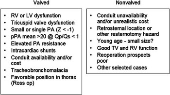
Decision making for right ventricular outflow tract reconstruction at primary or secondary operation for common arterial trunk and related anomalies is not always straightforward. We have summarised our own view, based on experience with valved and non-valved techniques (Fig 4). In general, if conduits are readily available, and if the health-care system supports their use in terms of cost, then both the short- and long-term results are likely to be better. Clearly, an effective counter-argument can be made, as non-valved repairs are routinely used by some excellent surgical teams. The majority of common arterial trunk survivors in Australia do have valved conduits that will eventually require replacement. Currently, in Australia, the price differential between valved allograft conduits, valved xenografts conduits, and mechanical valves is not significant. For comparison, in Australia, the Melody valved conduit implant system currently costs approximately six times the price of the surgically implantable Contegra conduit, although total hospital costs are considerably reduced as compared with surgery. Handmade conduits involving polytetrafluoroethylene tubes or autologous pericardium are of course much less expensive, and the availability issue is much less problematic.
Decision making for valved versus non-valved repair of circulatory arrest time and related anatomic situations: listed are factors that may influence the surgical team to take one approach over another. However, in practice, most surgeons apply one of the two strategies as a routine. A selective approach would be reasonable given that acceptable results can be obtained with either valved or non-valved repairs. RV = right ventricle; LV = left ventricle; PA = pulmonary artery; TV = tricuspid valve.

Technical strategy for conduit change
The goals for conduit change in common arterial trunk are straightforward, and include the following:
-
1. Safe sternal re-entry
-
2. Establishment of a competent unobstructed right ventricular to pulmonary arterial connection
-
3. Preservation of myocardium
-
4. Avoidance of coronary and phrenic nerve injury
-
5. Correction of associated cardiovascular lesions
-
6. Limitation of number of future operations
-
7. Facilitation of catheter-based or hybrid approach to future pulmonary valve replacement.
Although traditionally a 50-mmHg peak systolic gradient from right ventricle to pulmonary artery under rest conditions has been used as the basic indication for conduit revision, our current understanding suggests that there is no immutable indication for conduit replacement relating solely to actual degree of obstruction. Insufficiency can be even more subjective in this regard. The decision to reoperate is generally taken in the context of the functional status of the right ventricle, taking into account all features relating to the underlying anatomy and physiology. A full discussion of indications lies outside the scope of this paper, but information has been presented in detail elsewhere.Reference Tirilomis, Friedrich, Zenker, Seipelt, Schoendube and Ruschewski 39 , Reference Belli, Salihoğlu and Leobon 40 Briefly, adequate pre-operative diagnostic information is essential for planning a conduit change, whether performed surgically or interventionally. The need for specific haemodynamic and imaging studies is dictated by individual cardiac anatomy, and assessment usually requires some combination of catheter studies, echocardiography, computerised tomography, and magnetic resonance imaging. Contrast echocardiography to look for the presence of intracardiac shunts is an important part of the pre-operative work-up. Cassorla et alReference Cassorla, Miller-Hance, Rouine-Rapp, Reddy, Hanley and Silverman 41 looked at the problem of atrial septal defect detection in pre-operative patients of varying diagnosis, noting that such defects are extremely common in children under 1 year of age or under 10 kg, and that even transoesophageal echocardiography with bubble contrast is not 100% sensitive in detection. A high index of suspicion and reviews of prior operation reports is always warranted. For reoperations on univentricular hearts, there is probably no effective way to neutralise this particular risk. For the surgeon, lateral chest X-ray, computerised tomography, and magnetic resonance imaging are particularly useful in assessment of the retrosternal space and definition of the relationship of the conduit, the sternum, the right ventricle, and the aorta. If not known for certain from prior operations, the coronary anatomy should be specifically assessed with computerised tomography angiography or catheterisation and angiography to avoid coronary injury. There should be an assessment of the peripheral vascular status, which typically includes ultrasound interrogation of femoral, carotid, and jugular vessels, to define appropriate sites for possible peripheral cannulation.
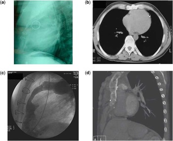
Resternotomy for any indication is a procedure that all experienced cardiac surgeons take seriously, the more so in the presence of cardiomegaly and cardiosternal symphysis, as may be encountered during conduit replacement for common arterial trunk. Many good techniques are available, and herein we describe our own preferences on the basis of 25 years of experience. Despite the fact that most reoperative conduit operations proceed without incident, there are certain cases that might present a higher risk than others, as shown in Figure 5.
Challenging resternotomy cases: ( a ) Hancock Dacron xenograft valved conduit embedded in the posterior sternal table; ( b ) obliteration of retrosternal space with cardiosternal symphysis; ( c ) conduit crossing midline retrosternally with compression. The degree of symphysis is underestimated on this cineangiogram, which highlights the lumen. This patient was about to undergo her 7th sternotomy and had sustained cardiac laceration at the previous two operations; and ( d ) severe sternal deformity and keloid formation with total cardiosternal symphysis. This patient had four prior sternotomies and Staphylococcal mediastinitis.

If right-sided cardiac laceration is encountered during resternotomy on a biventricular heart, rapid blood loss may follow. We follow the standard cardiac surgical teaching regarding the inadvisability of further dissection. Peripheral cannulation for cardiopulmonary bypass is performed while an assistant applies pressure to the chest wall to control bleeding. This usually allows the remaining dissection to be performed and the bleeding sites controlled.
Even with emergency peripheral cannulation and establishment of cardiopulmonary bypass, an empty beating heart has the potential to draw air across an interatrial communication, which can then be ejected to the aorta, causing cardiac and neurologic injury. Although we generally aim to close all interatrial communications at the time of primary operations, many surgeons elect to leave a small atrial septal defect for “protection” in the event of crisis situations involving right ventricular failure, diastolic dysfunction, pulmonary hypertension, etc. The benefit of such practice is debatable. In high-risk resternotomy patients, we would try to close an atrial septal defect with an occlusion device at the time of cardiac catheter study in order to eliminate this particular hazard during resternotomy.
Many surgeons attempt to reconstitute the pericardium at the time of conduit implantation. In some cases, the native pericardium can be advanced or meshed to extend it. More commonly, polytetrafluoroethylene membrane or other pericardial substitutes such as bovine pericardium, polyglycolate absorbable mesh, and silicone urethane polymers are used to facilitate the next sternotomy.Reference Loebe, Alexi-Meskhishvili, Weng, Hausdorf and Hetzer 42 , Reference Sakuma, Iguchi, Ikada and Tabayashi 43 Reconstitution with absorbable matrix – to be repopulated by host cells – is an intellectually appealing method.Reference Bel, Ricci and Piquet 44 However, the actual results of all pericardial reconstitution in terms of reducing the hazard of resternotomy vary significantly in the literature. The end points of such studies are sometimes vague, often involving subjective assessment of the severity of adhesions, histologic evaluation of explanted tissues, dissection time, and so forth. What this means from a technical point of view is difficult to interpret. It would be fair to say that polytetrafluroethylene membrane remains the gold standard when direct pericardial closure is not possible. In our own experience this technique probably has been quite useful in preserving a plane between heart and sternum, facilitating safe resternotomy, even if some of the epicardial landmarks tend to be blurred.
One of the more difficult anatomic situations for resternotomy is encountered when a conduit that crosses the midline retrosternally, as might follow classical – physiologic – conduit repair of discordant and transposition with ventricular septal defect and left ventricular outflow tract obstruction, or in children with situs and/or rotational abnormalites. In addition to being vulnerable to sternal compression at the time of primary operation, this sort of conduit can easily be entered during resternotomy, especially when it has been oversized. Another potentially vulnerable group includes some children with severe sternal deformity, either congenital or as a result of previous operations (Fig 5).
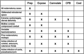
In Figure 6, we present some guidelines for resternotomy in high-risk cases. Our recommendations range from simple antiseptic preparation of peripheral vessels for possible cannulation – either cervical or femoral, depending on the size of the patient and pre-operative assessment – all the way to presternotomy peripheral cannulation and hypothermic cardiopulmonary bypass, as might be used for infected aortic aneurysms. Traditionally, femoral cannulation – or external iliac – is preferred for establishment of cardiopulmonary bypass without entering the chest. Axillary cannulation is sometimes used in adults. However, cervical cannulation using a low oblique incision – following the anterior sternomastoid border – can be very useful for rapid institution of cardiopulmonary bypass, especially in children under 20 kg, or in bigger patients with no femoral access. In our hands, this technique is much faster and more effective than femoral cardiopulmonary bypass, and there has been a long and very successful experience using the cervical approach for venoarterial extracorporeal membrane organisation cannulation in the intensive care unit. We described a technique for cervical cannulation for extracorporeal membrane organisation in 1991, which we also have adapted for cardiopulmonary bypass.Reference Karl, Iyer, Sano and Mee 45 The technique has yielded an excellent long-term patency for both vessels following direct repair, which is almost always possible at the time of decannulation. Avoiding right-sided internal jugular lines in children who may need cervical cannulation is a useful practice.
Guidelines for the degree of preparedness before resternotomy under various anatomic circumstances: Clearly, each case must be considered on its own merits, bearing in mind that peripheral cannulation takes time, even under non-emergent conditions, and that peripheral cannulation as a prophylatic measure in high-risk cases is safe and reproducible. CPB = cardiopulmonary bypass.

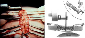
Figure 7 shows the resternotomy technique that we prefer, which is adapted from that of Stark and Pacifico.Reference Stark and Pacifico 46 Although no technique is 100% reliable, this one has been effective even in some very difficult anatomic situations. Following initial incision, the sternal wires used for sternal closure at prior operations are cut but left in situ. The ends are elevated and held on tension with wire drivers. Then a micro-oscillating saw is used to cut down to and just through the wires, thereby limiting the depth of the cut and protecting the heart from injury. Wire fragments are removed, and the sternal edges are elevated with bone hooks to facilitate liberation of small portions of the right atrium and aorta, no more than what is required for cannula insertion for cardiopulmonary bypass. The remainder of the dissection is done on cardiopulmonary bypass, using electrocautery, and limited to just what is needed to liberate the conduit, unless other intracardiac surgery is required. If the patient is already on cardiopulmonary bypass via peripheral cannulation, only the conduit itself needs to be dissected, otherwise the dissection is limited to the conduit and cannulation sites.
Stark technique for resternotomy: following initial incision, the sternal wires used for sternal closure at prior operations are cut but left in situ. The ends are elevated and held on tension with wire drivers. Then an oscillating saw is used to cut down to and just through the wires, thereby limiting the depth of the cut and protecting the heart from injuryReference Stark and Pacifico 46 (used by permission).

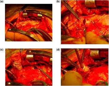
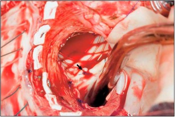
In the absence of an interatrial or interventricular communication, we aim to perform the entire operation at normothermia with a beating heart. However, if an atrial septal defect requires closure, this part of the procedure is performed before opening the conduit, using a brief period of induced ventricular fibrillation, eliminating the need for dissection of the aorta for cross-clamping. The conduit is then retracted medially with suture and excised completely, which is usually somewhat easier with Dacron conduits than with allografts (Fig 8). In either case, the transition point to native tissue is usually obvious. The branch pulmonary arteries are calibrated with Hegar dilators, and if no revision is required, distal and then proximal conduit anastomoses is performed. The distal end of the conduit is cut just above the valve commissures at a slight bevel, keeping the valve close to the pulmonary artery in the coronal plane to avoid compression following sternal closure. A simple running suture technique is used for both ends of the Contegra conduit. The proximal end can be cut as obliquely as required to eliminate the need for additional patch material.
Hancock 12-mm xenograft Dacron conduit explant 4 years after circulatory arrest time repair: ( a ) following limited dissection. The conduit is retracted medially with stay sutures. ( b ) In the absence of intracardiac shunts, the conduit is opened with beating heart normothermic cardiopulmonary bypass. ( c ) Conduit is excised back to native tissue. Taking care to avoid coronary injury. ( d ) Pulmonary arteries are calibrated to assess need for enlargement or intra-operative stent placement.

Surgical teams in Leiden and the Mayo clinic have reported experience with intra-operative pulmonary stents, a technique that we also like in Brisbane, especially for older patients with proximal or distal branch pulmonary artery stenosis who require conduit revision (Fig 9).Reference Bökenkamp, Blom, De Wolf, Francois, Ottenkamp and Hazekamp 47 , Reference Menon, Cetta, Dearani, Burkhart, Cabalka and Hagler 48 This technique is expedient as compared with a direct surgical approach, and is probably more effective in solving pulmonary artery problems that result from aortic compression or lack of space within the transverse sinus. In smaller patients, redilatable stents can be used. Intra-operative stent placement is gaining acceptance worldwide, as the surgical approach to branch pulmonary artery stenosis has been somewhat unreliable at best and may require a significant additional dissection – or even cardioplegic arrest and aortic transection.
Intra-operative pulmonary arterial (PA) stents: bare metal stent in the right PA, placed intra-operatively at the time of pulmonary valve implantation. Direct vision deployment of stents in the PA is technically straightforward and effective in overcoming PA stenosis related to compression in the transverse sinus. Anchoring of the stent with sutures is also possibleReference Menon, Cetta, Dearani, Burkhart, Cabalka and Hagler 48 (used by permission).

Sternal closure following conduit revision can be done with pericardial reconstitution as discussed above, and any peripheral vascular cannulation sites should be meticulously reconstructed. Post-operative intensive care management is usually straightforward.
Summary
Common arterial trunk patients today have a much improved survival probability over their counterparts from earlier eras, and the population of children who will require conduit revision is growing. It is incumbent upon the surgical and interventional teams to perform this revision in a safe and expedient manner, with the goal of limiting or reducing the need for future operations, and facilitating future catheter-based interventions. Attention to risk factors and team preparation will ensure this outcome. The future of revisional conduit surgery will see increasing use of catheter-based techniques.