INTRODUCTION
Schistosomiasis is one of the neglected diseases and is caused by blood flatworms of the genus Schistosoma. It currently afflicts ∼200 million people in 76 countries worldwide (Gryseels et al. Reference Gryseels, Polman, Clerinx and Kestens2006). The most prevalent species are Schistosoma mansoni, Schistosoma japonicum (S. japonicum) and Schistosoma haematobium. In China and South Asia, schistosomiasis japonica caused by S. japonicum is one of the most serious public health problems (Zhang et al. Reference Zhang, Zhu, Ward, Xu, Zhang, Guo, Zhao and Jiang2012b).
Schistosomes have a complex life cycle involving two different hosts: snails and humans. Larval schistosomes (cercariae) are released by intermediate hosts, freshwater snails, and subsequently invade their definitive hosts such as human or other mammals. Once in a mammalian host, cercariae develop into schistosomula and adult worms reside in the portal mesenteric system of the host for several years. A large number of eggs produced by paired male–female schistosomes not only results in a severely pathological reaction in the final host but is also responsible for disease dissemination (Andrade, Reference Andrade2009). Therefore, precise identification of infected reservoirs is crucial for the implementation of drug treatment for schistosomiasis control. Currently, discerning schistosome eggs by direct microscopy is considered to be a ‘gold’ standard for the diagnosis of schistosomiasis. However, the method is limited in its sensitivity, and is time consuming and a laborious procedure (Xu et al. Reference Xu, Gordon, Hu, Mcmanus, Chen, Gray, Ju, Zeng, Gobert, Ge, Lan, Xie, Jiang, Ross, Acosta, Olveda and Feng2012). Consequently, there is still great need for fundamental studies to identify novel diagnostic biomarkers and drug targets for schistosomiasis control.
MiRNAs are small, endogenous non-coding RNA molecules that regulate gene expression post-transcriptionally by targeting the 3′ untranslated region (3′ UTR) of messenger RNAs (Bartel, Reference Bartel2004). MiRNAs regulate genes involved in key biological processes such as development, differentiation, proliferation and apoptosis (Bushati and Cohen, Reference Bushati and Cohen2007). Recently it has been shown that significant levels of miRNA occur in extracellular human body fluids and these abundant and specific miRNAs in plasma or serum can serve as a novel class of biomarkers for cancer and other diseases (Schwarzenbach et al. Reference Schwarzenbach, Hoon and Pantel2011). In addition, cultured mammalian cells also export miRNAs into the extracellular environment (Valadi et al. Reference Valadi, Ekstrom, Bossios, Sjostrand, Lee and Lotvall2007). Schistosomes are also able to excrete certain proteins in the circulation of a final host to facilitate their parasitism (Hewitson et al. Reference Hewitson, Grainger and Maizels2009; Liao et al. Reference Liao, Yuan, Xiao, Liu, Lv, Zhao and Wu2011). Although numerous miRNAs have been identified in S. japonicum (Xue et al. Reference Xue, Sun, Zhang, Wang, Huang and Pan2008; Hao et al. Reference Hao, Cai, Jiang, Wang and Chen2010; Cheng and Jin, Reference Cheng and Jin2012), it remains unknown whether there are schistosome-specific miRNAs in the circulation of final hosts infected with S. japonicum.
In the present study, we applied the Illumina Solexa high-throughput sequencing (deep sequencing) to characterize schistosome-specific small RNA populations in the plasma of rabbits infected with S. japonicum, resulting in the identification of five schistosome-specific miRNAs. Then, four of the five schistosome-specific miRNAs identified were further confirmed by real-time RT-PCR.
MATERIALS AND METHODS
Animals and parasites
The animal study protocol was approved by the Animal Care and Use Committee of the Shanghai Veterinary Research Institute, Chinese Academy of Agricultural Sciences. The procedures involving animals were carried out in accordance with the guidelines of the Association for Assessment and Accreditation of Laboratory Animal Care International (AAALAC). The life cycle of S. japonicum (Anhui isolate, China) was maintained in New Zealand rabbits and Oncomelania hupensis in the Shanghai Veterinary Research Institute of China. Unless indicated otherwise, New Zealand rabbits and BALB/c mice were infected with approximately 1000 and 100 cercariae, respectively. Rabbit blood samples of about 10 mL from each rabbit were obtained from the carotid artery at the indicated date. There were four groups (uninfected, 16, 28 and 35 days post infection (dpi)) and four rabbits in each group for blood collection. The different stages of schistosomes (7, 14, 22 and 35 dpi) were collected from the infected rabbits through the perfusion of the hepatic portal system according to the method described by Yolles et al. (Reference Yolles, Moore, De Giusti, Ripsom and Meleney1947). Eggs were also isolated from the livers of infected rabbits.
Plasma preparation and RNA isolation
The New Zealand rabbits infected with S. japonicum cercariae were euthanased at 16, 28 and 35 dpi. Blood was collected into a 50 mL tube containing sodium citrate as anticoagulant and then the blood sample was centrifuged for 10 min at 1900 g at 4 °C. The supernatant was transferred into a fresh tube and then centrifuged for 10 min at 16 000 g at 4 °C. The supernatant was then equally divided into three parts for RNA isolation by using QIAamp Circulating Nucleic Acid kit (Qiagen, Germany), Trizol (Invitrogen, Grand Island, NY) and RNAiso plus (Takara, China), respectively. The isolated plasma RNAs were pooled and then verified with an Agilent 2100 Bioanalyzer (Agilent Technologies, Palo Alto, CA).
Small RNA libraries and DNA sequencing
Small RNA libraries were constructed using the TruSeq Small RNA Sample Prep Kit (Illumina, San Diego, CA) according to the manufacturer's protocol. Briefly, approximately 0·1 μg of pooled plasma RNA was subjected to polyacrylamide gel electrophoresis (PAGE) and then the 18–30 nt size range of RNA was enriched for purification. The purified small RNA was subjected to sequencing with an Illumina Genome Analyzer (Illumina) according to the manufacturer's instructions. Briefly, proprietary adapters were ligated to the 5′ and 3′ termini of these small RNAs, and the ligated small RNAs were then used as templates for cDNA synthesis. The cDNA was amplified with 22 PCR cycles to produce libraries that were sequenced using Solexa's proprietary sequencing-by-synthesis method. DNA sequencing was performed with an Illumina Genome Analyzer at the BGI (Beijing Genomics Institute, Shenzhen, China).
Bioinformatic analysis of S. japonicum small RNAs
Raw datasets produced by deep sequencing from each library were subjected to further analysis. Clean reads were obtained after removing the low quality reads, adaptor null reads, insert null reads, 5′ adaptor contaminants, reads with smaller than 18 nt, and reads with polyA tail. Adapter sequences were then trimmed from both ends of clean reads. All identical sequences were counted and merged as unique sequences, herein referred to as sequence tags.
To identify schistosome-specific small RNA populations in the plasma of S. japonicum-infected rabbits, we performed the following analyses: (1) the clean reads derived from the RNA isolated from the plasma of rabbits infected with S. japonicum were compared with those derived from uninfected rabbits for determining the specific small RNA populations associated with S. japonicum infection. (2) The specific small RNA populations associated with schistosome infection in P1 (the pooled RNAs from the total RNAs isolated from the plasma of schistosome-infected rabbits at 16, 28 and 35 dpi) and P2 (the pooled RNAs from the total RNAs isolated from the plasma of schistosome-infected rabbits at 16 and 28 dpi) datasets were mapped to the draft S. japonicum genome sequences (sjr2_scaffold.fasta, downloaded from ftp://lifecenter.sgst.cn:2121/nucleotide/corenucleotide) using SOAP (Short Oligonucleotide Alignment Program) (http://soap.genomics.org.cn). Only small RNAs that mapped perfectly to the S. japonicum genome were taken for further analysis. (3) Small RNAs in the two libraries that mapped perfectly to the S. japonicum genome were further filtered by mapping to the rabbit genome (http://asia.ensembl.org/Oryctolagus_cuniculus/,Version65) using the SOAP. If a small RNA mapped perfectly to both the S. japonicum genome and the rabbit genome, it was excluded from further annotation. Only a small RNA uniquely mapped to the S. japonicum genome was retained for further annotation.
For annotation of small RNAs, the small population was first categorized as rRNA, tRNA, snRNA (small nuclear RNA) and snoRNA (small nucleolar RNA) by matching against the sequences of non-coding RNAs collected in Rfam (Version 10.1) and the NCBI GenBank data (http://www.ncbi.nlm.nih.gov/) by using BLAST. Then, these unmatched small RNAs were further analysed against miRbase (Version 18) and GenBank for determining the known miRNA or homology miRNA. Finally, remaining small RNAs were further analysed to predict new miRNA using MIREAP (http://sourceforge.net/projects/mireap). Predicted hairpin-like structures were folded using RNAfold (Zuker, Reference Zuker2003).
Mouse plasma RNA isolation
BALB/c mice were infected with about 100 cercariae by inoculating abdominal skin surface with a moist paste. At 16 and 25 dpi, blood samples of 0·3 mL from each mouse were collected from the orbital sinus of infected and uninfected mice (at least 3 mice) into a 2 mL tube (sodium citrate as anticoagulant). The pooled blood samples were first spun for 10 min at 1900 g at 4 °C and then the supernatant was then centrifuged for 10 min at 16 000 g at 4 °C. The plasma RNA was isolated by using mirVana PARIS Kit (Invitrogen) according to the manufacturer's instructions. For the detection of miRNA abundance in the mice infected with different number of S. japonicum cercariae, BALB/c mice were infected with about 50 and 100 cercariae as described above, respectively.
Real-time RT–PCR and semi-quantitative RT–PCR analysis of schistosome-specific miRNAs from the plasma of schistosome-infected mice
A miScript II RT Kit (Qiagen) was used to reverse-transcribe RNA to cDNA. The 20 μL reverse-transcription reactions contained 12 μL eluted solution (approximate 60 ng of total RNA), 4 μL 5×miScript HiFlex Buffer, 2 μL 10×miScript Nucleics Mix and 2 μL miScript Reverse Transcriptase Mix. The temperature program was 60 min at 37 °C, 5 min at 95 °C and then held at 4 °C. Real-time PCR was performed using miScript SYBR Green PCR Kit (Qiagen) in a Mastercycler ep realplex (Eppendorf, Germany). The 20 μL PCR reaction included 2 μL of RT product (1:5 dilution), 10 μL 2×QuantiTect SYBR Green PCR Master Mix, 2 μL 10×miScript Universal Primer, 2 μL 10×miScript Primer Assay and 4 μL H2O. The reactions were incubated in a 96-well plate at 95 °C for 15 min, followed by 40 cycles of 94 °C for 15 s, 55 °C for 30 s and 70 °C for 30 s. The miScript Primers for Bantam, miR-3479, miR-10 and miR-0001 were ordered from Qiagen (Qiagen's proprietary). Mouse glyceraldehyde 3-phosphate dehydrogenase (GAPDH) was used as the internal control (Forward primer: 5′ CAT GGC CTT CCG TGT TCC TA3′; Reverse primer: 5′CCT GCT TCA CCA CCT TCT TGA T 3′). All reactions were run in triplicate. The 2−ΔCt method was used to calculate relative miRNA abundance.
For semi-quantitative RT–PCR, a stem-loop based RT-PCR method was used to analyse miRNA abundance. A stem-loop RT primer was used to reverse-transcribe mature miRNAs to cDNAs (Bantam, 5′ GTC GTA TCC AGT GCA GGG TCC GAG GTA TTC GCA CTG GAT ACG ACA CCA GC 3′; miR-10, 5′ GTC GTA TCC AGT GCA GGG TCC GAG GTA TTC GCA CTG GAT ACG ACC CAA AC 3′; miR-3479,5′ GTC GTA TCC AGT GCA GGG TCC GAG GTA TTC GCA CTG GAT ACG ACC AAG GC 3′). The 10 μL reverse-transcription reactions contained approximately 40 ng of total RNA, 2 μL 5×PrimeScript® Buffer (Takara), 0·5 μL PrimeScript® RT Enzyme Mix I (Takara), and 0·5 μL specific RT primer (20 μ m). The temperature program was 30 min at 42 °C, 5 min at 95 °C and then held at 4 °C. The semi-quantitative PCR was performed in a Mastercycler ep realplex (Eppendorf). The 20 μL PCR reaction included 2 μL of RT product (1 : 1 dilution), 1× SYBR Premix Ex Taq II (Takara), 0·5 μ m specific forward primer (bantam_F, 5′ ATC GTA CGT GGG TGA GAT CGC 3′; miR-10_F, 5′ ATC GTA CGT GGG AAC CCT GTA 3′; miR-3479_F, 5′ ATC GTA CGT GGG TAT TGC ACT 3′), 0·5 μ m common reverse primer (5′ GCA GGG TCC GAG GTA TTC 3′). Parallel, random primer (Takara) was also used to synthesize cDNA for determining the abundance of GAPDH (Forward primer: 5′ CAT GGC CTT CCG TGT TCC TA3′; Reverse primer: 5′CCT GCT TCA CCA CCT TCT TGA T 3′) as the control. The reactions were run in triplicate in a 96-well plate and incubated at 95 °C for 10 s, followed by 40 cycles of 95 °C for 5 s and 60 °C for 30 s. The PCR products were analysed on 2% agarose gels and images were captured under a UV light by using a digital camera.
Real-time RT–PCR analysis of the abundance of schistosome-specific miRNAs in different development stages of S. japonicum
Several stages of schistosomes were collected from the rabbits infected with S. japonicum at days 7, 14, 22 and 35 dpi, respectively. Isolation of eggs was performed as described (Cheng and Davis, Reference Cheng and Davis2007). Total RNA from different stages of schistosomes was isolated using Trizol (Invitrogen) following the manufacturer's instructions and real-time RT–PCR was performed. Briefly, total RNA was added to a reverse transcription reaction containing miScript Reverse Transcriptase Mix and miScript Nucleics Mix (Qiagen) incubated at 37 °C for 60 min and 94 °C for 5 min. Real-time PCR was performed by using miScript SYBR Green PCR Kit (Qiagen). Each PCR reaction contained 1 μL diluted cDNA (1 : 10 dilution) in a final volume of 25 μL containing 12·5 μL 2× QuantiTect SYBR Green PCR Master Mix, 2·5 μL 10×miScript Universal Primer and 1 μL miScript miRNA primers (Bantam, miR-3479 and miR-10 primers used as described above) in Mastercycler ep realplex (Eppendorf) using the following thermal cycling profile: 95 °C for 15 min, followed by 45 cycles of amplification (94 °C for 15 s, 57 °C for 30 s and 70 °C for 30 s). The abundance of S. japonicum nicotinamide adenine dinucleotide dehydrogenase (NADH) (forward primer: CGA GGA CCT AAC AGC AGA GG; reverse primer: TCC GAA CGA ACT TTG AAT CC) was used as an internal control for normalization. The 2−ΔCt method was used to calculate relative miRNA abundance.
Enzyme-linked immunosorbent assay
Around 400 mixed-sex living adult worms (28 days) were soaked in 1 mL of RPMI1640 (pH 7·4) in a 15 mL tube for 20 min at room temperature under ambient atmospheric conditions. Tubes were then posted vertically and the RPMI1640 supernatant was carefully transferred into a fresh tube and centrifuged at 800 g for 10 min at 4 °C to remove any potential worm debris. Proteins in the supernatants were precipitated with cold acetone. The protein pellet was dissolved in incubation buffer (carbonate/bicarbonate buffer, pH 9·6) for enzyme-linked immunosorbent assay (ELISA) analysis.
Incubation buffer containing proteins was incubated in 96-well microplates (200 ng/well) (Nunc Maxisorp, Thermo Fisher, Rockland, IL). After blocking with 1% bovine serum albumin (BSA) in phosphate-buffered saline with 0·05% Tween 20 (T-PBS) (T-PBS–1% BSA), the plates were incubated with primary antibodies (Ago1, Ago2 and Ago3), which were raised in our previous study (Chen et al. Reference Chen, Yang, Guo, Peng, Liu, Li, Lin and Cheng2010). The primary antibodies (0·1 mL) were diluted 1 : 500 in T-PBS–1% BSA and the secondary antibody (0·1 mL) was diluted 10 000-fold. Horseradish peroxidase (HRP)-conjugated Protein G (Rockland Inc., Gilbertsville, PA) served as the secondary antibody in this study and 3, 3′, 5, 5′-tetramethylbenzidine (KPL, Gaithersburg, MD) was used as the substrate for HRP. Optical density (OD) at 450 nm was determined using a microplate reader (MTP-500, Corona Electric, Tokyo, Japan). The ELISA reaction was performed with positive (the purified Ago 1 protein obtained in our previous study), negative (RPMI1640 medium only) and blank controls.
Immunohistochemistry
Immunohistochemistry was performed to determine the distribution of Ago protein in adult worms of S. japonicum. Briefly, S. japonicum adult worms (28 days) were fixed in 4% formaldehyde solution and maintained at 4 °C. The tissue sections were prepared by the paraffin section method. The sections were then deparaffinized with xylene and rehydrated through gradient ethanol immersion. Endogenous peroxidase activity was quenched by 0·3% (v/v) hydrogen peroxide in methanol for 20 min, followed by three 5-min washes with PBS. The sections were then blocked with 10% (w/v) BSA in PBS for 1 h, followed by overnight incubation at 4 °C with Ago2- and Ago3 antibodies (dilution 1 : 100) in PBS containing 3% (w/v) BSA. Negative control reactions were performed by replacing the primary antibody with pre-immune mouse serum. After three 5-min washes with PBS containing 0·02% (v/v) Tween-20, the sections were treated with biotinylated goat anti-mouse antibody for 20 min at room temperature, followed by three additional 5-min washes with PBST. Then the sections were incubated with streptavidin–horseradish peroxidase for 20 min at room temperature, followed by repeated washes, as described above. Reaction products were visualized with 3, 3-diaminobenzidine at room temperature for 5 min. Sections were rinsed with tap water, immediately dehydrated by sequential immersion in gradient ethanol and xylene, then mounted with permount on cover slips. Images were captured under a light microscope equipped with a digital camera.
RESULTS
Sequencing
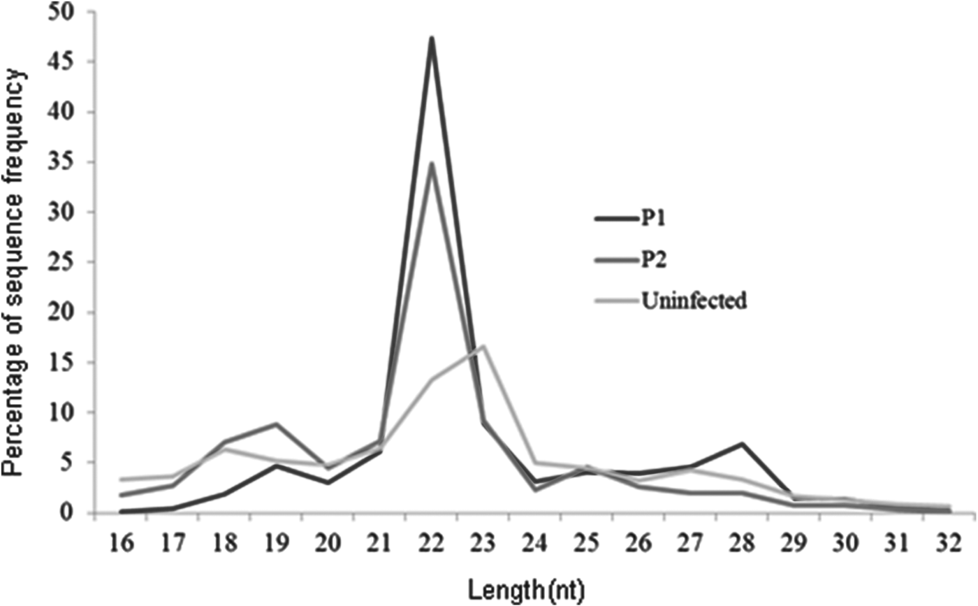
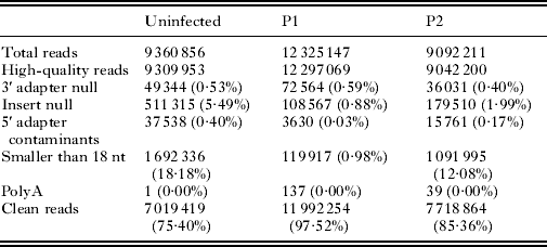
We obtained 7 019 419, 11 992 254 and 7 718 864 high-quality clean reads from UNI-, P1- and P2 library, respectively (Table 1). The majority of these small RNAs were shown to be between 21 and 23 nt in length (Fig. 1).
Size distribution of small RNAs in different rabbit plasma libraries. P1, the pooled RNAs from the total RNAs isolated from the plasma of schistosome infected rabbits at 16, 28 and 35 days post-infection; P2, the pooled RNAs from the total RNAs isolated from the plasma of schistosome-infected rabbits at 16 and 28 days post-infection; Uninfected, the pooled RNAs from the total RNAs isolated from the plasma of uninfected rabbits.

Statistics of small RNA sequences from the individual libraries
The percentage in parentheses means the percentage to the high-quality reads.
Bioinformatic analysis
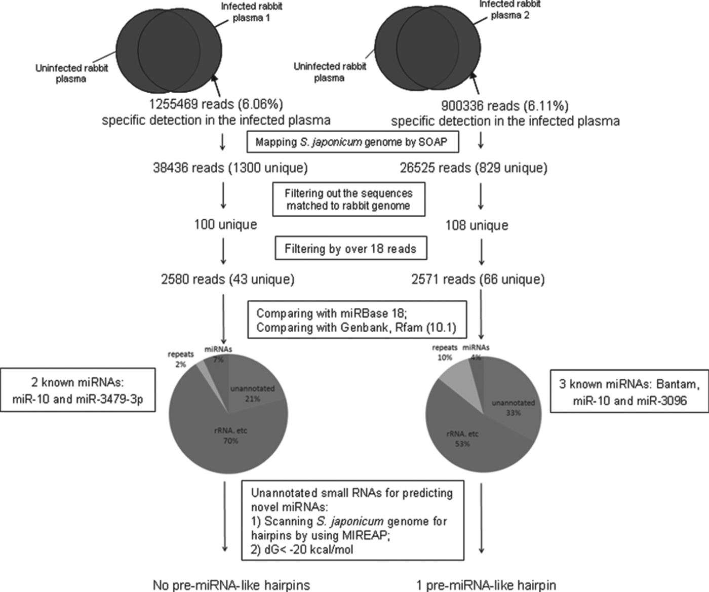
The results of our bioinformatic analysis (Fig. 2) indicated that the P1 and P2 libraries contained 1 255 469 and 900 336 specific small RNAs, respectively. Of these, 38 436 reads (3·06%, 1300 unique small RNAs) in the P1 library and 26 525 reads (2·95%, 829 unique small RNAs) in the P2 library were mapped perfectly to the S. japonicum genome. Further mapping of these two small RNA subsets to the rabbit genome revealed a total of 100 small RNAs in the P1 library and 108 in the P2 library that were S. japonicum-specific (Fig. 2). All of the small RNAs (over 18 sequence reads) are listed in Supplementary Table S1– in Online version only.
Workflow of bioinformatical analysis of small RNA datasets for the identification of schistosome-specific small RNA population in the plasma of schistosome-infected rabbits.
Bioinformatic characterization of schistosome-specific small RNAs over 18 reads indicated that 70% small RNAs in the P1 and 53% in the P2 were classified as ribosome RNA, tRNA or non-coding RNA. Then, the unmatched small RNAs in each library were further analysed against miRBase by using BLAST. The results indicated that 7% of these small RNAs in the P1 library and 4% in the P2 library matched to known schistosome miRNAs in the miRbase. These miRNAs included miR-10, miR-3479, Bantam and miR-3096 (Table 2). Finally, remaining small RNAs in the two libraries (21% in P1 and 33% in P2) were further analysed to predict novel miRNA candidates by applying the criteria described in Fig. 2. Only one pre-miRNA-like hairpin met the described criteria, suggesting that this small RNA may be a novel miRNA in S. japonicum, named miR-0001 (miRBase ID: sja-miR-8185) (Table 2). The putative precursor structure of the predicted miRNA is shown in Fig. 3.
The putative precursor structure of novel miRNA (miR-0001) in Schistosoma japonicum.
List of schistosome-specific miRNAs identified in the plasma of the rabbits infected with S. japonicum

a The sequences were obtained from our deep sequencing results.
Confirmation of schistosome-specific miRNAs identified by real-time RT–PCR and semi-quantitative PCR
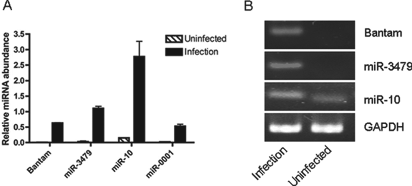
To confirm our findings from deep sequencing, we used real-time RT–PCR and stem-loop based semi-quantitative PCR to confirm the presence of these miRNAs in the plasma of S. japonicum-infected mice. Since human GAPDH mRNA is present in the circulation (Tsui et al. Reference Tsui, Ng and Lo2002), we therefore determined whether murine GAPDH is present in the circulation of mice. Our results indicated murine GAPDH was able to be repeatedly detected in the plasma of mice either by real-time RT–PCR or by regular PCR (Supplementary Figure S1– in Online version only). In addition, the abundance of GAPDH in the plasma did not change observably between infected and uninfected mice (Supplementary Figure S1). Consequently, we used GAPDH as an internal control to compare the expression of some of the newly identified miRNAs between infected mice and uninfected mice. Real-time RT–PCR analyses indicated that all miRNAs investigated (Bantam, miR-3479, miR-10 and miR-0001) were detected in the plasma of mice infected with S. japonicum (Fig. 4A). Although S. japonicum miR-10 was likely to be detectable in uninfected mice probably due to the high sequence identity (90% identity) with mouse miR-10, its expression was significantly increased in the plasma of S. japonicum-infected mice (Fig. 4A). Similar results were also observed using stem-loop based semi-quantitative PCR except for miR-0001 and miR-3096, which were not able to obtain significant PCR product (Fig. 4B).
Confirmation of schistosome-specific miRNAs identified by real-time RT–PCR and semi-quantitative PCR. (A) Real-time RT–PCR analysis of schistosome-specific miRNAs identified in the plasma of mice infected with Schistosoma japonicum cercariae. Data illustrate representative experiments and show the mean and standard error derived from triplicate experiments; (B) Semi-quantitative PCR analysis of schistosome-specific miRNAs identified in the plasma of mice infected with S. japonicum cercariae.
Correlation between the abundance of schistosome-specific miRNAs in the plasma of schistosome-infected mice and the number of S. japonicum cercariae inoculated
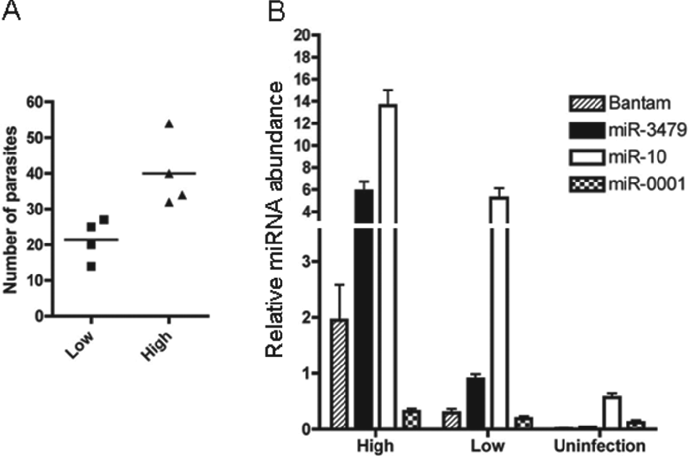
The abundance of schistosome-specific miRNAs was further investigated in mice inoculated with either 50 or 100 S. japonicum cercariae. At 26 dpi, the mice in each group were perfused to determine worm recovery. As expected, more worms were collected from the mice infected with relatively high numbers of S. japonicum cercariae (Fig. 5A). The mouse plasma in each group was pooled for RNA isolation, and the isolated plasma RNAs were subjected to real-time RT–PCR. As shown in Fig. 5B, all four miRNAs investigated were detected in plasma from mice infected with both low and high numbers of S. japonicum. In addition, the abundance of each of these four miRNAs significantly increased in the plasma of mice infected with relatively high numbers of S. japonicum cercariae as compared with plasma of mice infected with low numbers of S. japonicum, indicating that the abundance of schistosome-specific miRNAs in the plasma of a infected final host is related to the number of schistosomes present.
Real-time RT–PCR analysis of the abundance of schistosome-specific miRNAs in the plasma of mice infected with different number of Schistosoma japonicum cercariae inoculated. (A) The number of worms collected from the mice infected with different number of S. japonicum cercariae; (B) Real-time RT–PCR analysis of the abundance of schistosome-specific miRNAs in the plasma of mice infected with different numbers of S. japonicum cercariae. The plasma isolated from mice (n = 4) were pooled for RNA isolation. Data illustrate representative experiments and show the mean and standard error derived from triplicate experiments.
Schistosome Argonaute 2/3 may be an ES protein
To better understand the mechanism of schistosome miRNA secretion, we then determined whether schistosome Argonaute (Ago) protein, which is a part of the RNA-induced silencing complex, is an ES protein. In our previous study, we cloned and expressed three Ago proteins (Ago1, Ago2 and Ago3) in S. japonicum (Chen et al. Reference Chen, Yang, Guo, Peng, Liu, Li, Lin and Cheng2010) and then produced three polyclonal antibodies against these three Ago proteins. By using these antibodies in an ELISA, we found that S. japonicum Ago2 and Ago3 were present in the supernatant of parasite culture medium (Fig. 6A and B), suggesting that these Ago proteins are ES proteins. Then, we used immunohistochemistry to determine the locations of Ago 2/3 in adult schistosomes. As shown in Fig. 6C and D, Ago2 and Ago3 are located in the tegument of adult parasites although they are also ubiquitously distributed in the body of worms.
Analysis of the expression of Schistosoma japonicum Ago proteins. (A, B) ELISA analysis of S. japonicum Ago proteins in the supernatant of parasite culture medium. Data illustrate representative experiments and show the mean and standard error derived from triplicate experiments; (C, D) Immunohistochemistry analysis of Ago2/3 expression in the tegument of adult schistosomes. Arrows indicate schistosome tegument.
Expression profiles of schistosome-specific miRNAs identified in different stages of S. japonicum
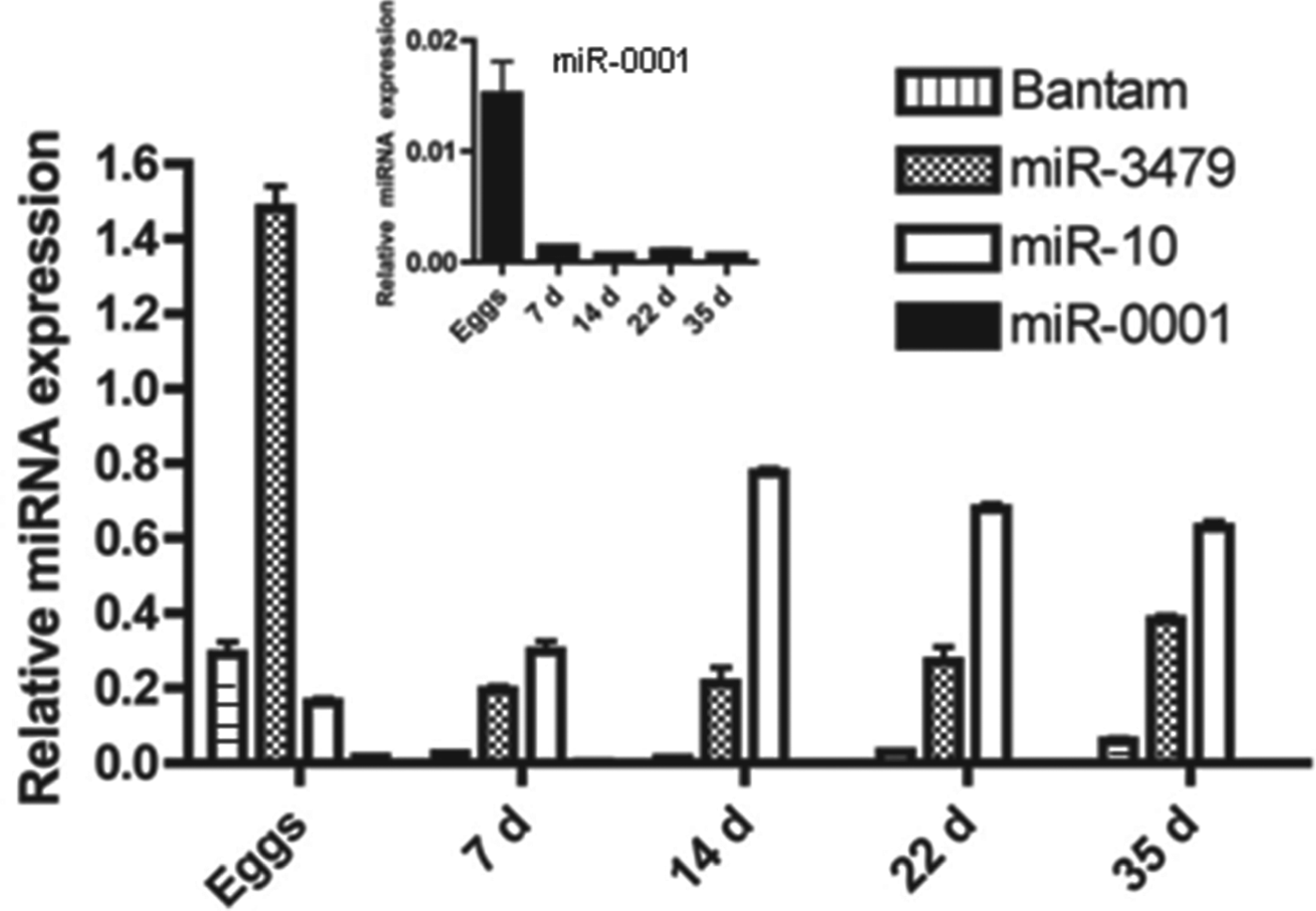
During their life in the final host, schistosomes undergo distinct morphological changes and physiological alternations. The abundance of an miRNA in different stages may imply its function in a specific stage. Consequently, we also investigated the abundance of Bantam, miR-3479, miR-10 and miR-0001 in several different stages in S. japonicum such as eggs, schistosomula (7 and 14 days) and adult worms (22 and 35 days) using real-time RT–PCR. As shown in Fig. 6, all of these miRNAs were detectable in all the stages of schistosomes investigated. Among them, bantam, miR-3479 and miR-0001 showed predominant expression in schistosome eggs, whereas miR-10 showed relatively high expression in 14-day schistosomula and adult worms (Fig. 7).
Expression profiles of schistosome-specific miRNAs in the different stages of Schistosoma japonicum. Data illustrate representative experiments and show the mean and standard error derived from triplicate experiments.
DISCUSSION
Circulating miRNAs have attracted considerable attention as novel biomarkers for diagnosing certain diseases and also as signal molecules for intercellular communications. Schistosomes as blood flukes live in blood vessels of their mammalian hosts. Identification of schistosome-specific miRNAs in the circulation of a final host may provide valuable clues not only for further developing novel biomarkers for the diagnosis of schistosomiasis but also for more deeply understanding host–parasite interactions. In the present study, we characterized a schistosome-specific small RNA population in the plasma of rabbits infected with S. japonicum cercariae using a deep sequencing method, resulting in the identification of five schistosome-specific miRNAs including four known miRNAs and one predicted novel miRNA.
Due to the small size and low abundance of small RNAs in the circulation, different procedures/reagents for RNA isolation may result in different small RNA populations (Kroh et al. Reference Kroh, Parkin, Mitchell and Tewari2010). To optimize yields of small RNAs during the processes of RNA isolation, we used three different procedures/reagents (QIAamp Circulating Nucleic Acid kit, Trizol and RNAiso plus) for plasma RNA isolation and then the RNA isolated from these three different procedures/reagents were combined for constructing small RNA libraries. Among several developmental stages of schistosomes in a final host, schistosomula are considered to be the most susceptible to the immune response and to anthelmintic drugs, whereas paired adult worms produce a large number of eggs, leading to a severe pathological reaction in the liver of the final host. Therefore, we mainly focused on schistosome-specific small RNAs in the circulation of a final host associated with schistosomula (16 days) and adult worms (28 and 35 days) and then mixed the plasma RNAs isolated from S. japonicum-infected rabbits related to the two stages for deep sequencing.
Schistosome infection may significantly alter the profile of circulating miRNAs in a final host. In the present study, our aim was to identify schistosome-specific small RNAs in the plasma of infected rabbits. To be valuable as biomarkers, these small RNAs should be (1) specifically present in the plasma of rabbits infected with S. japonicum and (2) map uniquely to the S. japonicum genome. The small RNAs associated with S. japonicum infection mapped to both the rabbit and the S. japonicum genomes were excluded since they could not be considered schistosome species-specific. In general, our computational analysis showed that there are similar numbers of schistosome-specific small RNAs in the two libraries (100 small RNAs in P1 and 108 small RNAs in P2) prepared in this study. However, we observed that the number of reads for these small RNAs was relatively low in both libraries (average reads are 60 for P1 and 39 for P2), suggesting that these schistosome-specific small RNAs are present in relatively low abundance in the plasma of the final host. The low abundance of schistosome-specific miRNAs in the plasma also led to only one S. japonicum miRNA (miR-10) being co-detected in the two libraries.
In the present study, we identified five schistosome-specific miRNAs in the plasma of schistosome-infected rabbits, including four known miRNAs (miR-10, miR-3479, miR-3096 and Bantam) and one predicted novel miRNA (miR-0001). To further confirm the expressions of these identified miRNAs, we used Qiagen miScript PCR System, which can efficiently discriminate between closely related miRNA isoforms, even when they differ by only a single base. Our results indicated all four miRNAs (miR-10, miR-3479, Bantam and miR-0001) investigated were able to be detected in the plasma of S. japonicum-infected mice. By contrast, there was low amplification of S. japonicum miR-10 in the plasma of uninfected mice, probably due to the fact that S. japonicum miR-10 shows a very high degree of sequence identity (90% identity) with its homologue in mice. Most importantly, three (Bantam, miR-3479 and miR-0001) of these four miRNAs validated by miScript PCR are schistosome specific, suggesting that these molecules may have potential as a novel class of biomarkers for the diagnosis of schistosomiasis. Due to lack of the miScript primer for miR-3096, we further included it for stem-loop RT–PCR analysis. Among all five miRNAs investigated, we were not able to obtain PCR products for miR-3096 and miR-0001. We deduce that this was likely due to the low quality of primers either for reverse transcription or for PCR, and/or low abundance of the miRNAs for stem-loop RT–PCR detection, and further research is needed for this problem. In addition, the stability of schistosome-specific miRNAs in the circulation of a final host remains to be characterized in a future study.
MiRNAs are currently understood to enter the circulation via two pathways: (1) passive leakage from broken cells and (2) active secretion by microvesicles and/or RNA-binding proteins such as high-density lipoprotein, Ago proteins and nucleophosmin 1 (Chen et al. Reference Chen, Liang, Zhang, Zen and Zhang2012). We were able to detect S. japonicum Ago2 and Ago3 in the supernatants of parasite cultures (Fig. 6). In addition, our immunohistochemistry results indicated that these Ago proteins may be located in the tegument of adult schistosomes (Fig. 6), implying that Ago2 and Ago3 may be secreted and/or excreted through schistosome tegument as are numerous other proteins secreted (Knudsen et al. Reference Knudsen, Medzihradszky, Lim, Hansell and McKerrow2005; Liu et al. Reference Liu, Cui, Hu, Feng, Wang and Han2009). It remains to be determined whether schistosome Ago2 and Ago3 are associated with some of the schistosome-specific miRNAs identified in the circulation of a final host. It is also necessary to clarify whether these circulating schistosome-specific miRNAs might also be by-products of dead/dying cells from the parasites.
Circulating miRNAs may functionally act as signal molecules in intercellular communication and then horizontally regulate target genes in the recipient cells (Zhang et al. Reference Zhang, Hou, Chen, Li, Zhu, Zhang, Li, Bian, Liang and Cai2012a). Among five S. japonicum-specific circulating miRNAs identified, Bantam miRNA has been shown to target a tumor-suppressor pathway and overexpression of Bantam led to cellular growth and the suppression of cellular death in Drosophila (Nolo et al. Reference Nolo, Morrison, Tao, Zhang and Halder2006). Moreover, human schistosomiasis is associated with increased risks for bladder (S. haematobium) and liver (S. mansoni, S. japonicum) cancers (Takemura et al. Reference Takemura, Kikuchi and Inaba1998; Gryseels et al. Reference Gryseels, Polman, Clerinx and Kestens2006). Consequently, it will be interesting to investigate whether schistosome Bantam miRNA is involved in the aetiology of the cancers in a final host that infected schistosomes.
In summary, we characterized pathogen-specific small RNA population in the plasma of a final host infected with S. japonicum using a deep sequencing method, resulting in the identification of five schistosome-specific miRNAs. Our findings may provide useful information not only for further developing novel biomarkers for the diagnosis of schistosomiasis but also for more deeply understanding host–parasite interaction.
FINANCIAL SUPPORT
The study was supported, in part or in whole, by the National Natural Science Foundation of China (Grant No. 30901068), Science and Technology Commission of Shanghai Municipality of China (Grant No.10PJ1412300) and Shanghai Talent Developing Foundation of China (Grant No.2009032).
SUPPLEMENTARY MATERIAL
To view supplementary material for this article, please visit http://dx.doi.org/10.1017/S0031182013000917.