Implications
Tail docking may have both short- and long-term welfare consequences for pigs. In addition to the short-term pain and distress caused by docking itself, long-term changes in pain processing may arise through abnormal nerve proliferation at the docking site. The present study found evidence of abnormal nerve proliferation in the tail stumps of pigs docked using both clippers and cautery iron, indicating the potential for long-term pain consequences with either method. However, as the degree of abnormal nerve proliferation did not differ between docking methods, no evidence for a longer-term welfare advantage of one method over the other was found.
Introduction
Tail biting, involving the destructive chewing of pen-mates’ tails, has been a recognised problem among commercially farmed pigs for the past century (Dougherty, 1976; European Food Safety Authority (EFSA), Reference Fisher and Boswick2007). It is an abnormal behaviour, the occurrence of which is thought to indicate reduced welfare amongst some or all pigs in an affected pen (Schroder-Petersen and Simonsen, Reference Scollo, Contiero and Gottardo2001; EFSA, Reference Fisher and Boswick2007; Stafford, Reference Sullivan2010). Tail-bitten pigs may experience severe welfare compromise as a result of tissue damage, haemorrhage, infection, abscesses and septicaemia, which, in severe cases, may lead to death (Schroder-Petersen and Simonsen, Reference Scollo, Contiero and Gottardo2001). Although management factors such as reduced stocking density and the provision of rooting material have been advocated to reduce tail-biting incidence (EFSA, Reference Fisher and Boswick2007; Sonoda et al., Reference Stafford2012; Taylor et al., Reference van der Avoort, Hovius, Selles, van Neck and Coert2012), tail docking remains the most widely adopted on-farm preventive strategy (EFSA, Reference Fisher and Boswick2007; Nannoni, Reference Nannoni, Valsami, Luca and Martelli2014).
Tail docking involves amputating some portion of the distal end of the tail, typically by severing with side clippers, or using a heated cautery iron to cut and cauterise in a single step (Sutherland and Tucker, Reference Sutherland, Bryer, Krebs and McGlone2011). Although there is evidence that tail docking reduces the frequency of tail biting among intensively farmed pigs (Hunter et al., Reference Kells, Beausoleil, Chambers, Sutherland, Morrison and Johnson2001; Sutherland et al., Reference Taylor, Parker, Mendl, Edwards and Main2009), the mechanisms underlying this phenomenon are not clear. Although the reduced length likely impairs the ability of pigs to grasp and manipulate the remaining tail, this probably does not sufficiently explain why tail-biting lesions are less common in ‘long’ docked pigs (tip only or distal third removed) than undocked pigs (Hunter et al., Reference Hunter, Jones, Guise, Penny and Hoste1999; Sutherland et al., Reference Taylor, Parker, Mendl, Edwards and Main2009; Scollo et al., 2015). It has been hypothesised that neuroanatomical changes in the tip of the docked tail result in hypersensitivity of the tail stump, stimulating protective behaviour towards this area (Simonsen et al., Reference Sonoda, Fels, Oczak, Vranken, Ismayilova, Guarino, Viazzi, Bahr, Berckmans and Hartung1991), although this has yet to be confirmed.
Following nerve transection, the proximal nerve stump seals off, forming a terminal swelling. Numerous fine projections (sprouts) arise from this, and attempt to connect with peripheral target tissues. When reconnection is prevented, as is the case with amputation, terminal swellings persist and nerve sprouts turn back on themselves forming tangled masses, or neuromas (Devor and Seltzer, Reference Done, Guise and Chennells1999), exacerbated by excessive fibrous tissue proliferation in the region (Foltán et al., 2008).
In humans, neural lesions such as neuromas have been associated with neuropathic pain following limb amputation and peripheral nerve injury (Lewin-Kowalik et al., Reference Morrison, Kells, Johnson and Hemsworth2006). Neuropathic pain is evoked through alterations in the structure and function of the somatosensory nervous system such that pain may occur spontaneously and responses to both noxious and innocuous stimuli are pathologically amplified (Costigan et al., 2009). However, whilst neural lesions are necessary precursors for neuropathic pain, not all neuromas are associated with alterations in pain perception (Costigan et al., 2009). In the human clinical literature, the reported prevalence of symptomatic (painful) neuromas subsequent to digital amputations of the hand is relatively low, ranging from 2.7% to 7.8% (van der Avoort et al. Reference Breward and Gentle2013; Fisher and Boswick, Reference Foltán, Klíma, Špačková and Šedý1983; Sullivan, Reference Sutherland, Bryer, Krebs and McGlone1985).
Terminal neuromas and irregular innervation of the tail stump have been reported in lambs (French and Morgan, Reference French and Morgan1992) and pigs (Simonsen et al., Reference Sonoda, Fels, Oczak, Vranken, Ismayilova, Guarino, Viazzi, Bahr, Berckmans and Hartung1991; Herskin et al., Reference Hunter, Jones, Guise, Penny and Hoste2015) following tail docking and in chickens following partial beak amputation (Breward and Gentle, Reference Costigan, Scholz and Woolf1985). In addition, both clinical and experimentally induced neuromas have been linked to neuropathic pain in animals (reviewed by Zimmermann, Reference Zimmermann2001). For example, abnormal spontaneous activity was recorded in afferent neurons in the region of neuroma formation following partial de-beaking in chickens and was accompanied by heightened thermal sensitivity and behavioural signs of persistent pain up to 6 weeks after amputation (Breward and Gentle, Reference Costigan, Scholz and Woolf1985). Likewise, Gross and Carr (Reference Herskin, Thodberg and Jensen1990) identified neuromas in the tail stump of six tail-docked dogs with histories of persistent autotomy, a behaviour considered indicative of chronic pain (Zimmermann, Reference Zimmermann2001). Thus, the presence of neuromas after amputation has the potential to cause neuropathic pain and impact detrimentally on the longer-term welfare of animals.
Although neuromas have previously been identified in the healed tail tips of pigs docked using both blunt trauma (Simonsen et al., Reference Sonoda, Fels, Oczak, Vranken, Ismayilova, Guarino, Viazzi, Bahr, Berckmans and Hartung1991; Done et al., Reference Dougherty2003) and cautery iron (Herskin et al., Reference Hunter, Jones, Guise, Penny and Hoste2015), the incidence of neuroma formation has not previously been compared between these methods. The aim of the present study was to compare neuroma development in healed tail tips from pigs docked using clippers and cautery iron, to determine whether docking method influenced neuroma formation.
Material and methods
Animals and housing
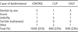
All experimental procedures were approved by the Rivalea Australia Animal Ethics Committee (protocol number 13B068C). The experiment was conducted at the Rivalea Australia Research and Innovation Unit, Corowa NSW, Australia between March and September 2014. The animals used in the study were part of a larger cohort also used to investigate mortality and tail damage. The cohort consisted of 40 healthy, viable sows (Large White×Landrace) that had the same farrowing date and their litters. The sows farrowed in individual farrowing crates. Three healthy, entire male piglets per litter were randomly assigned to one of three treatments, performed at 2 days of age: no tail docking (CONTROL); tail docking using clippers (CLIP); tail docking using cautery iron (CAUT). Allocation of pigs within a litter to each treatment was based upon computer-generated random selection, following confirmation of good health. Pigs were mixed at weaning (i.e. not kept in groups of similar tail length) and remained in a conventional (semi-slatted) housing system until market. Pig health and welfare was monitored daily. Pigs identified as having superficial wounds in a localised area, minimal bleeding and no evidence of bleeding or infection received first aid and were placed in a hospital pen before market. Pigs showing signs of severe infection, or with extensive tail damage, were euthanased (as per the Australian Pesticides and Veterinary Medicines Authority guidelines). The total number of piglet deaths and removals per treatment, along with causes, are shown in Table 1. There were no significant differences between treatments in the number of piglet deaths and piglet removals due to illness or injury (χ 2=0.09; P=0.95). Pigs were slaughtered at commercial premises at 21 weeks of age and their tails/tail stumps harvested for subsequent analysis.
Number of pig deaths and removals due to illness or injury among pigs with intact tails (CONTROL, n=40) and those tail docked using clippers (CLIP, n=40) or cautery iron (CAUT, n=40) from the full cohort of 120 pigs used in the wider study

Experimental procedures
Tail docking
Piglets were quietly picked up from their home pen and were held, supported under the arm of the technician with their hind area exposed. Piglets in the CONTROL treatment were held in the same manner for ~30 s and returned to their pen. Pigs in the CLIP treatment had their tails docked using clean, sanitised side clipper pliers (Bahco 2101G side cutter pliers; SNA Europe, Smithfield, NSW, Australia). Pigs in the CAUT treatment had their tails docked using a clean, sanitised gas operated cautery iron (Stericut® Tail Docker; Cotran Corporation, Portsmouth, RI, USA). In both docking treatments the tail was cut ~2 cm from the base, in between the first and second visible external vertebrae, and a chlorhexidine disinfectant spray was applied immediately.
Collection and preparation of tail tissue
Following slaughter, tails/tail stumps from 25 pigs per treatment were cut off at the root and individually fixed in 10% neutral buffered formalin (NBF) for 7 days. Samples were then removed, wrapped in formalin-soaked towels and sealed in individual plastic bags for transport to the laboratory. Upon receipt, samples were unpacked and submerged in 10% NBF for storage before processing within 4 weeks.
Histology and immunohistochemistry
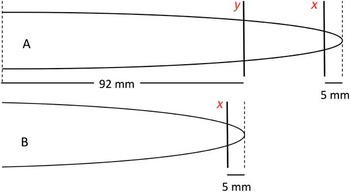
Preparation and staining of formalin-fixed samples was undertaken at Massey University, New Zealand. Before sectioning, all tail samples were examined for gross evidence of trauma that may be indicative of tail biting (tail lesions, scabs or scarring), and the tail length (mm) was measured and recorded. All tails collected at slaughter were cross-sectioned 5 mm from the distal tip, whilst control (undocked) tails were also cross-sectioned at a second location, 92 mm from the tail base (Figure 1). The average length of docked tails at the time of slaughter was 92 mm, thus cross-sections were prepared at this level in control tails in order to visualise normal tail neuroanatomy in the region equivalent to the site of amputation of docked tails.
Schematic diagram indicating sampling sites for histological analysis. Undocked tails (a) and docked tail stumps (b) collected at slaughter were cross-sectioned 5 mm from the distal tip (x). Undocked tails were additionally cross-sectioned 92 mm from the root (y), representing the mean length of all docked tails.

All tail sections were decalcified and dekeratinised in potassium hydroxide and Veet® (Reckitt Benckiser, Berkshire, UK) according to standard laboratory protocol, then processed through graded alcohols and xylene before being embedded in paraffin. The formalin-fixed, paraffin-embedded tissues were sectioned at 4 to 5 μm and stained with haematoxylin–eosin.
Where required, additional sections were stained with Masson’s trichrome and S100 to further optimise nerve visualisation and confirm neuroma presence. Tissue sections were deparaffinised, blocked for endogenous peroxidase and treated with primary antibodies towards S100 (polyclonal rabbit anti-S100; FLEX Anti-S100; Dako) as described by Nielsen et al. (Reference Schroder-Petersen and Simonsen2011).
Tail sections were examined under light microscope (Olympus BX51, Olympus Corporation, Tokyo, Japan) at 4, 20 and 40× magnification. Two pathologists (W. R. and S. H.), both blinded to treatment, examined sections independently using the same equipment, and assigned scores of 1, 2 or 3 as follows: 1=discrete well-organised nerve bundles (normal morphology); 2=moderate proliferation and disorganisation within fibrous connective tissue, affecting less than half the circumference of the tail; 3=marked proliferation to form almost continuous disorganised bundles or non-continuous enlarged bundles compressing the surrounding, densely fibrous, connective tissue. The presence of disorderly, proliferative nerve bundles surrounded by fibrous connective tissue (i.e. scores of 2 or 3) indicated neuroma formation.
Following initial independent examination of tail sections, scores were compared between pathologists. Where discrepancies were identified, both scorers re-examined the slides in question together and arrived at a consensus as to the appropriate classification before statistical analysis.
Statistical analysis
All statistical analyses were performed using SAS version 9.3.1 (SAS Institute Inc., Cary, NC, USA, 2012).
Inter-rater reliability of scores (based on initial independent scores) was determined using Cohen’s κ coefficient.
Non-parametric one-way ANOVA (Kruskal–Wallis test) was carried out to compare neuroanatomy scores between treatments. Where a significant treatment effect was found, post hoc pairwise comparison of mean scores was performed using the Wilcoxon–Mann–Whitney rank-sum test. In addition, Fisher’s exact test was performed to determine whether the distribution of scores differed from expected.
Results
Descriptive neuroanatomy
Tails harvested at slaughter (21 weeks of age)
Although 25 tails per treatment were targeted, difficulties with sample adhesion meant that histological scoring could only be performed on a total of 15 control (undocked), 19 clipper-docked and 18 cautery-docked pigs (n=52). In some cases, poor tissue adhesion meant that only part of the sample could be examined. Samples were only scored where the entirety of the cross-section could be visualised.
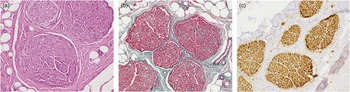
No abnormal nerve proliferation was observed in sections from the distal portion (section x, Figure 1) of control tails (Table 2; Figure 2). Gross evidence of tail-tip trauma (lesions, scabs or scarring) was observed in 5/15 control tails, with corresponding evidence of inflammation and/or scarring apparent on histological analysis. Neural histomorphology at the site approximating the point of tail docking (section y, Figure 1) was unremarkable, with discrete nerve fibres present and no signs of neural proliferation observed.
Control (undocked) pig tail sectioned at level equivalent to docking site, showing nerve histomorphology score=1. (a) Haematoxylin–eosin; (b) Masson’s trichrome; (c) S100 (all 20× magnification).

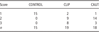
Summary of nerve histomorphology scores from cross-sections from the distal tail tip of undocked (CONTROL) clipper-docked (CLIP) and cautery-docked (CAUT) pigs

Score of 1=discrete well-organised nerve bundles (normal morphology); 2=moderate proliferation and disorganisation within fibrous connective tissue, affecting less than half the circumference of the tail; 3=marked proliferation to form almost continuous disorganised bundles or non-continuous enlarged bundles compressing the surrounding densely fibrous connective tissue. Scores of 2 or 3 indicate neuroma formation.
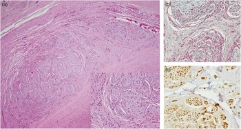
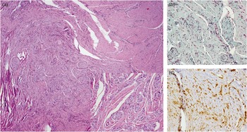
The average length of all docked tails (n=50) collected at slaughter was 92 (range 75 to 140) mm. Evidence of abnormal neural proliferation consistent with neuroma formation (i.e. score 2 or 3) was observed in 17/19 tails from pigs docked using clippers and 17/18 tails from pigs docked using cautery iron (Table 2; Figures 3 and 4).
Cross-section 5 mm from the tip of a docked pig tail, showing nerve histomorphology score=2. (a) Moderately disorganised proliferative nerve bundles of varying sizes, surrounded by thin, frequently loosely arranged layers of fibrous connective tissue. Haematoxylin–eosin, 4× magnification. Inset: 20× magnification at same site. (b) Masson’s trichrome (magnification 20×). (c) Small-to-medium nerve fibres stained brown with S100 (magnification 20×).

Cross-section 5 mm from the tip of a docked pig tail, showing nerve histomorphology score=3. (a) Poorly organised highly proliferative nerve bundles, often within dense fibrous connective tissue. Haematoxylin–eosin (magnification 4×). Inset: 20× magnification at same site. (b) Note numerous small proliferating nerves within connective tissue. Masson’s trichrome (magnification 20×). (c) Large number of small, disorganised, immunopositive nerve fibres. S100 (magnification 20×).

Inter-rater reliability
Initial independent examination of tissue sections revealed 83% agreement in the assigned scores. Inter-rater reliability was substantial (Cohen’s κ coefficient=0.73; 95% confidence interval 0.58 to 0.89). Following consultation and re-examination, consensus scores were reached for all samples before statistical comparisons being performed.
Comparison of neural histomorphology
Kruskal–Wallis analysis revealed a significant effect of treatment on neuroanatomy scores (χ 2=31.25; P<0.001). Post hoc tests revealed that mean neural histomorphology scores for both the CLIP and CAUT groups (Wilcoxon rank means=35.0 and 31.7, respectively) were higher than those in the CONTROL group (rank mean=9.5) (P<0.001). Although the percentage of tails scored as 3 (marked neural proliferation) was higher in pigs docked using clippers than those docked using cautery (42% v. 17%, respectively), mean nerve histomorphology score did not differ between the two docking methods (P=0.23). Fisher’s exact test likewise revealed no difference in the distribution of histomorphology scores between CAUT- and CLIP-docked pigs (P=0.16).
Discussion
The aim of the present study was to compare the neural histomorphology of healed tail tips of pigs docked using clippers and cautery iron, to determine whether docking method influenced the extent of neuroma formation.
Due to the often marked and haphazard degree of neural proliferation observed in the tips of docked tails, the number and/or size of individual neuromas could not be determined, as it was not clear whether large regions of proliferation resulted from the regeneration of single or multiple severed nerves. Therefore a unique descriptive scale was developed and used to compare neural morphology.
Evidence of neuroma formation, including the presence of disorganised excessive nerve proliferation within moderate to dense connective tissue, was observed in almost all tails docked using both clippers and cautery iron, whereas no evidence of neuromas was found in tail tips from control, undocked pigs, despite gross evidence of trauma due to tail biting in 5/15 tails. The incidence of neuroma formation in tails docked using hot cautery in the present study (94%) was higher than that reported by Herskin et al. (Reference Hunter, Jones, Guise, Penny and Hoste2015) (64%). This may, in part, be attributable to the fact that in the latter study pig tails were halved midsagitally before cross-sectioning one-half, whereas the present study examined cross-sections of the entire tail.
Given the reported associations between neuroma formation and the occurrence of neuropathic pain (Zimmermann, Reference Zimmermann2001; Lewin-Kowalik et al., Reference Morrison, Kells, Johnson and Hemsworth2006), the presence of neuromas in almost all of the tail stumps of pigs docked using clippers and cautery iron suggests that tail docking by either method has the potential to induce longer-term alterations in pain processing and attendant welfare compromise in pigs. This does, however, need to be confirmed before any robust conclusions about pig welfare can be drawn. This might be achieved, for example, by assessing thermal and mechanical nociceptive thresholds in the tail tip over the lifetime in commercial pigs with intact tails and those docked using clippers or cautery.
The present study indicates that there was no difference in the incidence of tail-tip neuroma formation between clipper- and cautery-docked pigs, suggesting that the longer-term pain consequences of tail docking using clippers and cautery may be similar. In their study of the anatomical and pathological consequences of tail docking using scissors or a turkey cautery de-beaker, Done et al. (Reference Dougherty2003) reported evidence of tail-tip neuroma formation in 100% of docked pig tails at 4 to 4.5 months, with undocked control pigs showing no evidence of neuromas.
Previous studies have indicated that hot cautery may induce less acute pain than blunt trauma docking methods. Both Sutherland et al. (Reference Sutherland and Tucker2008) and Morrison et al. (Reference Nielsen, Jensen and Leifsson2013) reported higher plasma cortisol concentrations in pigs docked using blunt trauma compared with those docked using cautery in the period immediately following docking. Further, pigs tail docked using cautery exhibited less electroencephalographic evidence of nociception than those docked using clippers (Kells et al., Reference Lewin-Kowalik, Marcol, Kotulska, Mandera and Klimczak2013). A comparison between control (undocked), clip- and cautery-docked pigs revealed no difference in tail-biting lesion scores between clipper- and cautery-docked pigs over the 7 weeks following docking (Sutherland et al., Reference Taylor, Parker, Mendl, Edwards and Main2009), suggesting the two methods do not differ in their ability to prevent tail biting. Therefore, if it can be confirmed that the longer-term welfare consequences do not differ between the two docking methods, the smaller acute pain responses previously identified may be cause for recommending the use of cautery over blunt trauma for tail docking pigs.
It must also be noted that the relatively small sample size examined (n=18 or 19 per group) may have reduced the ability of this study to discriminate between docking methods. In addition, the inclusion of male pigs only meant that any effects of sex could not be ascertained.
In conclusion, tail docking of pigs using either side clippers or cautery iron resulted in the formation of neuromas in the healed tail tip. The degree of abnormal nerve regeneration did not differ between docking methods, providing no evidence for a longer-term welfare advantage of one method over the other, at least in terms of the potential for alterations in pain processing.
Acknowledgements
The authors would like to acknowledge the assistance of Sarah Michael, Stuart Hunter, Saritha Gils and Evelyn Lupton in the preparation and analysis of histology samples. This study was undertaken with funding assistance from Australian Pork Limited.