Introduction
As the leading cause of preventable death in the world, tobacco smoking kills almost 6 million people each year (World Health Organization, 2011). The public health problems and associated economic costs motivate studies that investigate the biological bases of smoking behaviors. The majority of adult-dependent smokers started smoking during adolescence (US Department of Health and Human Services, 2014). About one-third of teenagers who become regular smokers will die prematurely from smoking-related diseases (Centers for Disease Control and Prevention, 1996). The current study evaluated early-life signs of developmental risk for smoking initiation by examining associations between genetic risk and brain structure in adolescents.
Smoking is thought to have a moderate to high heritability (Li, Cheng, Ma, & Swan, Reference Li, Cheng, Ma and Swan2003; Vink, Willemsen, & Boomsma, Reference Vink, Willemsen and Boomsma2005). Although several single nucleotide polymorphisms (SNPs) have been identified from genome-wide association studies conducted by large consortia (Liu et al., Reference Liu, Tozzi, Waterworth, Pillai, Muglia, Middleton and Marchini2010; Thorgeirsson et al., Reference Thorgeirsson, Gudbjartsson, Surakka, Vink, Amin, Geller and Stefansson2010; Tobacco Genetics Consortium, 2010), the effect of any single genetic variant is small, with the most significant SNPs accounting for 0.03% of the variance in smoking initiation (Tobacco and Genetics Tobacco Genetics Consortium, 2010) in comparison with 19–28% explained by the joint effect of all the measured DNA variants (Lubke et al., Reference Lubke, Hottenga, Walters, Laurin, de Geus, Willemsen and Boomsma2012). This suggested that the genetic basis of smoking behavior consists of a large number of genetic variants, each with a small contribution. Therefore, tests based on a polygenic risk score (PGRS) are more powerful than SNP-based tests as they jointly consider the weak effects of multiple associated SNPs (Dima & Breen, Reference Dima and Breen2015). This approach has also shown promise for uncovering the contribution of genetic burden to brain endophenotypes related to complex diseases such as schizophrenia (Liu et al., Reference Liu, Zhang, Cui, Qin, Tao, Li and Jiang2017) and Alzheimer's disease (Li et al., Reference Li, Zhang, Li, Liu, Qin, Yu and Jiang2018).
Studies have shown an association between the smoking PGRS correlates of tobacco risk in independent samples, including the amount of cigarette usage (Meyers et al., Reference Meyers, Cerda, Galea, Keyes, Aiello, Uddin and Koenen2013; Vink et al., Reference Vink, Hottenga, de Geus, Willemsen, Neale, Furberg and Boomsma2014) and the progression of smoking (Belsky et al., Reference Belsky, Moffitt, Baker, Biddle, Evans, Harrington and Caspi2013; Uhl et al., Reference Uhl, Walther, Musci, Fisher, Anthony, Storr and Rose2014). The smoking PGRS also predicts alcohol (Vink et al., Reference Vink, Hottenga, de Geus, Willemsen, Neale, Furberg and Boomsma2014) and cannabis phenotypes (Musci et al., Reference Musci, Fairman, Masyn, Uhl, Maher, Sisto and Ialongo2018; Vink et al., Reference Vink, Hottenga, de Geus, Willemsen, Neale, Furberg and Boomsma2014). However, how these genetic variants relate to the brain remains largely unknown, follow-up studies are needed to search for brain endophenotypes to bridge the gene–behavior gap between polygenic risk and smoking. Cortical volume is a suitable candidate endophenotype for the following reasons. First, recent neuroimaging meta-analyses have consistently reported smaller cortical volume in smokers compared with nonsmokers (Brody et al., Reference Brody, Mandelkern, Jarvik, Lee, Smith, Huang and London2004; Gallinat et al., Reference Gallinat, Meisenzahl, Jacobsen, Kalus, Bierbrauer, Kienast and Staedtgen2006; Sutherland et al., Reference Sutherland, Riedel, Flannery, Yanes, Fox, Stein and Laird2016). Second, cortical volume is highly heritable. Twin magnetic resonance imaging (MRI) reports have demonstrated considerable genetic effects on cortical volume (Joshi et al., Reference Joshi, Lepore, Joshi, Lee, Barysheva, Stein and Thompson2011). Third, recent twin studies have reported that the majority of the phenotypic overlap between smoking and cortical volume was of genetic origin (Prom-Wormley et al., Reference Prom-Wormley, Maes, Schmitt, Panizzon, Xian, Eyler and Neale2015). Hence, we chose cortical volume to test the effect of smoking PGRS in the current study.
In this study, we investigated the association between smoking PGRS and brain structure, then a mediation analysis was performed to test whether the effect of smoking PGRS on smoking behavior was mediated by regional gray matter volume. We addressed this issue using the IMAGEN cohort, a population-based European dataset of 14-year-old adolescents. Our analyses may increase our understanding of smoking pathogenesis and risk and shed light on the potential development of more effective measures of preventing tobacco use.
Methods
Samples
We analyzed data from the IMAGEN project, a well-characterized, longitudinal imaging genetics study that recruited participants through secondary schools at eight sites located in eight European centers (London, Nottingham, Dublin, Mannheim, Berlin, Hamburg, Paris, and Dresden) (Schumann et al., Reference Schumann, Loth, Banaschewski, Barbot, Barker and Buchel2010). Exclusion criteria included a personal history of serious medical, neurological, and/or psychiatric conditions (such as schizophrenia or bipolar disorder), neurodevelopmental disorders (such as autism), or an IQ of less than 70. A detailed description of the recruitment and assessment procedures, inclusion criteria, and further exclusion criteria has been published previously (Schumann et al., Reference Schumann, Loth, Banaschewski, Barbot, Barker and Buchel2010). Written informed consent was obtained from the parent or guardian and verbal assent was obtained from the adolescent. This study was approved by the local ethics committees at all participating sites.
Genotyping
Blood samples were collected at the local institute, and DNA was extracted from blood samples using the Gentra Puregene Blood Kit (Qiagen, Valencia, CA) according to the manufacturer's instructions. Genotype information was collected using the Illumina (Little Chesterford, UK) Human610-Quad Beadchip or Illumina Human660-Quad Beadchip in three batches. Then, quality control was implemented in PLINK (Purcell et al., Reference Purcell, Neale, Todd-Brown, Thomas, Ferreira, Bender and Sham2007) software (pngu.mgh.harvard.edu/wpurcell/plink/). SNPs with call rates of <98%, minor allele frequency <1%, or deviation from Hardy–Weinberg equilibrium (p ⩽ 1 × 10⁻⁴) were excluded. Individuals with an ambiguous sex code, excessive missing genotypes (failure rate >2%), or outlying heterozygosity (heterozygosity rate 3 standard deviations from the mean) were also excluded from the analyses. Identity by state similarity was subsequently calculated to estimate cryptic relatedness for individuals. Closely related individuals with identity by descent of >0.1875 were excluded. Then population stratification was examined by principal component analysis to identify (and exclude) individuals with divergent ancestry (from CEU). After quality control procedures, 477 245 SNPs in 1774 individuals were considered for the subsequent smoking PGRS calculation.
Polygenic risk score
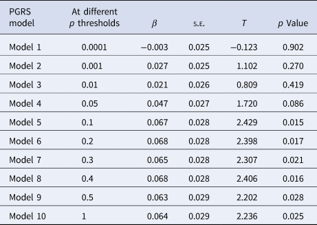
PGRS were calculated using the ‘score’ utility in PLINK based on publicly available results data from the genome-wide meta-analyses of smoking initiation within cohorts of the Tobacco and Genetics (TAG) Consortium (N = 74 053) (Tobacco Genetics Consortium, 2010). The PGRS was computed following the method developed by Purcell and colleagues (International Schizophrenia Consortium et al., Reference International Schizophrenia Consortium, Purcell, Wray, Stone, Visscher, O'Donovan and Sklar2009). To ensure that all SNPs in the PGRS model were independent, p value-informed clumping was performed (Vassos et al., Reference Vassos, Di Forti, Coleman, Iyegbe, Prata, Euesden and Breen2017; Xu et al., Reference Xu, Li, Qin, Jun Li, Zhuo, Liu and Yu2018), retaining the SNP with the smallest p value within a 500-kb window and excluding SNPs that were in linkage disequilibrium (r 2 < 0.25). Each variant (risk allele) was summed to create an aggregate smoking PGRS, and those variants with a larger effect size (indicated by the β-coefficient) were given a greater weight. Ten progressive p thresholds (see Table 2) including the commonly used ones (Lancaster et al., Reference Lancaster, Linden, Tansey, Banaschewski, Bokde and Bromberg2016; Ruderfer et al., Reference Ruderfer, Fanous, Ripke, McQuillin, Amdur and Kendler2014) were selected in the current study.
Smoking behavior
Smoking behavior was assessed through self-reports using a questionnaire adapted from the European School Survey Project on Alcohol and Other Drugs (ESPAD). Cigarette use was captured using the following question. The participants were asked ‘On how many occasions during your lifetime have you smoked cigarettes?’ To answer it, the following options were provided: 0, 1–2, 3–5, 6–9, 10–19, 20–39, 40 or more. To make the lower ranges, we grouped together 1–2, 3–5, and 6–9 occasions into a ‘1–9 occasions’ category and grouped together 10–19, 20–39, and 40 or more occasions into a ‘10 or more occasions’ category. Thus, we classified the participants into the following three groups, based on their lifetime use of cigarettes: ‘never users,’ ‘users on a few occasions,’ and ‘users on more occasions.’
MRI data acquisition and preprocessing
MRI data were acquired at eight IMAGEN assessment sites with 3T MRI scanners from various manufacturers (Siemens, Philips, General Electric, and Bruker). The scanning parameters were carefully chosen to minimize variation between scanners (Schumann et al., Reference Schumann, Loth, Banaschewski, Barbot, Barker and Buchel2010), and the same scanning protocol was used at all the sites. For anatomical reference, T1-weighted images were obtained using a 3-dimensional T1-weighted sequence based on the Alzheimer's Disease Neuroimaging Initiative protocol (http://www.loni.ucla.edu/ADNI/Cores/index.shtml). Anatomical images were visually inspected for motion artifacts, gross structural abnormalities.
The T1-weighted data were analyzed with the atlas-based FreeSurfer software (http://surfer.nmr.mgh.harvard.edu). The technical details of the cortical reconstruction and volumetric segmentation procedures have been described previously (Schilling et al., Reference Schilling, Kuhn, Paus, Romanowski, Banaschewski and Barbot2013). Briefly, the cortical surface was constructed through an automated procedure, involving segmentation of the white matter, intensity normalization, tessellation of the gray/white matter boundary, and automatic correction of topological defects (Dale, Fischl, & Sereno, Reference Dale, Fischl and Sereno1999; Fischl & Dale, Reference Fischl and Dale2000). After the initial surface model had been constructed, measures of cortical volume at each vertex over the whole cortex were computed by FreeSurfer. The volume was defined as the product of the thickness and the area of the surface layer equidistant between the outer and inner cortical surface. The thickness at each vertex was the shortest distance from the gray-white boundary to the pial surface on the tessellated surface (Fischl & Dale, Reference Fischl and Dale2000). The surface area was calculated as the average area of the tessellated triangles including that vertex. Then all the individual reconstructed cortical surfaces were aligned to an average template with a surface-based registration algorithm. The segmentation of the whole cortex of each subject was visually inspected. The visual quality control was carried out by 10 independent raters. The cortical volume maps were then smoothed using a Gaussian kernel (10 mm FWHM).
Statistical analysis
First, we investigated the association between the smoking polygenic score and tobacco use in our sample; linear regression models were used to investigate the associations between the smoking PGRS (independent variable; once for 1 of 10 progressive p thresholds) and smoking behavior (dependent variable), with gender, age, and testing sites included as covariates. Next, using a generalized linear model, vertex-by-vertex analyses were performed to estimate the association between the PGRS of smoking and the cortical volume at each vertex, controlling for gender, site, and intracranial volume. The analyses were corrected for multiple comparisons using the built-in Monte Carlo simulations (using the FreeSurfer program, mri_glmfit-sim, corrected for two spaces) (Genovese, Lazar, & Nichols, Reference Genovese, Lazar and Nichols2002). Namely, the uncorrected significant clusters were tested against an empirical null distribution of maximum cluster size built using synthesized Z-distributed data across 5000 permutations, producing cluster-wise probability (<0.05) that were fully corrected for multiple comparisons. Then, to explore the relationship between the structural correlates of smoking PGRS and smoking behavior, the cortical volume values of the significant cluster was extracted.
Mediation model
Then, a mediation model was set up, to test the mediated (indirect path from gene to smoking through the brain) and unmediated (direct path from gene to smoking) relationships between the PGRS and smoking behavior. The 95% confidence intervals were created using 10 000 bootstrap samples to test the significance of indirect effects. The PROCESS macro developed by Hayes (Reference Hayes2012) was used, the significance of paths at p < 0.05 was ascertained if zero was not included in the 95% CI.
Results
Sample characteristics
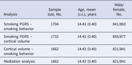
The sample used in these analyses included 1774 participants [of whom 899 (50.68%) were female; mean age = 14.43 ± 0.40 years]. Data on the participants, including their smoking behavior and the PGRS, are shown in Table 1.
Sample size for each polygenic risk profile score regression analysis

Smoking PGRS associated with smoking behavior
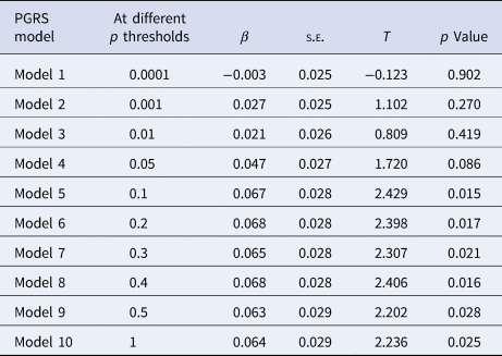
We found nominally significant positive associations between the smoking PGRS and the number of occasions of tobacco use at 6 out of 10 p thresholds. To ensure our result was not from false positives, multiple comparison corrections were used. As these is some overlap of contributing SNPs between PGRSs at different p thresholds, these PGRSs were not independent and the Bonferroni multiple testing correction would be overly conservative, so we used false discovery rate (FDR) multiple testing correction (in R) instead (Benjamini & Hochberg, Reference Benjamini and Hochberg1995). The PGRS at a p value threshold of 0.1 showed the most significant association (Table 2), indicating that it could best detect the associations with smoking behavior, and the association remained significant after correction (FDR-corrected p = 0.047). Therefore, we chose this PGRS (p threshold = 0.1) in our subsequent analysis, as was done in previous studies (Xu et al., Reference Xu, Li, Qin, Jun Li, Zhuo, Liu and Yu2018).
Association between smoking initiation PGRS and behavior
Cortical volume associated with smoking PGRS and smoking behavior
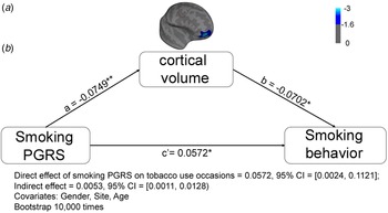
The entire cortex analysis revealed an inverse correlation between smoking PGRS and cortical volume of the right orbitofrontal cortex (OFC; corrected cluster-wise p = 0.007, Monte-Carlo correction, see Fig. 1a). No positive correlation was found between the smoking PGRS and the cortical volume. To explore the mediating role of cortical structure between polygenic risk and smoking behavior, the cortical volume of the orbitofrontal cluster resulting from the whole brain analysis of smoking PGRS was extracted. The cortical volume of the orbitofrontal cluster was significantly correlated with the number of occasions of tobacco use (T = −2.598, p = 0.009) when taking the covariates, gender, age, site, and eTIV into consideration. These suggested that subjects with a higher smoking PGRS had a smaller volume in the right OFC and that individuals with a smaller volume in the right OFC are likely to consume more cigarettes.
Smoking PGRS-cortical volume-tobacco use mediation model after controlling for gender and age. Path coefficients in the graph were standardized regression weights. CI, confidence interval. *p < 0.05, **p < 0.01, ***p < 0.001.

Mediation model
Given the three concurrent relationships between the genes, brain structure, and behavior, a path model was applied to examine whether the cortical volume of the OFC mediated the effect of the smoking PGRS on the number of occasions of tobacco use. The result revealed a significant indirect path from the smoking PGRS and tobacco use through the OFC volume (ab = 0.005, 95% CI = 0.0011–0.0128). This suggested that the effect of the smoking PGRS on tobacco use behavior was partially accounted for by PGRS-related variations in the OFC's volume (Fig. 1b).
Discussion
The current study found significant associations between the polygenic risk for smoking, the cortical volume of the OFC, and the number of occasions tobacco use in adolescents. Furthermore, a path analysis indicated that the cortical volume of the OFC mediated the association between the smoking PGRS and tobacco use. To our knowledge, this is the first smoking imaging genetics study that correlated a smoking PGRS with a brain structure. The smoking PGRS used here was based on 24 593 SNPs that predict smoking initiation status at a p < 0.10 threshold for SNP inclusion from a large genome-wide meta-analysis (Tobacco Genetics Consortium, 2010). The PGRS influenced smoking behavior in our sample, and further, could characterize individual variation in cortical volume in the OFC. This result supported the strategy of combining PGRS with imaging genetics to investigate the etiology of tobacco smoking.
One of the major findings of this study was the correlation of the smoking PGRS with cortical volume. Twin studies suggested that cortical volume is highly heritable (Blokland, de Zubicaray, McMahon, & Wright, Reference Blokland, de Zubicaray, McMahon and Wright2012) and is significantly genetically correlated with smoking behavior (Prom-Wormley et al., Reference Prom-Wormley, Maes, Schmitt, Panizzon, Xian, Eyler and Neale2015). These studies, together with previously reported gray matter volume abnormalities in smokers (Sutherland et al., Reference Sutherland, Riedel, Flannery, Yanes, Fox, Stein and Laird2016; Zhong et al., Reference Zhong, Shi, Shen, Dai, Zhu, Ma and Sheng2016), indicate that reduced cortical volume is a genetically modulated endophenotype of smoking. Studies have shown that cortical volume can be explained by neuronal density, neuronal size (Popescu et al., Reference Popescu, Klaver, Voorn, Galis-de Graaf, Knol, Twisk and Geurts2015), and the number of neurons (Carassiti et al., Reference Carassiti, Altmann, Petrova, Pakkenberg, Scaravilli and Schmierer2018). Therefore, more direct biological evidence, i.e. histological results, are required to explain the genetic contribution to cortical volume.
We observed a significant inverse correlation between smoking PGRS and cortical volume of the right OFC. Cortical volume within this cluster mediated the correlation of the polygenic risk for smoking and smoking behavior. These findings are in line with previous research implicating a critical role of the OFC in smoking. Structural meta-analyses studies have consistently reported reduced gray matter volume in the OFC among smokers compared with non-smokers (Sutherland et al., Reference Sutherland, Riedel, Flannery, Yanes, Fox, Stein and Laird2016; Zhong et al., Reference Zhong, Shi, Shen, Dai, Zhu, Ma and Sheng2016). Additionally, greater progressive volume loss in the OFC was displayed in elderly participants with a lifetime history of smoking (Durazzo, Insel, & Weiner, Reference Durazzo, Insel and Weiner2012). Besides, functional neuroimaging findings showed that alteration in OFC activity is involved in smoking craving (Hayashi, Ko, Strafella, & Dagher, Reference Hayashi, Ko, Strafella and Dagher2013). Moreover, pharmacological studies demonstrated high-affinity nicotinic receptors in the human OFC (Perry, Davila-Garcia, Stockmeier, & Kellar, Reference Perry, Davila-Garcia, Stockmeier and Kellar1999), increased high-affinity nicotinic receptors after smoking in humans (Perry et al., Reference Perry, Davila-Garcia, Stockmeier and Kellar1999), and an increased BOLD fMRI signal in the OFC after acute nicotine administration in rats (Bruijnzeel et al., Reference Bruijnzeel, Alexander, Perez, Bauzo-Rodriguez, Hall, Klausner and Febo2014). Therefore, our findings echo these structural (Sutherland et al., Reference Sutherland, Riedel, Flannery, Yanes, Fox, Stein and Laird2016; Zhong et al., Reference Zhong, Shi, Shen, Dai, Zhu, Ma and Sheng2016), functional MRI (Hayashi et al., Reference Hayashi, Ko, Strafella and Dagher2013), and pharmacological studies (Bruijnzeel et al., Reference Bruijnzeel, Alexander, Perez, Bauzo-Rodriguez, Hall, Klausner and Febo2014; Perry et al., Reference Perry, Davila-Garcia, Stockmeier and Kellar1999).
The negative associations of smoking PGRS and the right OFC volume and the mediating role of the OFC between the polygenic risk for smoking and tobacco use were further substantiated by the critical role of the OFC in impulse control and reward processing (Ruff & Fehr, Reference Ruff and Fehr2014) and that impulsivity and reward processing are the common traits linked to smoking (Liu, Lan, Wu, & Yan, Reference Liu, Lan, Wu and Yan2019). Studies applying morphometric imaging techniques have demonstrated a correlation of a smaller right OFC volume with increased impulsivity (Boes et al., Reference Boes, Bechara, Tranel, Anderson, Richman and Nopoulos2009; Matsuo et al., Reference Matsuo, Nicoletti, Nemoto, Hatch, Peluso, Nery and Soares2009), and the link of OFC lesions to risky decision-making (Studer, Manes, Humphreys, Robbins, & Clark, Reference Studer, Manes, Humphreys, Robbins and Clark2015). Besides, compared with nonsmokers, smokers have shown deficits in impulse control and reward processing, such as more steep delay discounting (Friedel, DeHart, Madden, & Odum, Reference Friedel, DeHart, Madden and Odum2014) and more reward sensitivity and less punishment sensitivity (Potts, Bloom, Evans, & Drobes, Reference Potts, Bloom, Evans and Drobes2014). These studies and our finding together indicated that individuals with a higher genetic risk for smoking had a smaller OFC volume, which may result in dysfunctions of reward and impulse control and might contribute to a greater risk for smoking initiation.
Intriguingly, the polygenic effect was only found in the right OFC, this result was not unexpected as the right OFC is often implicated in behavioral traits related to impulsivity, and as impulsivity is commonly associated with tobacco use (Flory & Manuck, Reference Flory and Manuck2009). For instance, a negative correlation between right OFC gray matter volume and trait impulsivity was observed in different samples (Crunelle et al., Reference Crunelle, Kaag, van Wingen, van den Munkhof, Homberg, Reneman and van den Brink2014; Matsuo et al., Reference Matsuo, Nicoletti, Nemoto, Hatch, Peluso, Nery and Soares2009). Besides, some neuroimaging (Ernst et al., Reference Ernst, Bolla, Mouratidis, Contoreggi, Matochik, Kurian and London2002) and brain-stimulation (Nejati, Salehinejad, & Nitsche, Reference Nejati, Salehinejad and Nitsche2018) studies suggested that the right OFC may be particularly critical to exert impulse control and therefore influence the risky taking behavior. Concerning the often-reported risk-taking propensity of smokers, our result suggests that a smaller right OFC volume in individuals with a higher smoking PGRS could indicate a deficit in impulse control and in turn more liability to smoke. Taken together, we believe that the right OFC volume represents a promising biomarker for delineating the brain structural substrate for genetic liability of smoking. It should be noted, however, that the hemispheric asymmetry in such associations is far from being firmly established. More systematic studies are needed to reproduce such lateralized effects and to uncover the underlying neurobiological mechanisms.
We note the following limitations of the current study. First, all of our samples are adolescents, given that this stage is particularly valuable to study as the majority of adult smokers (80–90%) start smoking during adolescence (Wittchen et al., Reference Wittchen, Behrendt, Hofler, Perkonigg, Lieb, Buhringer and Beesdo2008). However, the brain is still undergoing significant developmental changes (Mills et al., Reference Mills, Goddings, Herting, Meuwese, Blakemore, Crone and Tamnes2016), the association between brain structure and cigarette smoking was exacerbated by advancing age (Durazzo, Meyerhoff, & Nixon, Reference Durazzo, Meyerhoff and Nixon2013). Thus, analyses stratified for different age groups are needed to yield age-dependent effect of smoking on gray matter. Second, nicotine use was assessed by using self-report questionnaires in this study; future studies may benefit from biological measurements, such as serum nicotine levels, as an index of cigarette exposure. Previous studies have indicated a good correlation between the self-reported number of cigarettes smoked and plasma cotinine levels (Eliopoulos, Klein, & Koren, Reference Eliopoulos, Klein and Koren1996), and clear differentiation of serum cotinine levels between self-reported smokers and non-smokers (Binnie et al., Reference Binnie, McHugh, Macpherson, Borland, Moir and Malik2004). These studies consistently supported a significant relationship between cotinine level and self-reported smoking status. Given this, self-reported smoking measures in our study should remain credible. Third, the current study only investigates the genetic effect on the OFC structure. Previous studies have consistently reported that the development of the OFC structure is affected by the prenatal exposure to maternal cigarette smoking (Lotfipour et al., Reference Lotfipour, Ferguson, Leonard, Perron, Pike, Richer and Paus2009; Toro et al., Reference Toro, Leonard, Lerner, Lerner, Perron, Pike and Paus2008). Therefore, further studies should also consider the effect of the environment and the gene–environment interaction on the OFC structure.
In summary, this study provided the first evidence of interrelationships between the polygenic risk of smoking, OFC morphology, and tobacco use, with a mediating effect of the brain between the genes and behavior in a large sample of adolescents. Our findings suggest that OFC volume may be a promising endophenotype in smoking imaging genetic studies and that future studies of smoking gene–behavior associations should consider the brain mediation effect.
Supplementary material
The supplementary material for this article can be found at https://doi.org/10.1017/S0033291720002962
Acknowledgements
The authors appreciate the suggestions of Dr Gareth J. Barker from the Department of Neuroimaging, Institute of Psychiatry, Psychology & Neuroscience, King's College London. The authors also appreciate Rhoda E. and Edmund F. Perozzi, PhDs for English and content editing assistance.
Author contributions
G.S. and T.J.: conceptualized this research; T.B., A.L.W.B., E.B.Q., S.D., H.F., V.F., H.G., P.G., A.H., B.I., J.-L.M., E.A., F.N., D.P.O., T.P., L.P., S.H., J.H.F., M.N.S., H.W., and R.W.: collected data; J.L.: analyzed the data; J.L. and B.L.: wrote the manuscript. All authors, including the contributing members of the IMAGEN Consortium, revised the manuscript.
Financial support
This work received support from the following sources: the National Key Basic Research and Development Program (973) (Grant No. 2011CB707800), the Strategic Priority Research Program of the Chinese Academy of Sciences (Grant No. XDB02030300), the Natural Science Foundation of China (Grant Nos. 81000582, 31300934, 91132301, and 91232718), the European Union-funded FP6 Integrated Project IMAGEN (Reinforcement-related behaviour in normal brain function and psychopathology) (LSHM-CT-2007-037286), the Horizon 2020 funded ERC Advanced Grant ‘STRATIFY’ (Brain network based stratification of reinforcement-related disorders) (695313), ERANID (Understanding the Interplay between Cultural, Biological, and Subjective Factors in Drug Use Pathways) (PR-ST-0416-10004), BRIDGET (JPND: BRain Imaging, cognition Dementia and next generation GEnomics) (MR/N027558/1), Human Brain Project (HBP SGA 2, 785907), the FP7 project MATRICS (603016), the Medical Research Council Grant ‘c-VEDA’ (Consortium on Vulnerability to Externalizing Disorders and Addictions) (MR/N000390/1), the National Institute for Health Research (NIHR) Biomedical Research Centre at South London and Maudsley NHS Foundation Trust and King's College London, the Bundesministeriumfür Bildung und Forschung (BMBF grants 01GS08152; 01EV0711; Forschungsnetz AERIAL 01EE1406A, 01EE1406B), the Deutsche Forschungsgemeinschaft (DFG grants SM 80/7-2, SFB 940, TRR 265, and NE 1383/14-1), the Medical Research Foundation and Medical Research Council (grants MR/R00465X/1 and MR/S020306/1), and the National Institutes of Health (NIH) funded ENIGMA (grants 5U54EB020403-05 and 1R56AG058854-01). Further support was provided by grants from: ANR (project AF12-NEUR0008-01 – WM2NA, ANR-12-SAMA-0004), the Eranet Neuron (ANR-18-NEUR00002-01), the Fondation de France (00081242), the Fondation pour la Recherche Médicale (DPA20140629802), the Mission Interministérielle de Lutte-contre-les-Drogues-et-les-Conduites-Addictives (MILDECA), the Assistance-Publique-Hôpitaux-de-Paris and INSERM (interface grant), Paris Sud University IDEX 2012, the fondation de l'Avenir (grant AP-RM-17-013), the Fédération pour la Recherche sur le Cerveau; the National Institutes of Health, Science Foundation Ireland (16/ERCD/3797), U.S.A. (Axon, Testosterone and Mental Health during Adolescence; RO1 MH085772-01A1), and by NIH Consortium grant U54 EB020403, supported by a cross-NIH alliance that funds Big Data to Knowledge Centres of Excellence.
Conflict of interest
T.B. served in an advisory or consultancy role for Lundbeck, Medice, Neurim Pharmaceuticals, Oberberg GmbH, and Shire. He received conference support or a speaker's fee from Lilly, Medice, Novartis, and Shire. He has been involved in clinical trials conducted by Shire and Viforpharma. He received royalties from Hogrefe, Kohlhammer, CIP Medien, and Oxford University Press. The present work is unrelated to these relationships. The other authors, including the contributing members of the IMAGEN Consortium, declare no competing interests.