Introduction
The family Anostomidae comprises 15 genera and approximately 162 valid species of characiform fishes, widely distributed throughout South America (Froese & Pauly Reference Froese and Pauly2025; Gimênes Jr. & Rech Reference Gimênes and Rech2022). Among these genera, Leporinus Agassiz, 1829 exhibits remarkable species richness, with about 86 described species, including some of economic and ecological importance (Botero et al. Reference Botero, Lourenço and Pinto2023; Froese & Pauly Reference Froese and Pauly2025; Gimênes Jr. & Rech Reference Gimênes and Rech2022; Ohara et al. Reference Ohara, Lima, Salvador and Andrade2017). Representatives of the genus Leporinus are known to host a variety of taxa of metazoan parasites, with approximately 126 recorded species (Eiras et al. Reference Eiras, Takemoto and Pavanelli2010; Lehun et al. Reference Lehun, Hasuike, Silva, Ciccheto, Michelan, Rodrigues, Nicola, Lima, Correia and Takemoto2020; Sousa et al. Reference Sousa, Diniz and Yamada2026). These parasites encompass a broad diversity of taxonomic groups, notably Myxozoa, Monopisthocotyla, Digenea, Cestoda, Nematoda, Acanthocephala, Branchiura, Copepoda, and Isopoda (Eiras et al. Reference Eiras, Takemoto and Pavanelli2010; Lehun et al. Reference Lehun, Hasuike, Silva, Ciccheto, Michelan, Rodrigues, Nicola, Lima, Correia and Takemoto2020; Pavanelli et al. Reference Pavanelli, Takemoto and Eiras2013).
Monopisthocotylans stand out as typically the most abundant parasitic group in anostomid fishes (Dias et al. Reference Dias, Vieira, Camargo, Silva, Azevedo and Abdallah2017; Lehun et al. Reference Lehun, Hasuike, Silva, Ciccheto, Michelan, Rodrigues, Nicola, Lima, Correia and Takemoto2020; Martins et al. Reference Martins, Justo, Cardenas and Cohen2017; Sousa et al. Reference Sousa, Diniz, Carvalho, Lopes and Yamada2023; Wendt et al. Reference Wendt, Monteiro and Amato2018; Yamada et al. Reference Yamada, Bongiovani, Yamada and Silva2017). To date, 21 monopisthocotylan species have been reported from fish of the genus Leporinus (Cohen et al. Reference Cohen, Justo and Kohn2013; Dias et al. Reference Dias, Vieira, Camargo, Silva, Azevedo and Abdallah2017; Lehun et al. Reference Lehun, Hasuike, Silva, Ciccheto, Michelan, Rodrigues, Nicola, Lima, Correia and Takemoto2020; Sousa et al. Reference Sousa, Diniz, Yamada and Yamada2024; Thatcher Reference Thatcher2006; Yamada et al. Reference Yamada, Bongiovani, Yamada and Silva2017, Reference Yamada, Müller, Zago, Yamada, Ebert, Franceschini and Silva2023; Zago et al. Reference Zago, Yamada, Franceschini, Bongiovani, Yamada and Silva2017, Reference Zago, Yamada, Yamada, Franceschini, Bongiovani and Silva2020). This number is expected to increase in the coming years with descriptions of new species, as several taxa have been reported only to genus or family level (Hasuike et al. Reference Hasuike, Michelan, Quagliato, Brandão and Takemoto2023; Lehun et al. Reference Lehun, Hasuike, Silva, Ciccheto, Michelan, Rodrigues, Nicola, Lima, Correia and Takemoto2020; Oliveira et al. Reference Oliveira, Gonçalves, Ferreira, Pinheiro, Neves, Dias and Tavares-Dias2017; Sousa et al. Reference Sousa, Diniz, Yamada and Yamada2024; Wendt et al. Reference Wendt, Monteiro and Amato2018).
Mymarothecioides was proposed by Soares et al. (Reference Soares, Magalhaes, Silva, Carneiro, Barbosa, Costa and Domingues2019) to accommodate four monopisthocotylan species found on the gills of Hydrolycus armatus (Jardine, 1841) (Characiformes, Cynodontidae) from the Amazon basin: Mymarothecioides xinguensis Soares & Domingues, 2019 (type species), M. altamirensis Soares & Domingues, 2019, M. ararai Soares & Domingues, 2019, and M. germanoi Soares & Domingues, 2019. According to Soares et al. (Reference Soares, Magalhaes, Silva, Carneiro, Barbosa, Costa and Domingues2019), Mymarothecioides differs from Mymarothecium Kritsky, Boeger & Jégu, Reference Kritsky, Boeger and Jégu1996, its morphologically closest genus, mainly by the absence of eyes and the presence of a medio-ventral vaginal opening, which justified the transfer of Mymarothecium whittingtoni Kritsky, Boeger & Jégu, Reference Kritsky, Boeger and Jégu1996 originally described by Kritsky et al. (Reference Kritsky, Boeger and Jégu1996) from serrasalmids from the Amazon basin to Mymarothecioides as Mymarothecioides whittingtoni.
The incorporation of molecular markers, particularly Mitochondrial Region of Cytochrome C Oxidase subunit 1 (COI mtDNA) and Large Subunit ribosomal DNA (LSU rDNA) regions, into taxonomic studies of Dactylogyridae parasitising Neotropical fishes represents a significant methodological advancement in recent decades (Diniz et al. Reference Diniz, Saraiva, Sousa, Ebert, Yamada, Silva, Costa and Yamada2026; Gasques et al. Reference Gasques, Graca, Prioli, Takemoto and Prioli2016; Mendoza-Palmero et al. Reference Mendoza-Palmero, Blasco-Costa and Scholz2015; Yamada et al. Reference Yamada, Osaki-Pereira, Acosta, Ebert and Silva2024b, Reference Yamada, Sousa, Ebert, Diniz, Tavares-Dias, Silva and Yamada2025). The combination of these methods with morphological taxonomy has provided important contributions, including the discovery of cryptic species (Gasques et al. Reference Gasques, Graca, Prioli, Takemoto and Prioli2016), the revision of generic classification for some taxa (Acosta et al. Reference Acosta, Mendoza-Palmero, Silva and Scholz2019), and the redefinition of the taxonomic status of species previously considered incertae sedis (Santos-Neto & Domingues Reference Santos-Neto and Domingues2023). Thus, the integration of genetic analyses in taxonomic studies, both for describing new species and re-evaluating known taxa, proves essential for elucidating phylogenetic relationships among monopisthocotylans (Diniz et al. Reference Diniz, Saraiva, Sousa, Ebert, Yamada, Silva, Costa and Yamada2026; Santos-Neto & Domingues Reference Santos-Neto and Domingues2024; Yamada et al. Reference Yamada, Sousa, Ebert, Diniz, Tavares-Dias, Silva and Yamada2025; Zago et al. Reference Zago, Yamada, Yamada, Franceschini, Bongiovani and Silva2020). In this context, the present study describes two new species of Monopisthocotyla of the genus Mymarothecioides found parasitising the gills of anostomid fishes from the Jaguaribe River and Paraná River basins in Brazil. Furthermore, we provide the first molecular characterisation (LSU rDNA and COI mtDNA) for a member of the genus Mymarothecioides.
Materials and methods
Fish and collection procedures
In northeastern Brazil, 35 specimens of Leporinus piau Fowler, 1941 were collected between June 2022 and June 2024, in the Lima Campos Weir (6°22’59.99" S, 38°58’0.01" W), municipality of Icó. Between January and May 2023, 47 specimens of Leporinus taeniatus Lütken, 1875 were collected in the Salgado River (6°42’41.4" S, 38°57’30.6" W), municipality of Lavras da Mangabeira, both located in the Jaguaribe River basin, state of Ceará. The capture and transportation of fish were authorised by SISBIO (Biodiversity Information and Authorization System; license no. 61328-2), and all procedures were conducted in accordance with the Ethics Committee for Animal Experimentation of the Universidade Regional do Cariri (CEUA no. 00165/2018.1). In southern Brazil, 59 specimens of Leporinus lacustris Amaral Campos, 1945 were collected between June 2023 and June 2024, in the Upper Paraná River floodplain (22°30’–22°00’S, 53°00’–53°30’W), near the municipality of Porto Rico, Paraná River basin, state of Paraná. All collections were carried out under the Long-Term Ecological Research Program (PELD) in the floodplain of the upper Paraná River, in compliance with the authorisation of the Ethics Committee on Animal Use of the Universidade Estadual de Maringá (CEUA no. 5560221024) and the SISBIO collection license (no. 52596-6).
Parasitological procedures
After capture, fish specimens were individually stored in plastic bags, frozen, and taken to the laboratory. During necropsy, the gills of the fish were removed, placed in Petri dishes with tap water, and checked for monopisthocotylans under a stereomicroscope. All monopisthocotylans found were collected and placed in vials containing 70% ethanol. For the morphological characterisation, some parasites were mounted on slides with Gray and Wess’s or Hoyer’s medium for the study of the sclerotised structures. For analyses of the internal organs, some specimens were fixed in 5% formalin, stained with Gomori’s trichrome, and mounted in Balsam of Canada (Eiras et al. Reference Eiras, Takemoto and Pavanelli2006). For molecular characterisation, some freshly killed hosts were examined in situ, and some specimens of the monopisthocotylans were collected and placed on a slide containing a drop of 0.9% saline solution and identified under a microscope. Afterwards, these specimens were placed in 99% molecular-grade ethanol for subsequent molecular analyses.
The morphology and morphometry of the parasites were analysed using an optical microscope equipped with a computerised system for image analysis with phase contrast (Zeiss Axioscope 5; Zeiss Axiocam 208 colour, Germany). Illustrations were made using a drawing tube mounted on a Leica DM750 microscope with phase-contrast optics and vectorised using the Inkscape 1.4 software. Measurements (all in micrometres) represent straight-line distances between extreme points and are expressed as the mean followed by the range and the number of specimens measured in parentheses; body length includes the haptor. Measurements of the sclerotised structures (bars, anchors, hooks, and copulatory complex) were performed according to Soares et al. (Reference Soares, Magalhaes, Silva, Carneiro, Barbosa, Costa and Domingues2019), and the numbering and distribution of hook pairs followed Mizelle and Price (Reference Mizelle and Price1963). The authorship of the new taxa followed Recommendation 50.1 (Identity of authors) of the International Code of Zoological Nomenclature. Parameters of infestation of parasites (prevalence and mean intensity of infestation) followed Bush et al. (Reference Bush, Lafferty, Lotz and Shostak1997). Type specimens and the hologenophore (Pleijel et al. Reference Pleijel, Jondelius, Norlinder, Nygren, Oxelman, Schander, Sundberg and Thollesson2008) were deposited in the Helminthological Collection of the Instituto Oswaldo Cruz (CHIOC), state of Rio de Janeiro, Brazil.
Molecular characterisation and delimitation
To extract DNA from the parasites, the DNeasy® Blood and Tissue Kit (QIAGEN®) was used, following the protocol suggested by the manufacturer. A ProFlex™ 3×32-well thermal cycler was used to carry out the Polymerase Chain Reaction (PCR). The DNA amplification reaction consisted of 1 U of Taq DNA polymerase (5 U/μL, Invitrogen), Tris-KCl (20 mM Tris-HCl pH 8.4; 50 mM KCl), 1.87 mM MgCl2, 0.1 mM of each Deoxynucleotide Triphosphates (dNTP), 4 μM of each primer, DNA template (10 ng), and Milli-Q water for a final volume of 23 μL.
Specific primers for the COI mtDNA and LSU rDNA regions were used to amplify the parasites’ DNA.
Specific primers for COI mtDNA were COI_Mono_5: 5′-TAATWGGTGGKTTTGGTAA-3′, COI_Mono_3: 5′-AATGCATMGGAAAAAAACA-3′, and COI_Mono_int3: 5′-ACATAATGAAARTGAGC-3′ (using a protocol adapted from Plaisance et al. Reference Plaisance, Rousset, Morand and Littlewood2008). Primary PCR amplifications (25 μL, using COI_Mono_5 and COI_Mono_3 primers) were performed with 2 μL of DNA extract; afterwards, we employed nested PCR (25 μL, using COI_Mono_5 and COI_Mono_int3) using 1 μL of the primary PCR product. Thermal cycling was performed with an initial denaturation for 3 min at 94°C, followed by 40 cycles for primary PCR and 35 cycles for nested PCR (30 s at 94°C, 30 s at 44°C, 1 min at 72°C for the primary PCR and 30 s at 94°C, 30 s at 45°C, 2 min at 72°C for the nested PCR, with a final extension of 7 min at 72°C).
The fragments of the LSU rDNA gene were amplified using the primers 382F (5′-AGC TGG TGG AGT CAA GCT TC-3′) and 1289R (5′-TGC TCA CGT TTG ACG ATC GA-3′), using a protocol adapted from Yamada et al. (Reference Yamada, Müller, Zago, Yamada, Ebert, Franceschini and Silva2023). Thermal cycling was performed with an initial denaturation for 5 min at 95°C, followed by 35 cycles (30 s at 95°C, 30 s at 56°C, 2 min at 72°C, with a final extension of 10 min at 72°C).
The PCR products were checked on a 1% agarose gel, and the size of the fragments obtained was estimated using a marker of known molecular weight (100 bp DNA Ladder, Invitrogen 0.5 μg/μL). The products obtained from DNA amplification were purified following the protocol described by Rosenthal et al. (Reference Rosenthal, Coutelle and Craxton1993). Sequencing reactions were carried out using the BigDye Terminator kit. Sequencing was performed by a private company using an AB-3500 automated sequencer.
The sequences obtained were edited and aligned by ClustalW (Thompson et al. Reference Thompson, Higgins and Gibson1994) using the BioEdit (Hall Reference Hall1999) and MEGA 11 (Tamura et al. Reference Tamura, Stecher and Kumar2021) programs, respectively. MEGA11 software was used to obtain the p-distance and the gene tree using the maximum likelihood method and 1,000 bootstrap resampling. Sequences of Mymarothecium viatorum Boeger, Piasecki & Sobecka, 2002 obtained from public genetic databases were compared with the newly generated sequences of Mymarothecioides spp. Gyrodactylus salaris was used as the outgroup.
The same sequences were used to carry out molecular species delimitation tests. For this, we selected three molecular species delimitation methods that differ methodologically to compare the congruencies between them and assess the best hypothesis that fits our data set: the Poisson Tree Process (PTP), performed on the server https://species.h-its.org/ptp/ as a tree-based method (Zhang et al. Reference Zhang, Kapli, Pavlidis and Stamatakis2013), the Automatic Barcode Gap Discovery (ABGD) based on the barcode gap, and the Assemble Species by Automatic Partitioning (ASAP) based on genetic distance (Puillandre et al. Reference Puillandre, Brouillet and Achaz2021); both available at https://itaxotools.org/download.html.
Access to the genetic heritage of the specimens was authorised through the Sistema Eletrônico Nacional de Gestão do Patrimônio Genético e do Conhecimento Tradicional Associado (SisGen) under the number AF0DE15, and the sequences obtained were deposited in GenBank: PX751182 to PX751185 (COI mtDNA) and PX741069 to PX741071 (LSU rDNA).
Results
Taxonomic summary
Class Monopisthocotyla Brabec, Salomaki, Scholz & Kuchta, 2023
Subclass Polyonchoinea Bychowsky, 1937
Order Dactylogyridea Bychowsky, 1937
Family Dactylogyridae Bychowsky, 1933
Subfamily Ancyrocephalinae Bychowsky, 1937
Mymarothecioides Soares & Domingues, 2019
Mymarothecioides leporini n. sp. Sousa, Hasuike, Diniz, Takemoto & Yamada
(Figure 1a–h)
Syn: Dactylogyridae gen. sp. 3 by Sousa et al. (Reference Sousa, Diniz, Yamada and Yamada2024)
Type-host: Leporinus piau Fowler, 1941 (Characiformes: Anostomidae)
Type locality: Lima Campos Weir, municipality of Icó, state of Ceará, Brazil (6°22’59.99" S, 38°58’0.01" W).
Other hosts: Leporinus taeniatus Lütken, 1875; Leporinus lacustris Amaral Campos, 1945 (Characiformes: Anostomidae)
Other localities: Salgado River, municipality of Lavras da Mangabeira, state of Ceará, Brazil (6°42’41.4" S, 38°57’30.6" W); Upper Paraná River, municipality of Porto Rico, state of Paraná, Brazil (22°30’–22°00’S, 53°00’–53°30’W).
Site of infestation: Gill filaments.
Infestation parameters: L. piau: Prevalence: 71.43% (25 infected fish of 35 analysed); total number of parasites: 112; mean intensity: 4.48 ± 4.78; range of intensity: 1–19. L. taeniatus: Prevalence: 23.4% (11 infected fish of 47 analysed); total number of parasites: 20; mean intensity: 1.82 ± 0.87; range of intensity: 1–4. L. lacustris: Prevalence: 20.3% (12 infected fish of 59 analysed); total number of parasites: 13; mean intensity: 1.1 ± 0.2; range of intensity: 1–2.
Specimens deposited: Holotype CHIOC 41124; paratypes CHIOC 41125a, 41126a-d, 41128, 41129a; paragenophore CHIOC 41125b, 41127; voucher CHIOC 41129b-c, 41130-41134. .
Molecular sequence data (Lima Campos Weir): PX741070 and PX741071 (LSU rDNA).
Molecular sequence data (Upper Paraná River): PX741069 (LSU rDNA) and PX751185 (COI mtDNA).
ZooBank registration: (urn:lsid:zoobank.org:act:425CAFFA-E23D-4926-A4DE-67EDA775DD07)
Etymology: The specific name designation leporini refers to the generic name of the type-host.
Description: (based on 15 specimens: two stained with Gomori’s trichrome and 13 clarified with Gray and Wess’s medium) Body robust, fusiform, total length 608 (395–923; n = 11), total width at level of germarium 219 (136–339; n = 15) (Figure 1a). Cephalic margin tapered; cephalic lobes poorly developed; four bilateral pairs of head organs, with rod-shaped secretion; cephalic glands unicellular, posterolateral to the pharynx. Eyes absent; accessory chromatic granules present in cephalic area. Pharynx subspherical, 55 (41–63; n = 15) long, 55 (35–74; n = 15) wide. Testis elongated, 78 (47–128; n = 9) long, 43 (25–56; n = 9) wide. MCO, 51 (42–55; n = 15) long, robust, spoon-shaped, expanded at the distal portion; base of MCO with sclerotised flange (Figure 1b). Accessory piece composed of two straight rods: a larger unit with distal portion with subterminal flap with hook and thumb; and a smaller unit with a pointed distal portion (Figure 1b). Germarium bacilliform, 129 (91–168; n = 13) long, 62 (37–113; n = 13) wide. Vagina single, muscular, dextral, opening at level of vitelline commissure. Seminal receptacle sigmoid. Vitelline follicles dense, distributed along the trunk. Peduncle long; haptor trapezoidal, 111 (73–146; n = 15) long, 165 (128–198; n = 15) wide. Anchors similar, each with divergent roots. Ventral anchor, outer 26 (21–49; n = 15) long, inner 16 (14–18; n = 15) long; base 21 (18–23; n = 15); root tips covered with sclerotised cap variable in shape; superficial root elongated; deep root elongated, heavily sclerotised on distal extremity; slightly curved shaft, point; point extending to level of tip of superficial root (Figure 1e). Dorsal anchor, outer 26 (19–37; n = 15) long, inner 17 (15–24; n = 15) long; base 20 (15–22; n = 15); root tips covered with sclerotised cap variable in shape; superficial and deep root elongated; slightly curved shaft and point; point extending to level of tip of superficial root (Figure 1f). Ventral bar slightly curved rod, with ornamented anteromedial process, 51 (45–59; n = 15) long, 7 (5–9; n = 14) wide including projection (Figure 1c). Dorsal bar, 46 (37–54; n = 14) long, 3 (2–5; n = 14) wide, slightly curved with flared ends (Figure 1d). Hooks similar in shape, with upright thumb, rounded, slightly curved shaft; short point; shank divided into two units, proximal unit inflated, distal unit compressed; hook pairs I and V, 18 (14–21; n = 15) long (Figure 1g); hook pairs II, III, IV, VI, and VII, 21 (17–24; n = 15) long (Figure 1h); filamentous hook loop at approximately half of shank length. Eggs, Mehlis’ glands, prostatic reservoir, uterus, and ootype not observed.
Mymarothecioides leporini n. sp. from Leporinus piau. (a) whole mount (composite, ventral view), (b) copulatory complex, (c) ventral bar, (d) dorsal bar, (e) ventral anchor, (f) dorsal anchor, (g) hook pairs I and V, (h) hook pairs II–IV, VI–VII.

Figure 1. Long description
The illustration consists of eight panels labeled a through h.
Panel a shows the whole mount in ventral view, featuring an elongated body with a cephalic region containing lobes, a central digestive and reproductive system, and a posterior haptor with hooks. A scale bar indicates 200 micrometers.
Panel b depicts the copulatory complex, a curved tubular structure with a 30 micrometer scale bar.
Panel c shows the ventral bar, a horizontal rod with a small central protrusion.
Panel d shows the dorsal bar, a simple curved rod. A 20 micrometer scale bar is positioned between panels c and d.
Panel e illustrates the ventral anchor, a hook-like structure with labels for Base, Outer, and Inner margins. A 20 micrometer scale bar is adjacent.
Panel f shows the dorsal anchor, similar in shape to the ventral anchor but with a slightly different base morphology.
Panel g displays hook pairs I and V, which are small, thin sclerotized hooks.
Panel h displays hook pairs II through IV, VI, and VII, which are similar in structure to those in panel g but slightly larger.
Remarks: The new species is allocated to Mymarothecioides due to the presence of the generic characteristics proposed by Soares et al. (Reference Soares, Magalhaes, Silva, Carneiro, Barbosa, Costa and Domingues2019), such as broad and arched MCO articulated with the accessory piece, with a subterminal hooked process; presence of an anteromedial projection on the ventral bar; and absence of eyespots. Among the five known species, Mymarothecioides leporini n. sp. closely resembles M. ararai; both species have MCO with expanded distal portion and sclerotised caps on the anchor roots. However, the new species can be easily distinguished from all congeners by possessing the following combination of characters: MCO spoon-shaped expanded at distal portion; accessory piece composed of two straight rods: a larger unit with distal portion with subterminal flap with hook and thumb, and a smaller unit with a pointed distal portion; anchors with elongate roots; and a ventral bar with an ornamented process. The presence of an anteromedial process on the ventral bar is a diagnostic characteristic of the genus. However, in the new species, the process is highly ornamented, forming undulations similar to ‘fingers of a glove’, whereas in its congeners the process is simple, varying only in size.
Mymarothecioides paranaensis n. sp. Hasuike, Sousa, Diniz, Yamada & Takemoto
(Figure 2a–h)
Type-host: Leporinus lacustris Amaral Campos, 1945 (Characiformes: Anostomidae).
Type locality: Upper Paraná River, municipality of Porto Rico, state of Paraná, Brazil (22°30’–22°00’S, 53°00’–53°30’W).
Site of infestation: Gill filaments.
Infestation parameters: Prevalence: 10.2% (6 infected fish of 59 analysed); total number of parasites: 7; mean intensity: 1.1 ± 0.5; range of intensity: 1–2.
Specimens deposited: Holotype CHIOC 41122a; paratypes CHIOC 41122b-c, 41123a; hologenophore CHIOC 41123b-d.
Molecular sequence data: PX751182, PX751183, PX751184 (COI mtDNA).
ZooBank registration: (urn:lsid:zoobank.org:act:6879B00A-B5B8-4904-AA5F-5C22FFAA4CC3)
Etymology: The specific name is derived from the Paraná River (type locality).
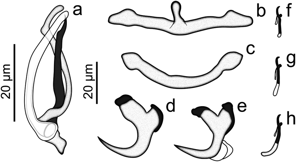
Description: (based on 5 specimens clarified with Hoyer’s medium) Body robust, fusiform, total length 301 (n = 1), total width at level of germarium 156 (n = 1). Cephalic lobes poorly developed; four bilateral pairs of head organs, with rod-shaped secretion; cephalic glands unicellular, posterolateral to the pharynx. Eyes absent; accessory chromatic granules present in cephalic area. Pharynx subspherical, 32 (n = 1) long, 30 (n = 1) wide. MCO 39 (n = 1) long, a sclerotised arcuate tube with rounded tip; base possessing a rectangular posterior flap (Figure 2a). Accessory piece composed of two elongated units: a hook-shaped unit; and another unit consisting of a sigmoid rod, with a rounded distal portion (Figure 2a). Germarium elongated, 89 (n = 1) long, 31 (n = 1) wide. Vaginal aperture marginal, dextral. Vitelline follicles dense. Peduncle long, expanded posteriorly; haptor trapezoidal, 79 (n = 1) long, 125 (n = 1) wide. Anchors similar, each with divergent roots covered with sclerotised cap; slightly curved shaft; elongate point extending beyond tip of superficial root. Ventral anchor, outer 31 (29–34; n = 5) long, inner 18 (14–21; n = 5) long, base 23 (22–24; n = 5); root tips covered with sclerotised cap, variable in shape (Figure 2d). Dorsal anchor, outer 31 (29–33; n = 4) long, inner 19 (17–21; n = 4) long, base 21 (19–23; n = 4); root tips covered with sclerotised cap variable in shape (Figure 2e). Ventral bar with anteromedial elongate projection, flared ends, 61 (44–69; n = 4) long, 18 (16–23; n = 4) wide including projection (Figure 2b). Dorsal bar 56 (53–60; n = 4) long, 6 (5–7; n = 4) wide, slightly U-shaped rod with flared ends (Figure 2c). Hooks similar in shape, with upright thumb; rounded, slightly curved shaft; short point; shank divided into two units; hook pairs I and V, 13 (11–15; n = 5) long (Figure 2f); hook pairs II, III, and IV, 17 (15–20; n = 4) long (Figure 2g); hook pairs VI and VII, 21 (17–24; n = 4) long (Figure 2h); filamentous hook loop extended at approximately 1/3 of shank length. Testis, prostatic reservoir, uterus, seminal receptacle, eggs, Mehlis’ glands, and ootype not observed.
Sclerotised structures of Mymarothecioides paranaensis n. sp. from Leporinus lacustris. (a) copulatory complex, (b) ventral bar, (c) dorsal bar, (d) ventral anchor, (e) dorsal anchor, (f) hook pairs I and V, (g) hook pairs II, III, and IV, and (h) hook pairs VI–VII.
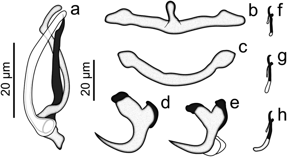
Figure 2. Long description
The drawing consists of eight panels labeled a through h.
* Panel a: Located on the far left, it shows a large, elongated copulatory complex oriented vertically. It features two intertwined tubular structures, one shaded dark and one light, with a 20 micro-meter scale bar to its left.
* Panel b: At the top center, a horizontal ventral bar with a small vertical protrusion in the middle and rounded ends.
* Panel c: Below panel b, a curved dorsal bar with thickened, bulbous ends.
* Panel d: Below panel c, a ventral anchor with a thick base, a sharp curved point, and a dark-shaded cap at the proximal end.
* Panel e: To the right of panel d, a dorsal anchor similar in shape to the ventral anchor but with a more pronounced bifurcated base and dark shading on both proximal tips. A 20 micro-meter scale bar is positioned between panels a and b/c.
* Panel f: At the top right, a small vertical hook with a curved tip and a loop near the base.
* Panel g: Below panel f, a slightly larger vertical hook with a similar curved tip and a partially shaded shaft.
* Panel h: At the bottom right, a third vertical hook with a dark-shaded middle section of the shaft.
Remarks: Mymarothecioides paranaensis n. sp. closely resembles M. ararai and Mymarothecioides leporini n. sp. These are the only species in the genus that possess an accessory piece composed of two units, and anchors with divergent roots, covered with sclerotised caps. The accessory piece, consisting of two long units, is observed in Mymarothecioides paranaensis n. sp. and M. ararai, while in Mymarothecioides leporini n. sp. the accessory piece presents two units of different sizes (one small and the other long). When comparing the morphology of the MCO, we find that Mymarothecioides paranaensis n. sp. has an MCO formed by an arched, sclerotised tube with a rounded tip; ax-shaped at the distal portion, small grooves at the edges of the distal opening in M. ararai; spoon-shaped and expanded at the distal portion in Mymarothecioides leporini n. sp. Furthermore, the new species and Mymarothecioides leporini n. sp. possess hook of different sizes, whereas in M. ararai the hooks are similar in size.
Molecular characterisation and delimitation
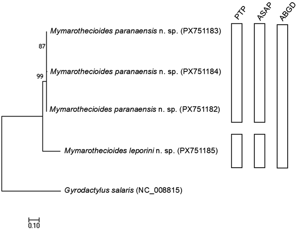
After editing and alignment, partial sequences of the COI mtDNA, with 605 base pairs (bp), were obtained for Mymarothecioides paranaensis n. sp. and Mymarothecioides leporini n. sp‥ Partial sequences of the LSU rDNA regions, with 615 bp, were obtained for Mymarothecioides leporini n. sp‥ It was not possible to amplify Mymarothecioides paranaensis n. sp. for this region due to amplification failure and, consequently, a lack of genetic material due to limited sample size. Mymarotheciuoides paranaensis n. sp. were grouped into a distinct clade from those identified as Mymarothecioides leporini n. sp‥ The PTP and ASAP species delimitation tests corroborate this separation, since each of the species was identified as a distinct molecular operational taxonomic unit (MOTU), and this separation did not occur through the ABGD test. Furthermore, the distance between the different species in this study ranged from 17.8% to 18.7% (Table 1).
Genetic distance matrix calculated by the p-distance model between the sequences obtained in the present study for Mymarothecioides paranaensis n. sp. and Mymarothecioides leporini n. sp. for the COI mtDNA region. All values are represented in percentages (%)
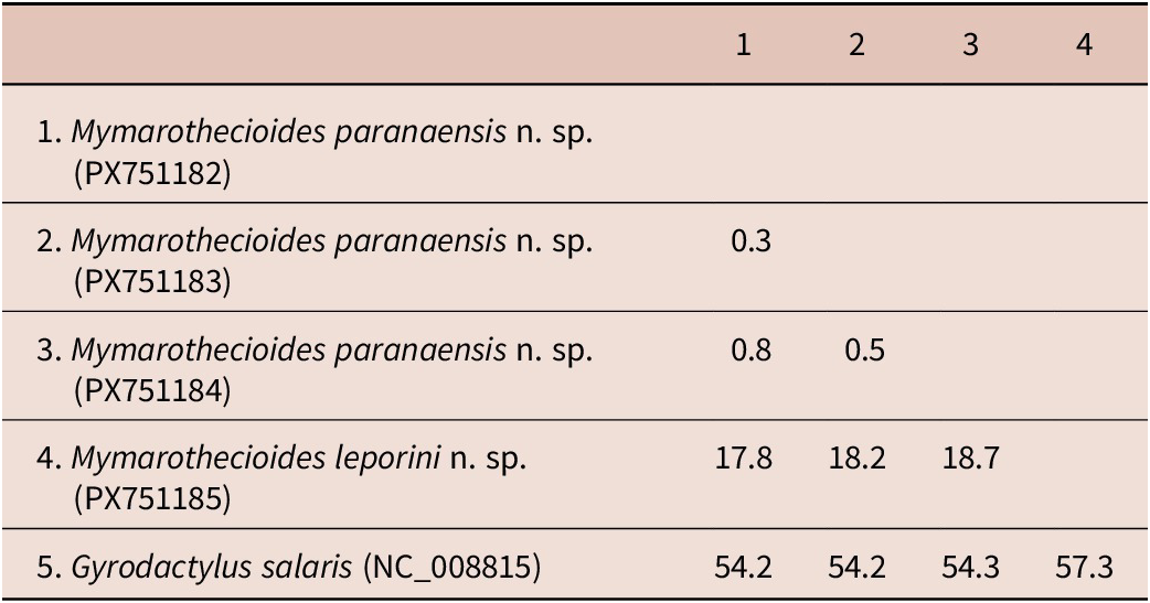
Table 1. Long description
The table presents a genetic distance matrix with five rows and four numbered columns. The values represent percentages.
Row 1: Mymarothecioides paranaensis n. sp. (P X 7 5 1 1 8 2) serves as the baseline for column 1.
Row 2: Mymarothecioides paranaensis n. sp. (P X 7 5 1 1 8 3) shows a distance of 0.3 compared to sample 1.
Row 3: Mymarothecioides paranaensis n. sp. (P X 7 5 1 1 8 4) shows distances of 0.8 compared to sample 1 and 0.5 compared to sample 2.
Row 4: Mymarothecioides leporini n. sp. (P X 7 5 1 1 8 5) shows significantly higher distances of 17.8 compared to sample 1, 18.2 compared to sample 2, and 18.7 compared to sample 3.
Row 5: Gyrodactylus salaris (N C 0 0 8 8 1 5) shows the highest divergence with distances of 54.2 compared to sample 1, 54.2 compared to sample 2, 54.3 compared to sample 3, and 57.3 compared to sample 4.
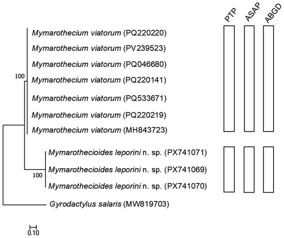
Moreover, in the analysis carried out for the LSU rDNA marker (Figure 4), the sequences of Mymarothecium viatorum used in the analyses grouped into a distinct clade, different from the Mymarothecioides leporini n. sp. clade in the present study. For this marker, the three delimitation tests separated the clades as distinct MOTUs. For the sequences obtained in this study, the distance ranged from 0 to 0.7%, with PX741071 being the most distant, while when compared to the sequences available in the database, the distance ranged from 28.1% to 62.3% (Table 2).
Genetic distance matrix calculated by the p-distance model between the sequences obtained in the present study for Mymarothecioides leporini n. sp. and sequences available in GenBank for Mymarothecium viatorum for the LSU rDNA region. All values are represented in percentages (%)
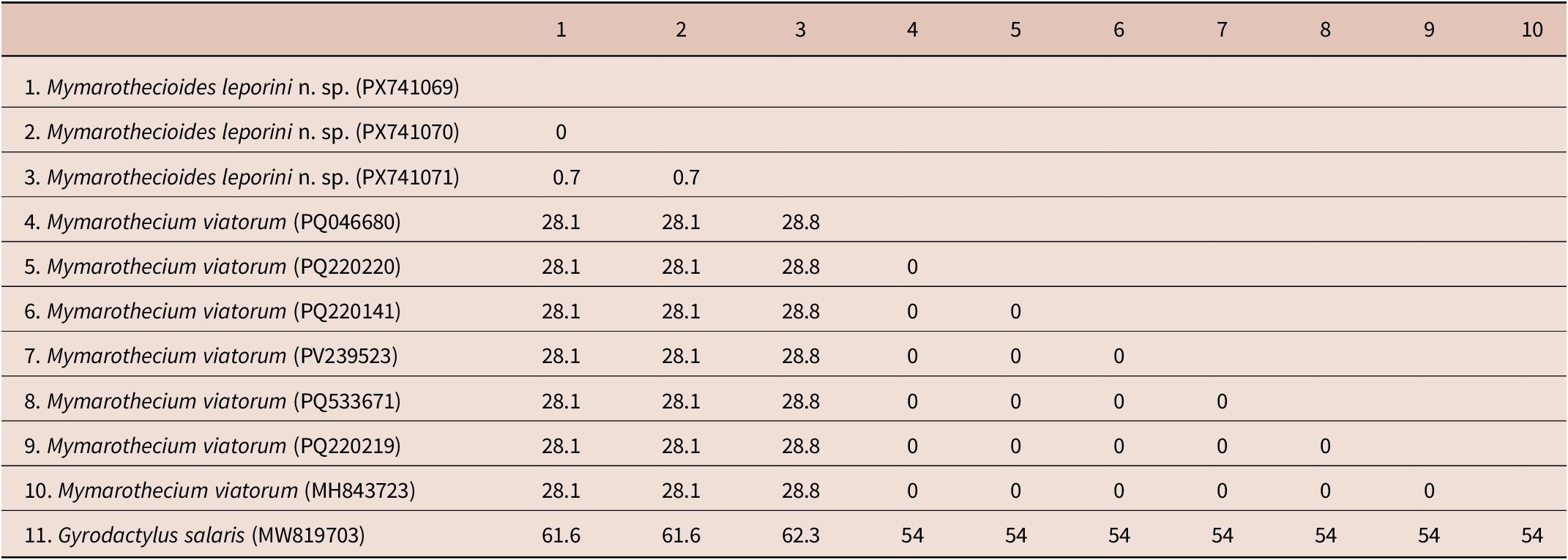
Table 2. Long description
The matrix compares 11 sequences. Rows and columns 1 through 3 are Mymarothecioides leporini n. sp. (P X 7 4 1 0 6 9, P X 7 4 1 0 7 0, P X 7 4 1 0 7 1). Rows and columns 4 through 10 are Mymarothecium viatorum (P Q 0 4 6 6 8 0, P Q 2 2 0 2 2 0, P Q 2 2 0 1 4 1, P V 2 3 9 5 2 3, P Q 5 3 3 6 7 1, P Q 2 2 0 2 1 9, M H 8 4 3 7 2 3). Row 11 is Gyrodactylus salaris (M W 8 1 9 7 0 3).
Data points:
* Between M. leporini sequences 1 and 2, the distance is 0 percent. Sequence 3 has a 0.7 percent distance from sequences 1 and 2.
* Between all M. viatorum sequences (4 through 10), the distance is 0 percent.
* Between M. leporini (1-2) and all M. viatorum (4-10), the distance is 28.1 percent.
* Between M. leporini (3) and all M. viatorum (4-10), the distance is 28.8 percent.
* Gyrodactylus salaris (11) shows the highest distances: 61.6 percent from M. leporini (1-2), 62.3 percent from M. leporini (3), and 54 percent from all M. viatorum sequences.
Discussion
Currently, five genera of monopisthocotylans are known to parasitise the gills of anostomid fishes in Brazil: Jainus Mizelle, Kritsky & Crane, 1968; Mymarothecioides; Tereancistrum Kritsky, Thatcher & Kayton, 1980; Trinibaculum Kritsky, Thatcher & Kayton, 1980; and Urocleidoides Mizelle & Price, 1964 (Cohen et al. Reference Cohen, Justo and Kohn2013; Dias et al. Reference Dias, Vieira, Camargo, Silva, Azevedo and Abdallah2017; Hasuike et al. Reference Hasuike, Michelan, Quagliato, Brandão and Takemoto2023; Sousa et al. Reference Sousa, Diniz, Yamada and Yamada2024). Hasuike et al. (Reference Hasuike, Michelan, Quagliato, Brandão and Takemoto2023) reported the first occurrence of parasites of the genus Mymarothecioides in the gills of hosts from the family Anostomidae. Soares & Domingues, Reference Soares, Magalhaes, Silva, Carneiro, Barbosa, Costa and Domingues2019 proposed the genus Mymarothecioides and characterised the genus as having: (1) eyes absent, though it may possess accessory chromatic granules; (2) gonads overlapping; (3) male copulatory organ (MCO) articulated with an accessory piece; (4) an accessory piece with a subterminal hook-like process; (5) haptor armed with two pairs of anchors, one pair of bars, and seven pairs of hooks, arranged in Ancyrocephalinae distribution; (6) a ventral bar with an anteromedial projection; and (7) parasitism on the gills of characiform fishes.
Considering the two new species described in the present study, Mymarothecioides paranaensis n. sp. occurred only on L. lacustris from the Upper Paraná River, state of Paraná, while Mymarothecioides leporini n. sp. showed a wider geographic distribution, occurring in L. lacustris from the Upper Paraná River and in L. piau and L. taeniatus from two rivers in the state of Ceará. The specimens of Mymarothecioides leporini n. sp. recovered from the three host species examined in the present study are morphologically similar and have similar measurements (see Table 3). Furthermore, specimens of Dactylogyridae gen. sp. 3 found in L. piau by Sousa et al. (Reference Sousa, Diniz, Yamada and Yamada2024) are considered synonymous with Mymarothecioides leporini n. sp‥
Comparative measurements (in micrometres) of specimens of the Mymarothecioides leporini n. sp. (Monopisthocotyla: Dactylogyridae) on species of fishes of the family Anostomidae (Characiformes) in the Neotropical region
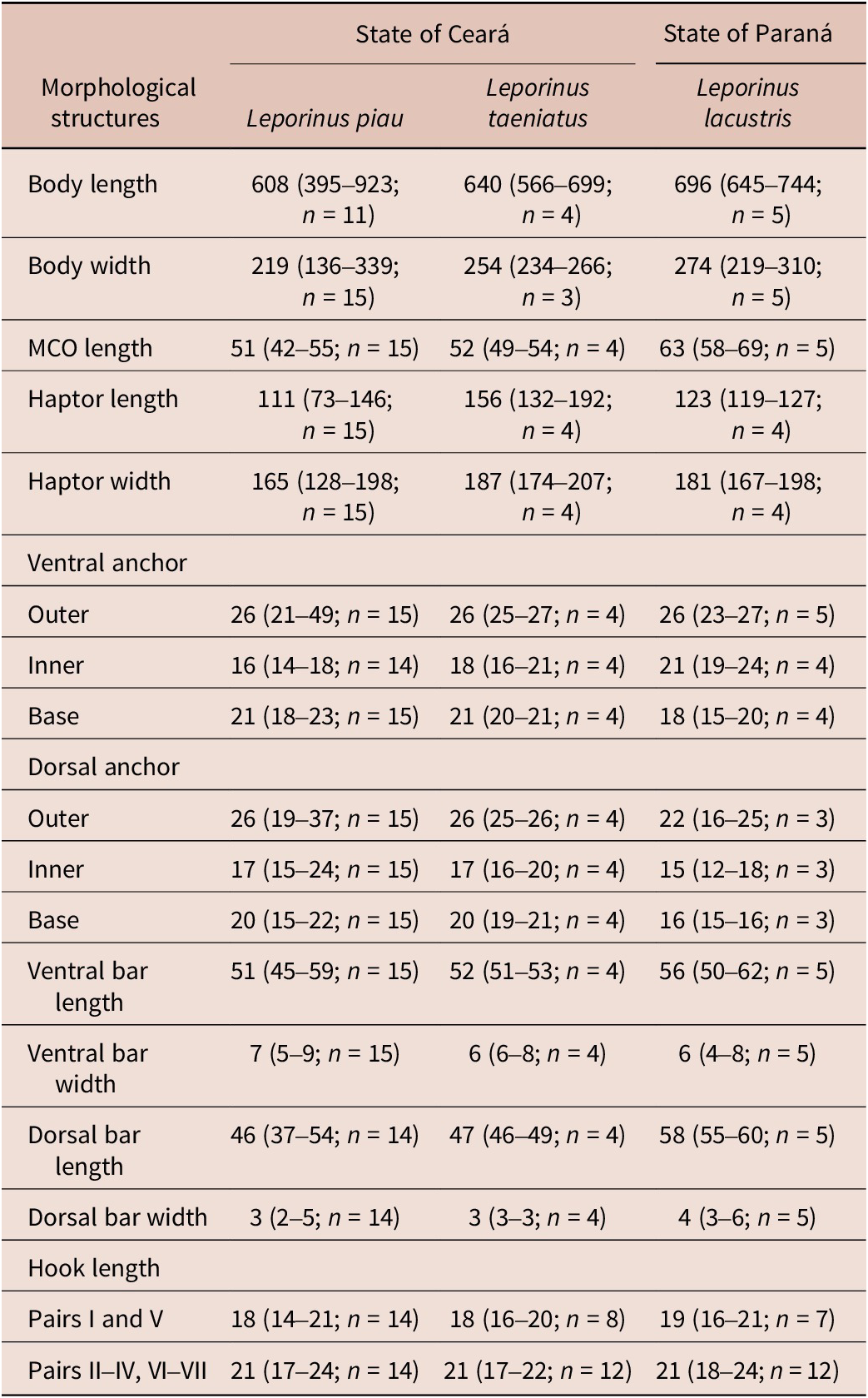
Table 3. Long description
The table compares measurements in micrometres for specimens found on Leporinus piau, Leporinus taeniatus, and Leporinus lacustris. Each entry includes the mean, range in parentheses, and sample size n.
* Body length: L. piau 608 (395–923, n=11); L. taeniatus 640 (566–699, n=4); L. lacustris 696 (645–744, n=5).
* Body width: L. piau 219 (136–339, n=15); L. taeniatus 254 (234–266, n=3); L. lacustris 274 (219–310, n=5).
* M C O length: L. piau 51 (42–55, n=15); L. taeniatus 52 (49–54, n=4); L. lacustris 63 (58–69, n=5).
* Haptor length: L. piau 111 (73–146, n=15); L. taeniatus 156 (132–192, n=4); L. lacustris 123 (119–127, n=4).
* Haptor width: L. piau 165 (128–198, n=15); L. taeniatus 187 (174–207, n=4); L. lacustris 181 (167–198, n=4).
* Ventral anchor (Outer, Inner, Base): L. piau (26, 16, 21); L. taeniatus (26, 18, 21); L. lacustris (26, 21, 18).
* Dorsal anchor (Outer, Inner, Base): L. piau (26, 17, 20); L. taeniatus (26, 17, 20); L. lacustris (22, 15, 16).
* Ventral bar length: L. piau 51; L. taeniatus 52; L. lacustris 56.
* Dorsal bar length: L. piau 46; L. taeniatus 47; L. lacustris 58.
* Hook length (Pairs I and V): L. piau 18; L. taeniatus 18; L. lacustris 19.
* Hook length (Pairs II–IV, VI–VII): L. piau 21; L. taeniatus 21; L. lacustris 21.
The analysis of the COI mtDNA region and species delimitation tests confirm that specimens identified as Mymarothecioides leporini n. sp. are distinct from those identified as Mymarothecioides paranaensis n. sp. Mymarothecioides leporini n. sp. is represented as a distinct MOTU (Figure 3), with a genetic distance ranging from 17.8% to 18.7% (Table 1). Such divergence is typical between different monopisthocotylan species (Hasuike et al. Reference Hasuike, Scorsim, Arjona, Bellay, Oliveira and Takemoto2024, Reference Hasuike, Scorsim, Arjona, Amaral, Damacena-Silva, Araújo, Bellay, Oliveira and Takemoto2025; Santos-Neto & Domingues Reference Santos-Neto and Domingues2023; Zago et al. Reference Zago, Yamada, Yamada, Franceschini, Bongiovani and Silva2020). In contrast, specimens of Mymarothecioides leporini n. sp. exhibit high similarity in the LSU rDNA region, indicating that these specimens belong to the same species (Figure 4; Table 2). Furthermore, it is important to note that it was not possible to amplify Mymarothecioides paranaensis n. sp. for this region due to amplification failure and, consequently, a lack of genetic material. However, future studies including molecular analysis can provide a better understanding of the relationships between species of Mymarothecioides.
Gene tree constructed using the maximum likelihood method with 1,000 bootstrap resampling for the COI mtDNA molecular marker. Gyrodactylus salaris was used as an outgroup, and the nucleotide substitution model used was Hasegawa–Kishino–Yano (HKY). The molecular operational taxonomic units (MOTUs) delimited by the PTP, ASAP, and ABGD methods are represented in the bars on the right-hand side of the gene tree.
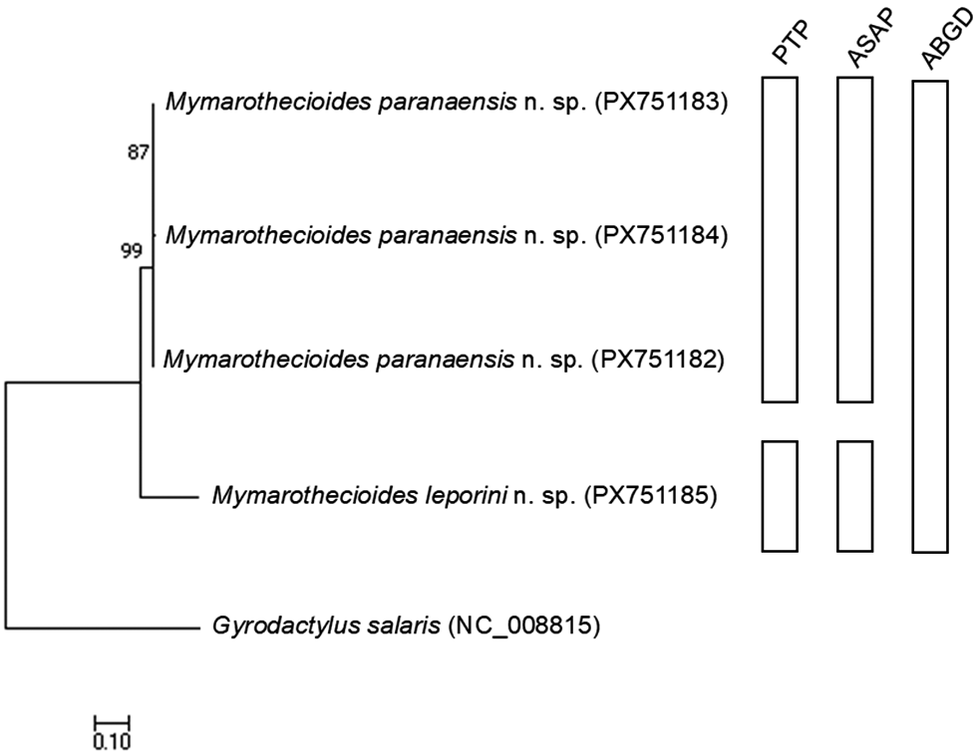
Figure 3. Long description
The phylogenetic tree begins on the left with a root leading to Gyrodactylus salaris (N C underscore 0 0 8 8 1 5) at the bottom. Above this, a major clade splits into two branches. The lower branch leads to Mymarothecioides leporini n. sp. (P X 7 5 1 1 8 5). The upper branch further divides into three closely related lineages of Mymarothecioides paranaensis n. sp. with accession numbers P X 7 5 1 1 8 3, P X 7 5 1 1 8 4, and P X 7 5 1 1 8 2. Bootstrap values of 87 and 99 are marked at the internal nodes. To the right of the species names are three columns of vertical bars. The first column, labeled P T P, and the second, labeled ASAP, both show two distinct bars: a long top bar encompassing the three M. paranaensis samples and a shorter bottom bar for M. leporini. The third column, labeled A B G D, shows a single continuous vertical bar spanning all four Mymarothecioides samples. A scale bar at the bottom left indicates a genetic distance of 0.10.
Gene tree constructed using the maximum likelihood method with 1,000 bootstrap resampling for the LSU rDNA molecular marker. Gyrodactylus salaris was used as an outgroup, and the nucleotide substitution model used was Tamura 3-parameter (T92). The molecular operational taxonomic units (MOTUs) delimited by the PTP, ASAP, and ABGD methods are represented in the bars on the right-hand side of the gene tree.
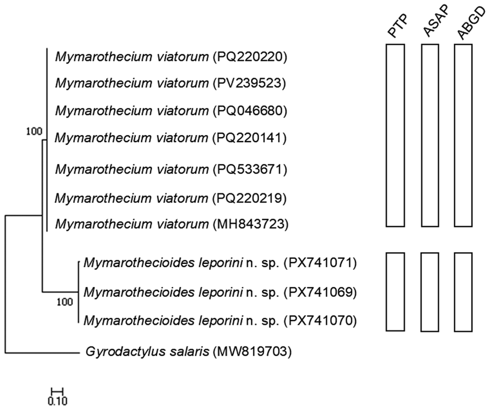
Figure 4. Long description
A phylogenetic tree diagram with a scale bar of 0.10 at the bottom left. The tree branches from a root on the left toward species names on the right.
* The outgroup at the bottom is Gyrodactylus salaris (M W 8 1 9 7 0 3).
* A major node with a bootstrap value of 100 leads to two main clades.
* The lower clade contains three specimens of Mymarothecioides leporini n. sp. with accession codes P X 7 4 1 0 7 1, P X 7 4 1 0 6 9, and P X 7 4 1 0 7 0.
* The upper clade, also supported by a bootstrap value of 100, contains seven specimens of Mymarothecium viatorum with accession codes P Q 2 2 0 2 2 0, P V 2 3 9 5 2 3, P Q 0 4 6 6 8 0, P Q 2 2 0 1 4 1, P Q 5 3 3 6 7 1, P Q 2 2 0 2 1 9, and M H 8 4 3 7 2 3.
To the right of the species names are three columns of vertical bars labeled P T P, A S A P, and A B G D. Each column contains two distinct bars: a long top bar corresponding to all Mymarothecium viatorum specimens and a shorter bottom bar corresponding to all Mymarothecioides leporini n. sp. specimens, indicating consistent M O T U delimitation across all three methods.
Studies on fish parasite fauna in Brazil’s semi-arid region remain scarce, particularly regarding monopisthocotylans, with only 40 species previously recorded (Alexandre & Yamada Reference Alexandre and Yamada2022; Diniz et al. Reference Diniz, Sousa, Carvalho and Yamada2022, Reference Diniz, Sousa, Yamada and Yamada2025, Reference Diniz, Saraiva, Sousa, Ebert, Yamada, Silva, Costa and Yamada2026; Falkenberg et al. Reference Falkenberg, Lima, Yamada, Ramos and Lacerda2024; Pereira et al. Reference Pereira, Yamada, Acosta and Yamada2026; Silva et al. Reference Silva, Silva and Yamada2021b, Reference Silva, Sousa, Diniz, Carvalho, Yamada, Pilarski, Tavares, Valladão, Dotta, Lizama and Takemoto2023, Reference Silva, Yamada and Yamada2026; Sousa et al. Reference Sousa, Diniz, Yamada and Yamada2024; Yamada et al. Reference Yamada, Diniz, Sousa, Yamada and Tavares-Dias2024a, Reference Yamada, Sousa, Ebert, Diniz, Tavares-Dias, Silva and Yamada2025). This number remains low compared to other Brazilian basins, such as the Amazon and Paraná (Cohen et al. Reference Cohen, Justo and Kohn2013; Lehun et al. Reference Lehun, Hasuike, Silva, Ciccheto, Michelan, Rodrigues, Nicola, Lima, Correia and Takemoto2020; Thatcher Reference Thatcher2006). However, this study, alongside recent research in the semi-arid region, highlights its potential for new taxonomic discoveries (Diniz et al. Reference Diniz, Sousa, Yamada and Yamada2025; Silva et al. Reference Silva, Dias, Silva and Yamada2021a, Reference Silva, Silva and Yamada2021b; Yamada et al. Reference Yamada, Diniz, Sousa, Yamada and Tavares-Dias2024a, Reference Yamada, Sousa, Ebert, Diniz, Tavares-Dias, Silva and Yamada2025). Therefore, we describe two new Mymarothecioides spp. parasitising the gills of anostomid fishes from two distinct Brazilian river basins. Based on morphological and molecular characterisation, we increase the number of valid species in the genus to seven. Our molecular analysis, combined with morphological traits, contributes new data to the growing genetic library of Dactylogyridae, enabling future studies to explore and evaluate phylogenetic relationships among its included genera.
Acknowledgements
The authors are grateful to the Núcleo de Pesquisa em Limnologia Ictiologia e Aquicultura – NUPÉLIA-UEM for providing the infrastructure, and to Legado verdes do Cerrado – Reserva Votorantim for all their support and help during the collections.
Author contributions
Writing the manuscript and methodology, W.B.B.S., W.T.H., P.O.F.Y., and M.F.B.G.D.; vector illustration, W.B.B.S. and P.O.F.Y.; morphometric analysis, M.F.B.G.D.; molecular data and phylogenetic inferences, B.S., A.V.O., and R.M.T.; review and editing, M.T.-D., R.M.T., and F.H.Y. All authors have read and approved the published manuscript version.
Financial support
The study received partial support through research grants from the Coordenação de Aperfeiçoamento de Pessoal de Nível Superior (CAPES) – W.B.B.S. (88887.704478/2022-00); Fundação Cearense de Apoio ao Desenvolvimento Científico e Tecnológico (FUNCAP) – P.O.F.Y. (FPD-0213-00301.01.01/23); M.F.B.G.D. (BMD-0008-02422.01.09/23); and Conselho Nacional de Desenvolvimento Científico e Tecnológico (CNPq) – for the productivity research grant awarded to M.T.-D. (301911/2022-3), R.M.T. (307921/2022-0), and F.H.Y. (304502/2022-7 and 174814/2023-2).
Competing interests
The authors declare they have no competing interests.
Ethical standards
All animal procedures were performed in full compliance with the Ethics Committee for Animal Experimentation (CEUA/URCA no. 00165/2018.1).