Evidence regarding the persistence of Ebola virus (EBOV) in the hospital environment and the role of cleaning procedures on virus stability is lacking. Some studies have suggested that EBOV can remain viable on surfaces for several days under favorable conditions.Reference Sagripanti, Rom and Holland 1 – Reference Fischer, Judson, Miazgowicz, Bushmaker, Prescott and Munster 3 Other studies have assessed the contamination within the patient environment; viable virus was absent but EBOV RNA was detected, in some cases, even following decontamination.Reference Bausch, Towner, Dowell, Kaducu, Lukwiya and Sanchez 4 , Reference Youkee, Brown, Lilburn, Shetty, Brooks and Simpson 5 Conversely, in another recent report, all environmental swabs collected from a EBOV patient’s room were negative for Ebola virus.Reference Wu, Wang, Yang, Zheng and Sha 6
Currently, no studies have been published reporting EBOV transmission through the environment or through fomites, if not visibly contaminated with body fluids. 7 However, guidelines strongly support a high level of precautions in environmental cleaning. 7
At the Lazzaro Spallanzani National Institute for Infectious Diseases (INMI), Rome, Italy, 2 EBOV patients were admitted. To assess contamination of the patient care environment and to evaluate the efficacy of cleaning procedures, swab samples were collected from different surfaces and fomites in patient rooms.
METHODS
Cleaning Procedures at INMI for EBOV Patient Rooms
According to internal protocols, daily cleaning of occupied patient rooms and management of spills of body fluids were performed using wipes and mops saturated with 0.5% sodium hypochlorite; the adequacy of daily cleaning was not assessed. After patient discharge, careful terminal cleaning of all room surfaces, reusable equipment, and everything downward to the floor was performed using manual scrubbing with wipes and mops saturated with 0.5% sodium hypochlorite, as suggested by international guidelines. 7
Summary of Clinical Courses
On November 25, 2014, the first EBOV patient was hospitalized in the high isolation unit (HIU) at INMI.Reference Petrosillo, Nicastri, Lanini, Capobianchi, Di Caro and Antonini 8 On December 4, his condition worsened and he was transferred to the intensive care high isolation unit (IC-HIU), where he remained for 7 days. On December 11, he returned to the HIU, and he was discharged on January 2, 2015. Profuse vomiting and diarrhea dominated the clinical course; EBOV was detectable in this patient’s blood until day 25 and in his urine until day 35 from admission.
The second patient was admitted on May 13, 2015, in the HIU. In the meanwhile, no patients were admitted to this room. The patient was discharged on June 10; intensive care was not required. EBOV was detectable in this patient’s blood until day 9 and urine until day 21.
Sample Collection and Timing
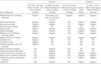
Sampling activities were performed wearing the same personal protective equipment used for medical care. Environmental samples were collected by slowly swiping the nylon-flocked swabs (Copan Italia, Italy) on the surfaces horizontally, vertically, and diagonally ≥10 times, then the swabs were re-suspended in polypropylene tubes containing 0.5 mL phosphate-buffered saline. The samples were immediately transported to the BSL-4 laboratory, which is located in the same compound. The sites and timing of the sampling are summarized in Table 1.
Results of Environmental Samples Collected from Patient Rooms at the Lazzaro Spallanzani National Institute for Infectious Diseases

NOTE. IC-HIU, intensive-care, high-isolation unit; HIU, high-isolation unit; PCR, polymerase chain reaction; i.v., intravenous; ND, not done.
a Different sites of floor and frequently touched surfaces, 1 sample for each site.
b Patient with negative viremia for EBOV but PCR-positive in other body fluids (ie, urine, sputum and material from oropharyngeal and rectal swabs).
c All floors were made of polyvinyl chloride.
d In both cases, the culture of viable virus on VERO E6 cells resulted negative. According to the clinical records, areas where PCR results were positive were intensively contaminated by vomit during the patient stay.
e Sampled on the day of discharge, without any previous cleaning.
Laboratory Testing
After inactivation in BSL 4, the samples were extracted and tested for the presence of EBOV genomes using a commercial quantitative real-time RT-PCR specific for filoviruses, targeting the L gene (RealStar Filovirus Screen RT-PCR Kit 1.0, Altona Diagnostics, San Francisco, CA). This test was approved by the World Health Organization for United Nations procurement. 9 PCR-positive samples were tested for the presence of infectious viral particles. Confluent monolayers of Vero E6 cells in T-25 flasks were inoculated with 0.1 mL of undiluted sample in 1 mL of minimum essential medium (MEM). After absorption at 37°C for 1 h, excess inoculum was removed, and complete medium with 2% fetal calf serum (FCS) was added to the flasks. Cells were observed each day for signs of cytopathic effect (CPE). If no CPE appeared, sequential passages were performed every 5 days by detaching and co-culturing with fresh cells, which were observed for another 5 days. At 20 days after inoculation, the cells were removed from the flask by scraping and both supernatants and cells were tested for the presence of EBOV-RNA.
RESULTS
Results are detailed in Table 1. The 2 swabs collected from the HIU floor after patient 1 was discharged were positive by PCR. The first swab was taken at the bedside and the other was taken under a table used by the patient; both zones had been heavily contaminated by bloody vomit and had been routinely cleaned at that time. Cultures on Vero cells of these samples were negative. PCR-positive areas were sampled again after terminal cleaning, and all tested PCR-negative. All other swabs for patients 1 and 2 were negative.
According to our findings, the daily cleaning of patient rooms with 0.5% hypochlorite was sufficient to remove EBOV in most cases. However, viral genetic material was found in areas where heavy contamination had occurred. Imperfect cleaning and intensive contamination by bloody vomit may have contributed to this finding.
The absence of viable EBOV suggests that 0.5% sodium hypochlorite is able to kill the virus, although a natural decay of infectivity after 6 days is possible.Reference Bibby, Casson, Stachler and Haas 10 Viral genetic material was not found in the same areas after terminal cleaning, and notably, all samples from the IC-HIU collected after terminal cleaning were negative despite the occurrence of contamination. This result supports the importance of a careful and extensive terminal cleaning.
The timing of sampling was not standardized and was not concordant between the 2 patients. Although convenience timing for sampling was used, this should not have affected the findings. These findings derive from the observations of only 2 patients in a high-level isolation setting, and the remarks suggested should be applied only in similar settings. Further studies are needed to assess the efficacy of these procedures in different sites and conditions.
ACKNOWLEDGMENTS
We thank Gabriella De Carli, MD, for English editing and suggestions. Other members of the Ebola INMI Task Force are Battisti, Nazario Bevilacqua, Gianluigi Biava, Evangelo Boumis, Ilaria Caravella, Stefania Cicalini, Pierangelo Chinello, Francesca Colavita, Alessanda Coppola, Angela Corpolongo, Loredana De Marchis, Nicola De Marco, Fabio Di Gianbattista, Vincenzo Galati, Gabriele Garotto, Paolo Giacomini, Mario Guiducci, Daniela Imola, Daniele Lapa, Francesco Nicola Lauria, Antonio Marasco, Luisa Marchioni, Andrea Mariano, Micaela Maritti, Antonella Marzolini, Lorena Martini, Alessandro Mercuri, Emanuele Nicastri, Paola Nieddu, Silvia Ondedei, Nicola Petrosillo, Serena Quartu, Silvia Rosati, Antonio Russo, Fabrizio Taglietti, Damiano Travaglini, Maurizio Vescovo, Laura Vincenzi, and Laura Vitolo.
The opinions expressed by the authors contributing to this journal do not necessarily reflect the opinions of the Centers for Disease Control and Prevention or the institutions with which the authors are affiliated. Written informed consent was obtained from the patients for publication of this study and any accompanying material.
Financial support: This work was supported by the Ministero della Salute, Italia-Ricerca Corrente, Istituti di Ricovero e Cura a Carattere Scientifico.
Potential conflicts of interest: All authors report no conflicts of interest relevant to this article.