Introduction
Parisite-(Nd) is a new member of the calcium–rare earth element (Ca–REE) fluorocarbonates. So far, four mineral species have been discovered in this mineral series (Donnay and Donnay, Reference Donnay and Donnay1953). Bastnäsite, REECO3F and synchysite, CaREE(CO3)2F are considered as the end-members of this polysomatic mineral series and parisite, CaREE2(CO3)3F2, and röntgenite, CaREE2(CO3)3F2 are considered as intermediate members (Ni et al., Reference Ni, Hughes and Mariano1993; Wu et al., Reference Wu, Meng, Pan, Yang and Li1998; Zeug et al., Reference Zeug, Nasdala, Ende, Habler, Hauzenberger, Chanmuang, Škoda, Topa, Wildner and Wirth2021). Rare earth elements easily replace each other in mineral crystal structures because of their similar properties. Cerium is the most common rare element in Ca–REE fluorocarbonates and correspondingly all the Ce-dominant analogues of the series, bastnäsite-(Ce), röntgenite-(Ce), synchysite-(Ce) and parisite-(Ce), have been found in Nature. In addition, La-dominant analogues: bastnäsite-(La) and parisite-(La); Nd-dominant analogues: bastnäsite-(Nd) and synchysite-(Nd); and Y-dominant analogues: bastnäsite-(Y) and synchysite-(Y) have also been reported (Smith et al., Reference Smith, Stone, Ross and Levine1960; Vainshtein et al., Reference Vainshtein, Pozharitskaya and Turanskaya1961; Mineev et al., Reference Mineev, Lavrischeva and Bykova1970; Scharm and Kühn, Reference Scharm and Kühn1983; Miyawaki et al., Reference Miyawaki, Yokoyama and Husdal2013; Menezes Filho et al., Reference Menezes Filho, Chaves, Chukanov, Atencio, Scholz, Pekov, da Costa, Morrison, Andrade, Freitas, Downs and Belakovskiy2018). Parisite-(Nd) is the Nd-dominant analogue of parisite-(Ce) and parisite-(La).
Parisite-(Ce) has generated considerable interest because of its economic importance as a primary source rare earth element for industrial use (Morteani, Reference Morteani1991). Parisite-(Ce), as one of the most common rare earth minerals, was first reported by J.J. Paris in 1835 from the emerald mine at Muzo in Colombia, South America (Warren and Palache, Reference Warren and Palache1911). It occurs as an accessory mineral in differentiated alkali massifs, granite pegmatites and REE-rich carbonatites. It can be found in many localities, for example the carbonatite orebody of Mountain Pass, California (Castor, Reference Castor2008), the carbonatite complex Amba Dongar, India (Doroshkevich et al., Reference Doroshkevich, Viladkar, Ripp and Burtseva2009), the alkaline granite–syenite pegmatites of the Mount Malosa pluton in Malawi (Guastoni et al., Reference Guastoni, Kondo and Nestola2010), the ultramafic lamprophyre–carbonatite complex near Delitzsch, Germany (Seifert et al., Reference Seifert, Kämpf and Wasternack2000), and the Bayan Obo Fe–Nb–REE deposit, Inner Mongolia, China (Zhang and Tao, Reference Zhang and Tao1986). The composition of specimen from Muso mine, Colombia (NMNH no. 122145) used for structure refinement (Ni et al., Reference Ni, Post and Hughes2000) is reported as Ca1.02(Ce0.94La0.50Nd0.37Pr0.10Sm0.05)Σ1.96(CO3)3F1.51. The crystal structure of parisite-(Ce) was once controversial. In an early study, its structure was defined as hexagonal symmetry with space group R3, a = 7.18 Å and c = 8.41 Å (Donnay and Donnay, Reference Donnay and Donnay1953). However, the work of Ni et al. (Reference Ni, Post and Hughes2000), refined the crystal structure of parisite-(Ce) to monoclinic symmetry with space group C2/c or Cc and a unit cell of a = 12.305(2), b = 7.1053(5), c = 28.250(5) Å, and β = 98.257(14)°. Parisite-(La) only discovered in the Mula mine, Brazil, has been approved as a new mineral (IMA2016-031) with chemical composition of Ca0.98(La0.83Nd0.51Ce0.37Pr0.16Sm0.04Y0.03)Σ1.94C3.03O8.91F2.09 and crystal structure of monoclinic (pseudo-trigonal), space group: C2, Cm, or C2/m, a = 12.356(1) Å, b = 7.1368(7) Å, c = 28.299(3) Å, β = 98.342(4)°, V = 2469.1(4) Å3 and Z = 12 (Menezes Filho et al., Reference Menezes Filho, Chaves, Chukanov, Atencio, Scholz, Pekov, da Costa, Morrison, Andrade, Freitas, Downs and Belakovskiy2018). Parisite-(Nd) was once reported in Bayan Obo deposit in the literature (Zhang and Tao, Reference Zhang and Tao1986), but only REE assemblage information was provided, and no complete chemical compositions and crystal structure were given. Hence it was not officially recognised in the official list of minerals from the Commission on New Minerals, Nomenclature and Classification of the International Mineralogical Association (IMA-CNMNC) (Jambor et al., Reference Jambor, Burke, Ercit and Grice1988).
Our investigation means that parisite-(Nd) has now been approved as a new mineral species by the IMA-CNMNC (IMA 2024-013, symbol Pst-Nd, Fan et al., Reference Fan, Yu, Gu, Zeng and Chen2024). Type material is deposited in the collections of the National Research Center for Geoanalysis, Baiwanzhuang Street 26, Beijing, People’s Republic of China (holotype), catalogue Number NRCGA2024001, and the Geological Museum of China, Yangrouhutong Street 15, Xicheng District, Beijing, People’s Republic of China, catalogue number GMCTM2024003 (cotype).
Occurrence
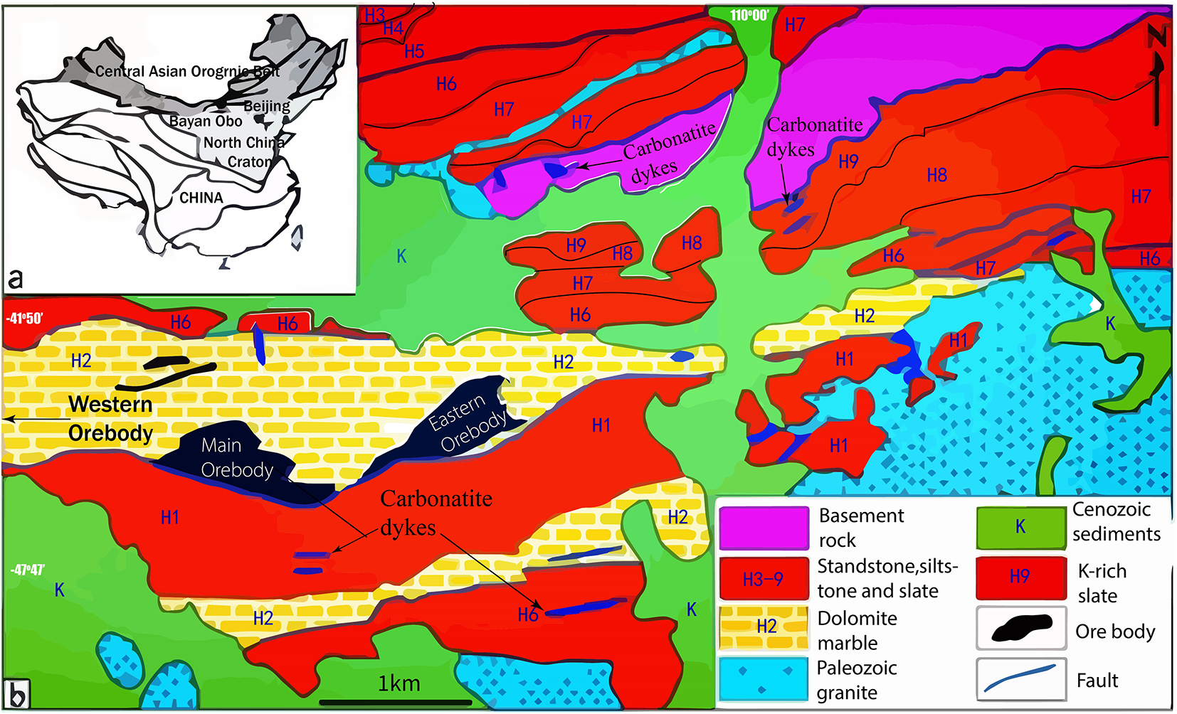
Parisite-(Nd) was found in dolomitic marble in the Bayan Obo Fe–Nb–REE deposit, 130 km north of the city of Baotou, Inner Mongolia, China. The Bayan Obo deposit is hosted by Palaeo- to Mesoproterozoic sedimentary rocks of the Bayan Obo Group (Nie et al., Reference Nie, Jiang, Su and Wang2002; Liu et al., Reference Liu, Song, Kynicky, Yang, Chen and Tang2022). It comprises six formations: Dulahala Formation (H1-H3), Jianshan Formation (H4-H5), Halahuoqite Formation (H6-H8), Bilute Formation (H9-H10), Baiyinbaolage Formation (H11-H12), and Hujiertu Formation (H13-H18) (Ling et al., Reference Ling, Liu, Williams, Teng, Yang, Ding, Wei, Xie, Deng and Sun2013; Yang et al., Reference Yang, Lai, Pirajno, Liu, Mingxing and Sun2017) (Fig.1).
(a) The location and (b) geological sketch map of the Bayan Obo REE–Nb–Fe deposit (modified from Ling et al., Reference Ling, Liu, Williams, Teng, Yang, Ding, Wei, Xie, Deng and Sun2013; Liu et al., Reference Liu, Song, Kynicky, Yang, Chen and Tang2022).

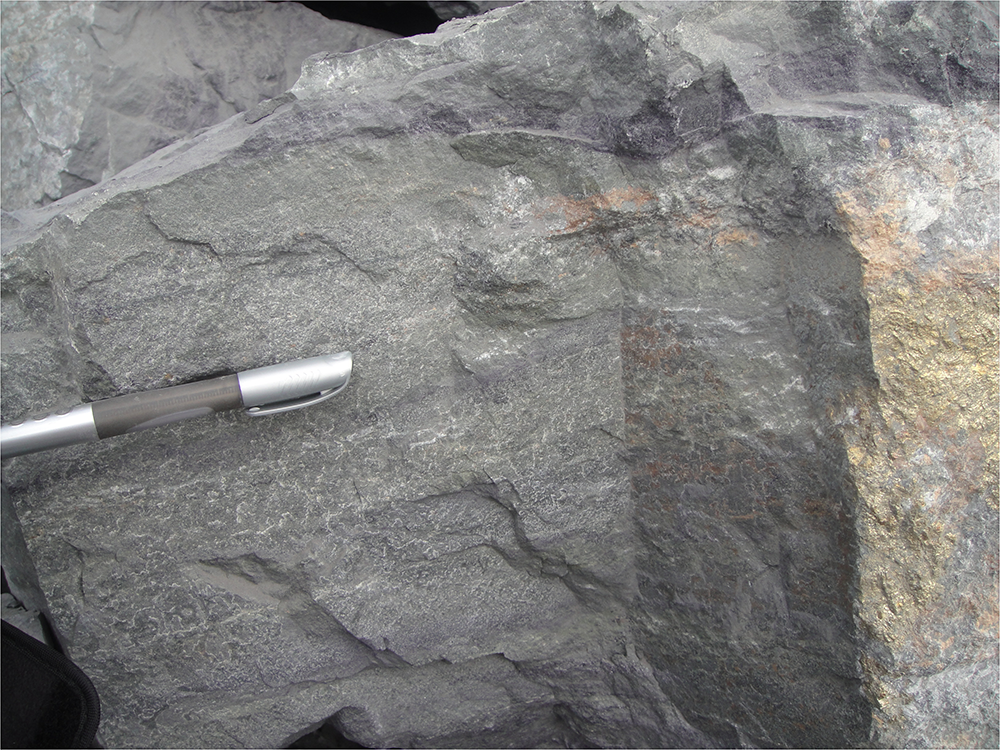
The specimens were collected from magnetite ores within the H8 formation in the eastern part of the mine (109°57.666ʹE, 41°47.951ʹN) (Fig. 2). Parisite-(Nd) was associated with calcite, aegirine, magnetite, hematite, fluorite, riebeckite, bastnäsite-(Ce), baryte, aeschynite-(Ce), aeschynite-(Nd), monazite, and parisite-(Ce). The ore-forming mechanism of the Bayan Obo deposits is still controversial. There are many different viewpoints on mineralisation age including (1) Mesoproterozoic to Neoproterozoic, 1360–789 Ma; (2) Caledonian, 485–386 Ma; or (3) Hercynian, 277–255 Ma (Yuan, Reference Yuan2012; Xie et al., Reference Xie, Qu, Yang, Liang, Zhong, Wang, Xia and Li2019). The models on the genesis of the Bayan Obo REE deposit include: (1) genetically related to carbonatite magmatism; (2) hydrothermal alteration of carbonate sedimentary rocks; and (3) carbonatite volcanic eruptions (Ren et al., Reference Ren, Zhang and Zhang1994; Smith, Reference Smith2007; Zhu et al., Reference Zhu, Sun and Pan2015; Yang et al., Reference Yang, Lai, Pirajno, Liu, Mingxing and Sun2017). The formation of REE mineralised dolomite might be related to interactions and reactions between the carbonatite magmas and/or associated fluids with sedimentary carbonate rocks, with the REE-bearing carbonatite magmas having undergone an intense fractionation enrichment process (Yang et al., Reference Yang, Song, Liu, Zhu, Kynicky and Chen2024). Parisite-(Nd) might be a product of bi-metasomatism Ca–Mg carbonate rock and/or carbonatite, and Na-, F-rich postmagmatic hydrothermal solutions.
The outcrop on which the new mineral parisite-(Nd) was found from the main ore of Bayan Obo REE–Nb–Fe ore deposit.

Appearance, physical and optical properties
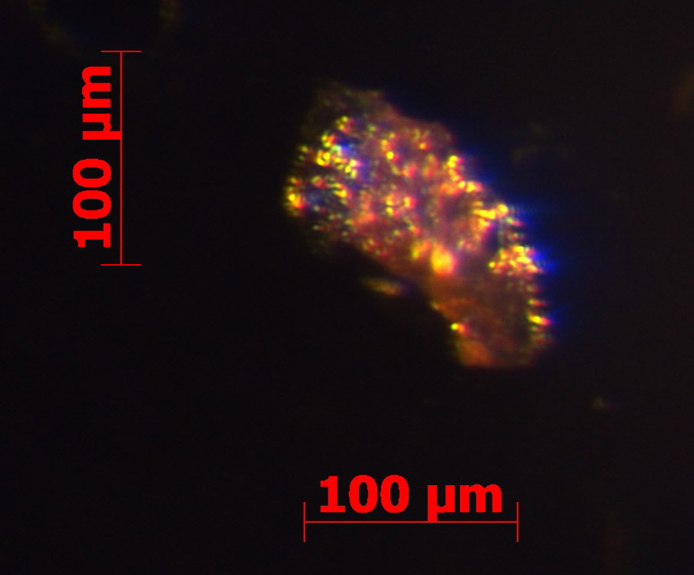
Parisite-(Nd) occurs as subhedral to anhedral irregular grains from 0.02 mm to 1 mm in maximum dimension. The a:b:c ratio calculated from unit-cell parameters is 1:0.5774:2.3088. Parisite-(Nd) has thin, tabular and hexagonal crystal habits, but most of the grains are broken and fractured. Parisite-(Nd) is transparent and has a yellowish-brown colour (Fig. 3), with a pale yellow streak. It displays vitreous to resinous lustre. It has no fluorescence under shortwave (254nm) and longwave (366nm) ultraviolet radiation. The Mohs hardness is between 4 and 5, which is estimated from comparison with fluorite and parisite-(Ce). Cleavage is distinct on pseudo-{001} and parting was not observed. The fracture is uneven, or conchoidal, and the mineral tenacity is brittle. Density was not measured due to the limited crystals available and nanometric crystal size. The calculated density is 4.357 g/cm3 on the basis of empirical formula and unit-cell volume from powder X-ray diffraction (XRD) data.
A microscopic view of yellowish-brown parisite-(Nd) from specimen number BZ-3-6.

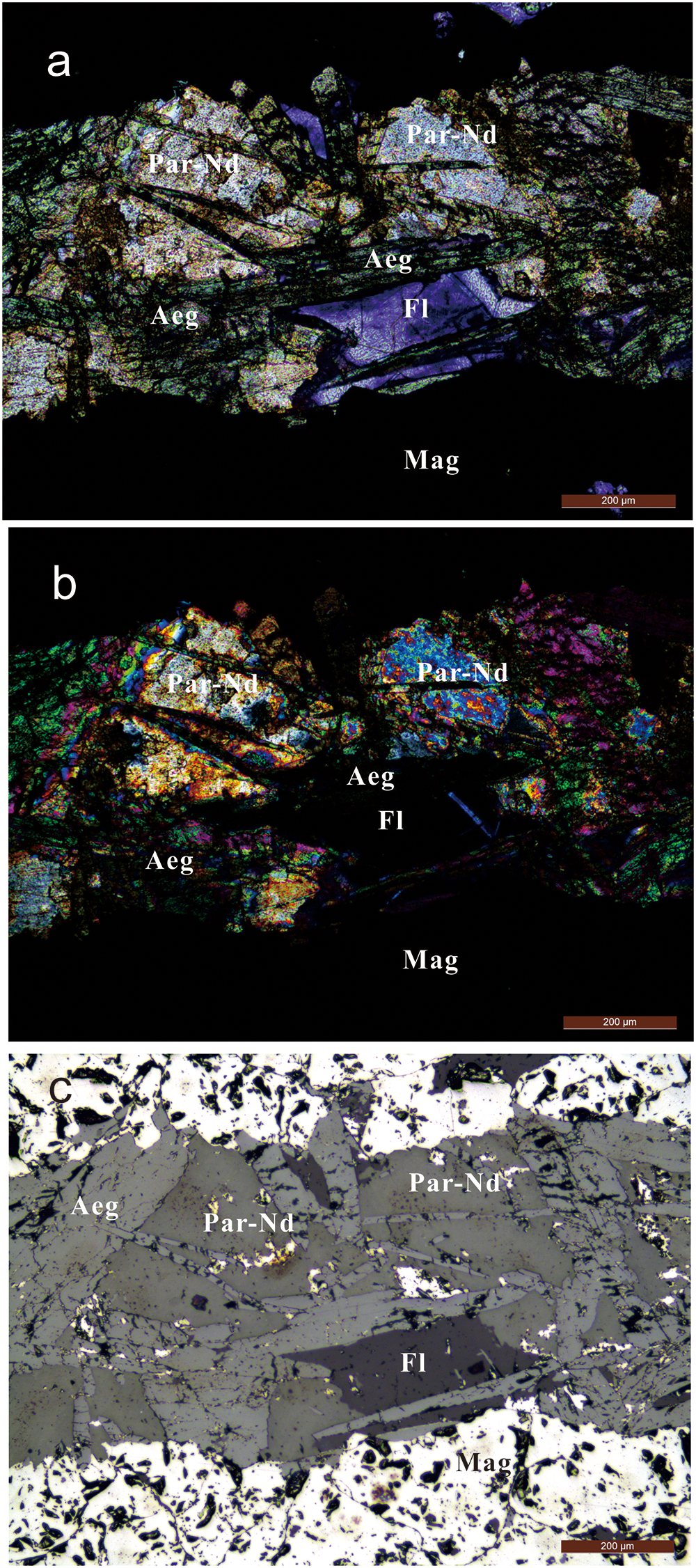
Under the polarising microscope (Fig. 4), parisite-(Nd) is colourless to brownish yellow in transmitted light with weak pleochroism. The other main optical characteristics of parisite-(Nd) are parallel extinction, interference colour from third-order bluish yellow to fourth-order white, uniaxial and optically positive. In transmitted light parisite-(Nd) is colourless, pseudo-uniaxial (+), ω = (1.679) and ε = (1.754) (the value not determined, data referenced from Zhang and Tao, Reference Zhang and Tao1986). The pleochroism is invisible.
Photomicrographs of parisite-(Nd) (Par-Nd) associated with fluorite (Fl), aegirine (Aeg) and magnetite (Mag). (a) Transmitted light; (b) cross polarised light; and (c) reflected light.

Raman spectroscopy
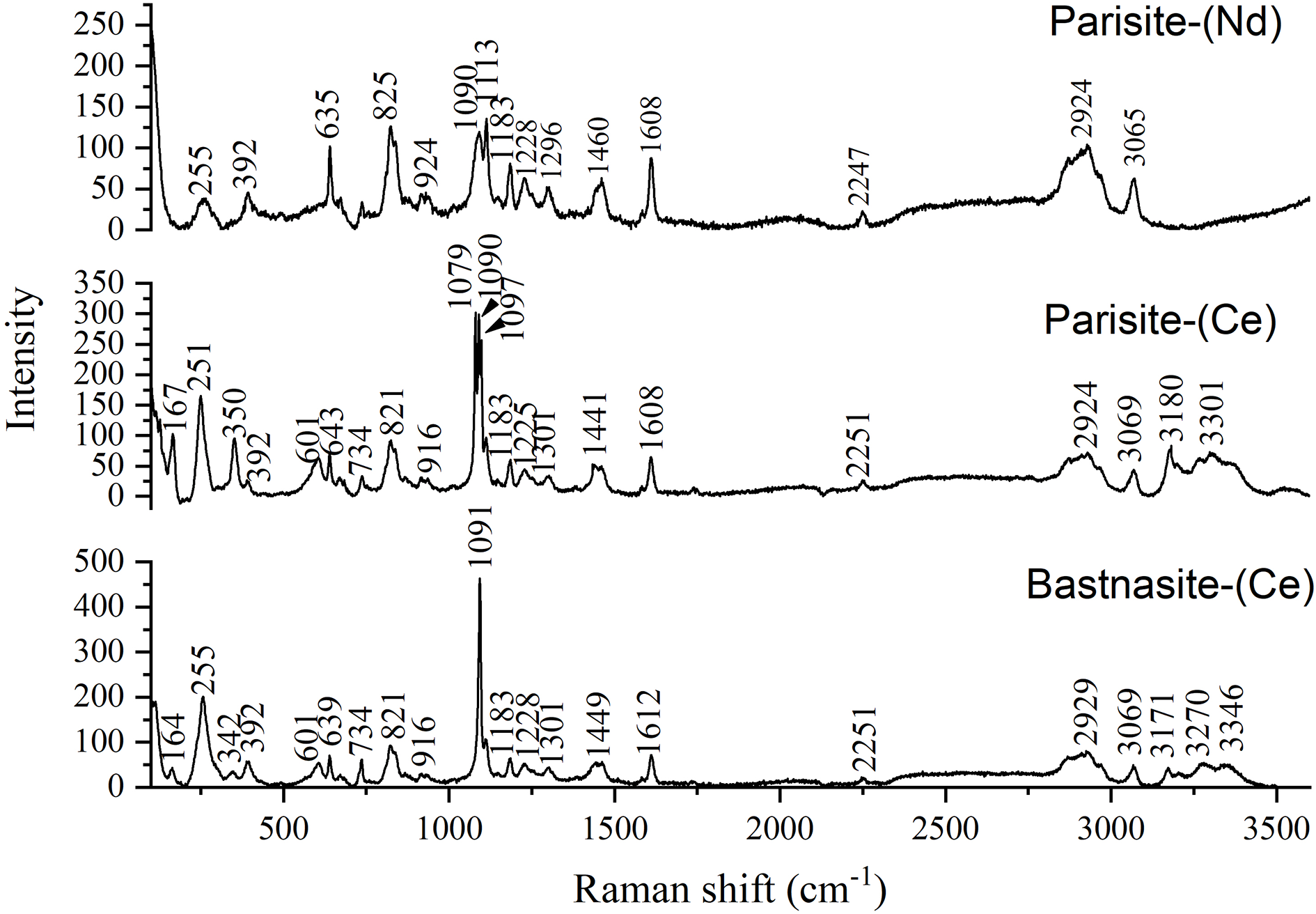
The Raman spectrum of parisite-(Nd) between 100 and 3600 cm–1 was recorded using a Renishaw inVia Reflex system, using a solid-state laser with a wavelength of 532 nm and a thermoelectric cooled CCD detector, with 1 cm–1 resolution and a spot size of 2 µm, and was compared with associated bastnäsite-(Ce) and parisite-(Ce) (Fig. 5).
Raman spectra of parisite-(Nd) in comparison with parisite-(Ce) and bastnäsite-(Ce).

Strong and sharp peaks of parisite-(Nd) are observed at 1113, 1090, 825, 635 and 1608 cm–1 and moderate to weak bands centred at 255, 392, 739, 924, 1183, 1228, 1296, 1640, 2247, 2924 and 3065 cm–1. The Raman spectrum of parisite-(Nd) is similar to the spectra of parisite-(Ce) and bastnäsite-(Ce), but the vibration of CO32– show some differences. The CO32– complex exhibits four internal vibrational modes: (1) a symmetric stretching vibration (ν1); (2) an out-of-plane bending vibration (ν2); (3) a doubly degenerate asymmetric stretching vibration (ν3); and (4) an in-plane bending vibration (ν4) (White, Reference White and Farmer1974). The most intense Raman band is assigned to a ν1 vibration at ∼1100 cm–1. The vibration spectrum of the (CO3)2– in bastnäsite-(Ce) is relatively symmetrical and the ν1 Raman scattering peak is located at 1091cm–1. The ν1 Raman band is split into three bands at ∼1079 cm–1, ∼1090 cm–1 and ∼1097 cm–1 in parisite-(Ce), while two bands at ∼1090 cm–1 and ∼1113 cm–1 in parisite-(Nd). These differences of ν1 Raman spectrum of fluorinated carbonates are probably dependent on the chemical compositions (Frost and Dickfos, Reference Frost and Dickfos2007). The bands assigned to the ν2(CO3)2– vibration are at ∼924 cm–1 and ∼825cm–1 in parisite-(Nd), which is a little higher than the band positions at ∼916 cm–1 and ∼821 cm–1 in parisite-(Ce) and bastnäsite-(Ce). The parisite-(Nd) spectrum shows the ν3(CO3)2– vibration band at ca. 1460 cm–1 in comparison with 1441 cm–1 in parisite-(Ce) and 1449 cm–1 in bastnäsite-(Ce). The bands in the range of 600∼740 cm–1 are assumed to be the ν4 bending mode. A sharp intensity band is observed at 635 cm–1 in parisite-(Nd), and two bands are observed at ∼601 cm–1 and ∼643 (639) cm–1 in parisite-(Ce) and bastnäsite-(Ce). A weaker peak at ∼739 cm–1 or ∼734 cm–1 is observed in three samples. The positions of the ν4 bands perhaps show the divergence of the ionic radius of the Nd and Ce cation in the carbonate structure. A number of low wavenumber bands are observed for parisite-(Nd) at ∼392 cm–1 and ∼255 cm–1. These two bands and another two bands around 350 (342) cm–1 and 167 (164) cm–1 are also observed in the Raman spectra of parisite-(Ce) and bastnäsite-(Ce). These bands are described as lattice modes involving halogen and F (Frost and Dickfos, Reference Frost and Dickfos2007). The Raman spectrum of all three fluorocarbonate minerals in the 2800 to 3500 cm–1 spectral range is reported in Fig. 5. Raman bands are observed at 2924 and 3065 cm–1. These bands are attributed to water stretching vibrations (Frost et al., Reference Frost, López, Scholz, Xi and Belotti2013). For parisite-(Nd) OH units are likely to replace F in a complex structure.
Chemical composition
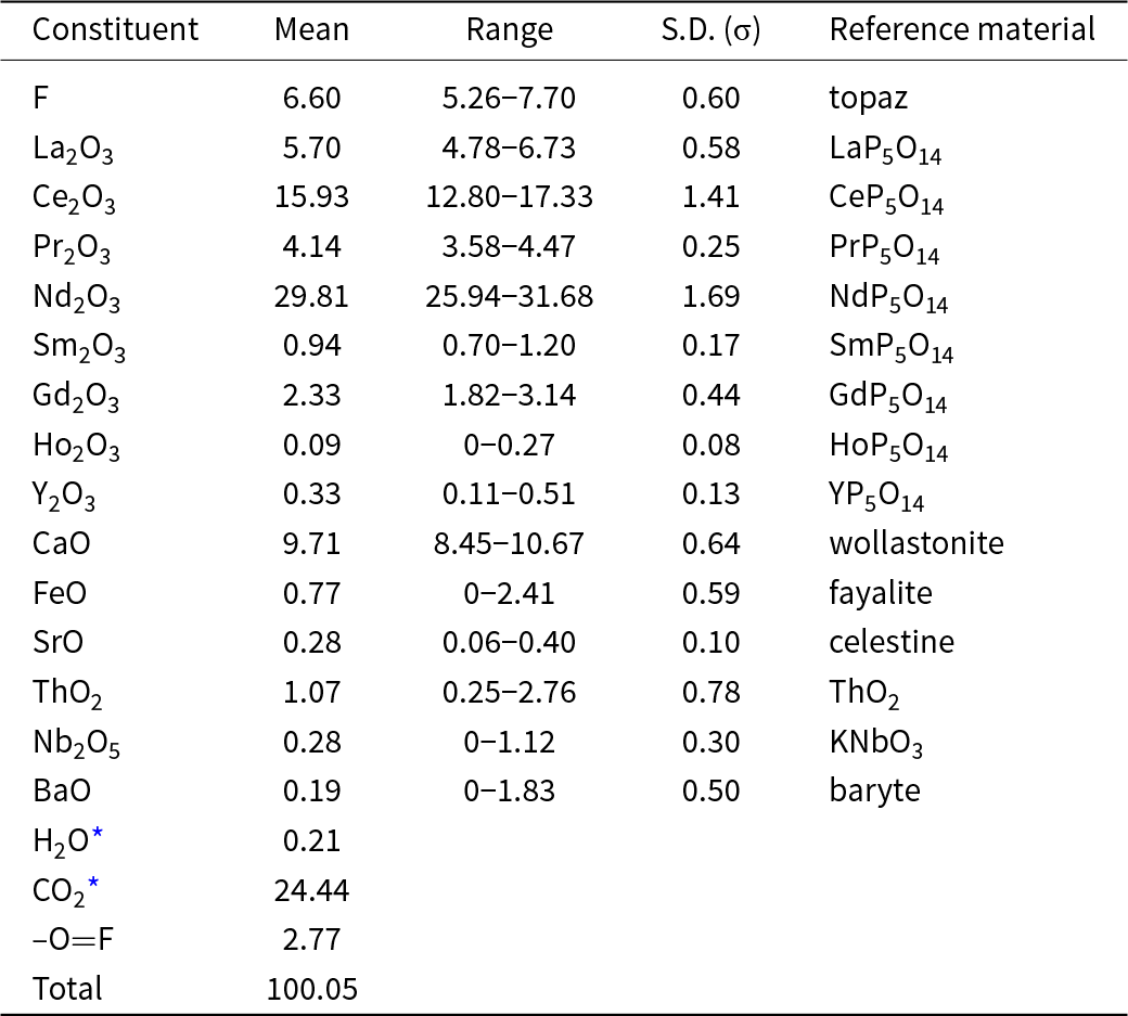
The chemical composition of parisite-(Nd) was studied with a JXA-iHP200F electron microprobe, operating in wavelength-dispersive mode with excitation voltage of 15 kV, specimen beam current 20 nA, and 5 μm beam diameter. The analytical data from 13 spots with ranges and standards employed are given in Table 1. There was insufficient material for a direct determination of CO2 and H2O. Thus, the calculation of the empirical formula was done in the following steps: (1) calculation of the REE site cations/Ca site cations ratio; (2) when REE site cations/Ca site cations = 2, then normalisation of the empirical formula to 3 cations (REE site cations + Ca site cations); and (3) calculation of the OH content (in atoms per formula unit) from the charge balance considering 3 CO32– groups and the calculated F content. The CO2 was calculated to 24.44 wt.% for charge neutrality. The empirical formula is (Ca0.945Fe0.058Sr0.015Ba0.007)Σ1.025(Nd0.967Ce0.529La0.191Pr0.137Gd0.070Sm0.029Th0.022Y0.016Nb0.011Ho0.003)Σ1.975(CO3)3F1.893OH0.023. The simplified formula is Ca(Nd,Ce,La,Pr,Gd)2(CO3)3F2. The ideal formula is CaNd2(CO3)3F2, which requires (wt.%) CaO 10.26%, Nd2O3 61.56%, F 6.95%, CO2 24.16%, O=F–2.93, total 100 wt.%. The ratio of rare earth to calcium is close to 2:1. In this composition the neodymium has a very dominant role compared to the dominant rare earth elements in parisite-(Ce) and parisite-(La).
Compositional data (in wt.%) for parisite-(Nd)

* Notes: H2O and CO2 from formula calculation; S.D. – standard deviation.
Crystallography
The crystal structure of parisite-(Nd) sample could not be determined by single-crystal X-ray studies due to the nature of the nanometric crystal aggregate and its structural complexity. Therefore, the powder XRD data and selected-area electron diffraction (SAED) were used for the crystallographic characterisation.
Powder X-ray diffraction data were collected with a Rigaku Xtalab Synergy single-crystal diffractometer (CuKα radiation) in Gandolfi powder mode at 50 kV and 1 mA. The unit-cell parameters were refined using the program by Holland and Redfern (Reference Holland and Redfern1997). A monoclinic unit cell of parisite-(Ce) by Ni et al. (Reference Ni, Post and Hughes2000) was referenced to index the diffraction spots. Unit cell parameters refined from the powder data are as follows: monoclinic symmetry with space group Cc (# 9), a = 12.3283(13), b = 7.1185(4) Å, c = 28.4633(37) Å, β = 98.529(14)°, V = 2470.28(42) Å3 and Z = 12.
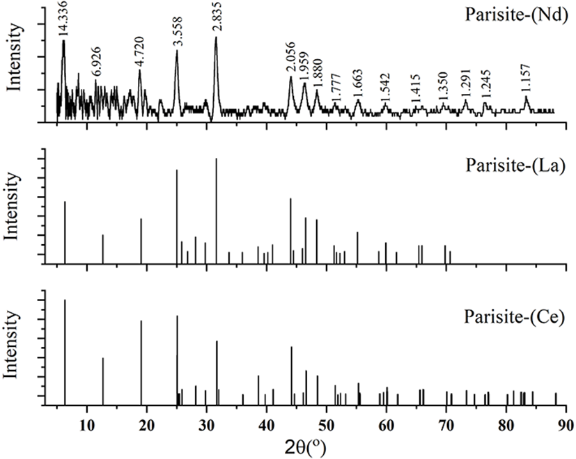
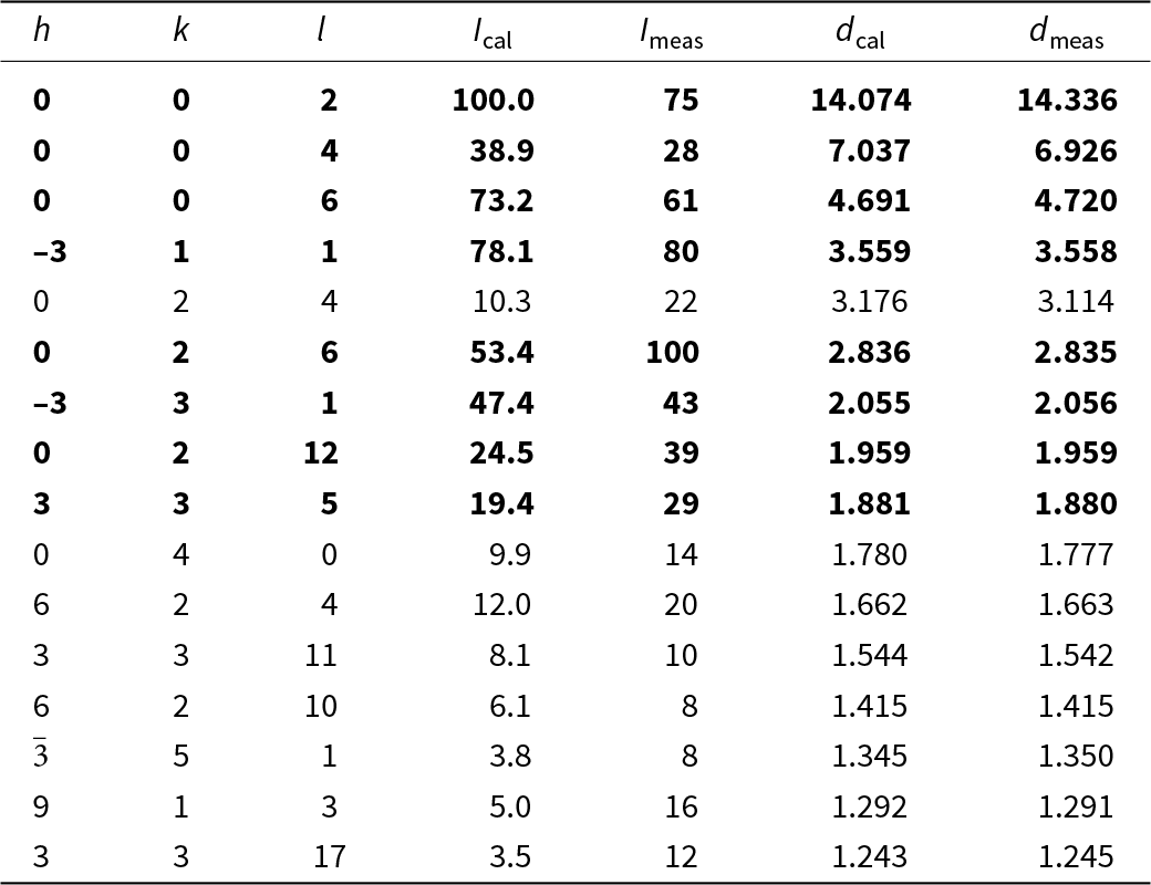
The complete powder X-ray diffraction data of parisite-(Nd) is shown in Table 2 and compared with parisite-(Ce) and parisite-(La) in Fig. 6. Based on the obtained XRD patterns of parisite-(Nd), the results show that these values are in agreement with the measured d-spacings for parisite-(Ce) (Supplementary Table 1), with most of the absolute values of difference <0.02 Å. The diagnostic reflections for parisite are (d-values in Å): 14.336, 6.926, 4.720, 3.558, 2.835, 2.056, 1.959 and 1.880. The interplanar spacings in the c axis direction of parisite-(Nd) have some difference with those of parisite-(Ce) due to the substitution of Nd to Ce.
Powder X-ray diffraction patterns of parisite-(Nd) from this work compared with parisite-(Ce) (Ni et al., Reference Ni, Post and Hughes2000) and parisite-(La) (Menezes Filho et al., Reference Menezes Filho, Chaves, Chukanov, Atencio, Scholz, Pekov, da Costa, Morrison, Andrade, Freitas, Downs and Belakovskiy2018).

Powder X-ray diffraction data (d in Å, I in %) for parisite-(Nd)

Note: The strongest lines are given in bold.
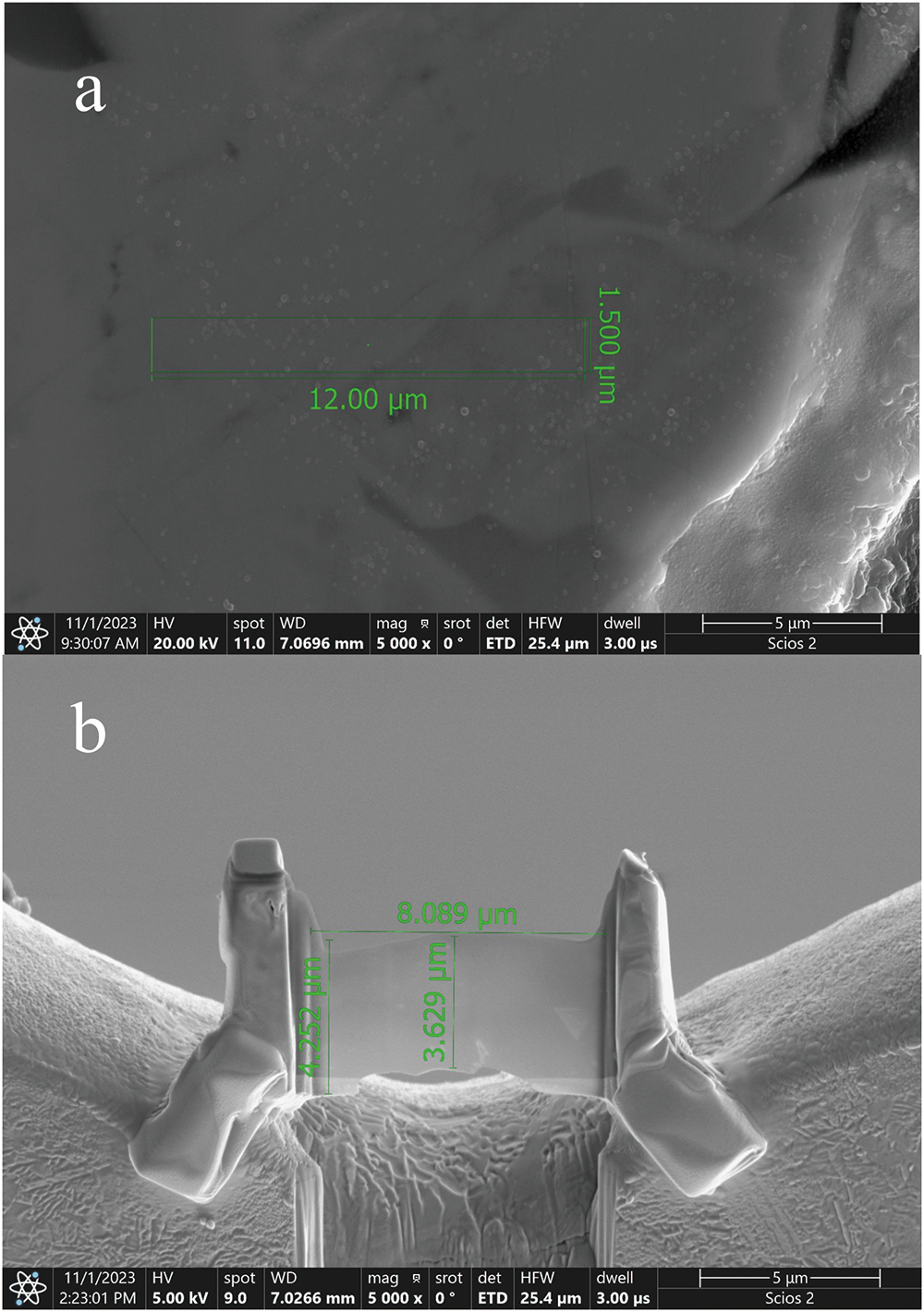
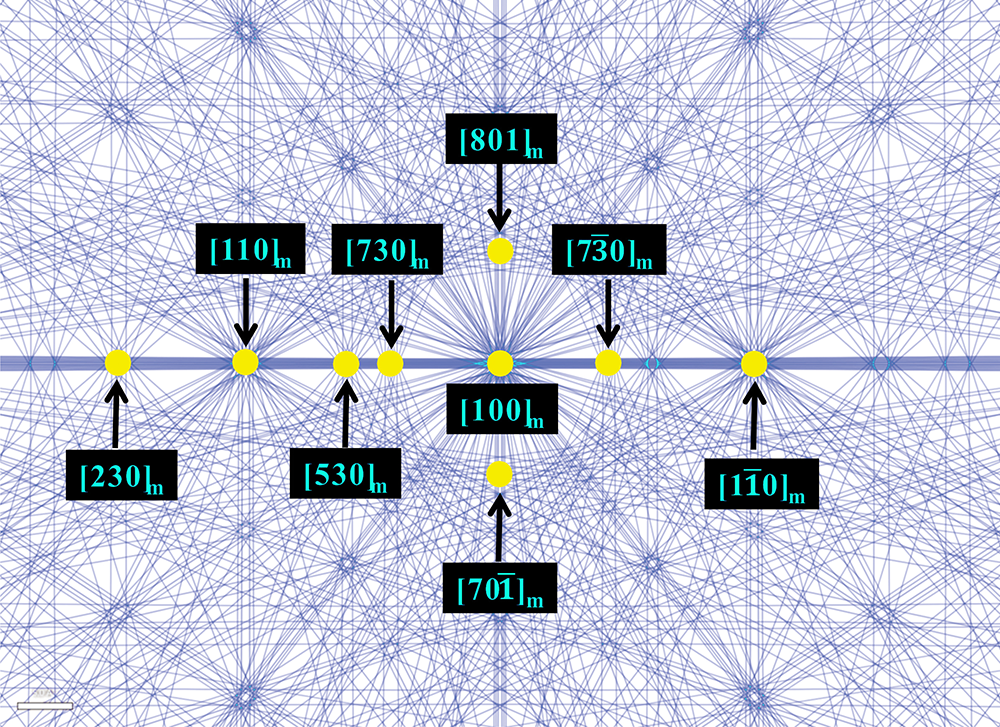
For transmission electron microscopy (TEM) studies, two perpendicular TEM foils of ∼80 nm thickness were prepared with a FEI Scois 2 focus ion beam (FIB) device at Beijing SHRIMP Center, Institute of Geology, Chinese Academy of Geological Sciences, and analysed by a JEM–2100 (HR) Transmission Electron Microscope equipped with a double-tilt holder, a Gatan digital camera, and an INCA Energy TEM100 energy-dispersive spectroscopy instrument at the Institute of Mineral Resources, Chinese Academy of Geological Sciences, operated at 200 kV. Images of one foil and its location in polished section before cutting are shown in Fig. 7. Before TEM testing on parisite-(Nd), the TEM instrument underwent camera constant calibration using a standard polycrystalline gold sample. Following calibration, the camera constant was determined to be Lλ = 9.26 mm·Å. Six different SAED patterns were acquired along two distinct Kikuchi lines within the same area of one TEM foil and simulated in Kikuchi maps using the SingleCrystal program (Figs 8 and 9), while three different SAED patterns (Fig. 10) were captured along a Kikuchi line in another TEM foil.
Images of TEM foil of parisite-(Nd) by FIB sampling for SAED analysis. Details: (a) the cut area; and (b) the size and morphology after ion thining.

Kikuchi map of parisite-(Nd) simulated using SingleCrystal software (Palmer, Reference Palmer2015). The labelled Kikuchi poles in the diagram correspond to the zone axes of SAED patterns in Fig. 9.

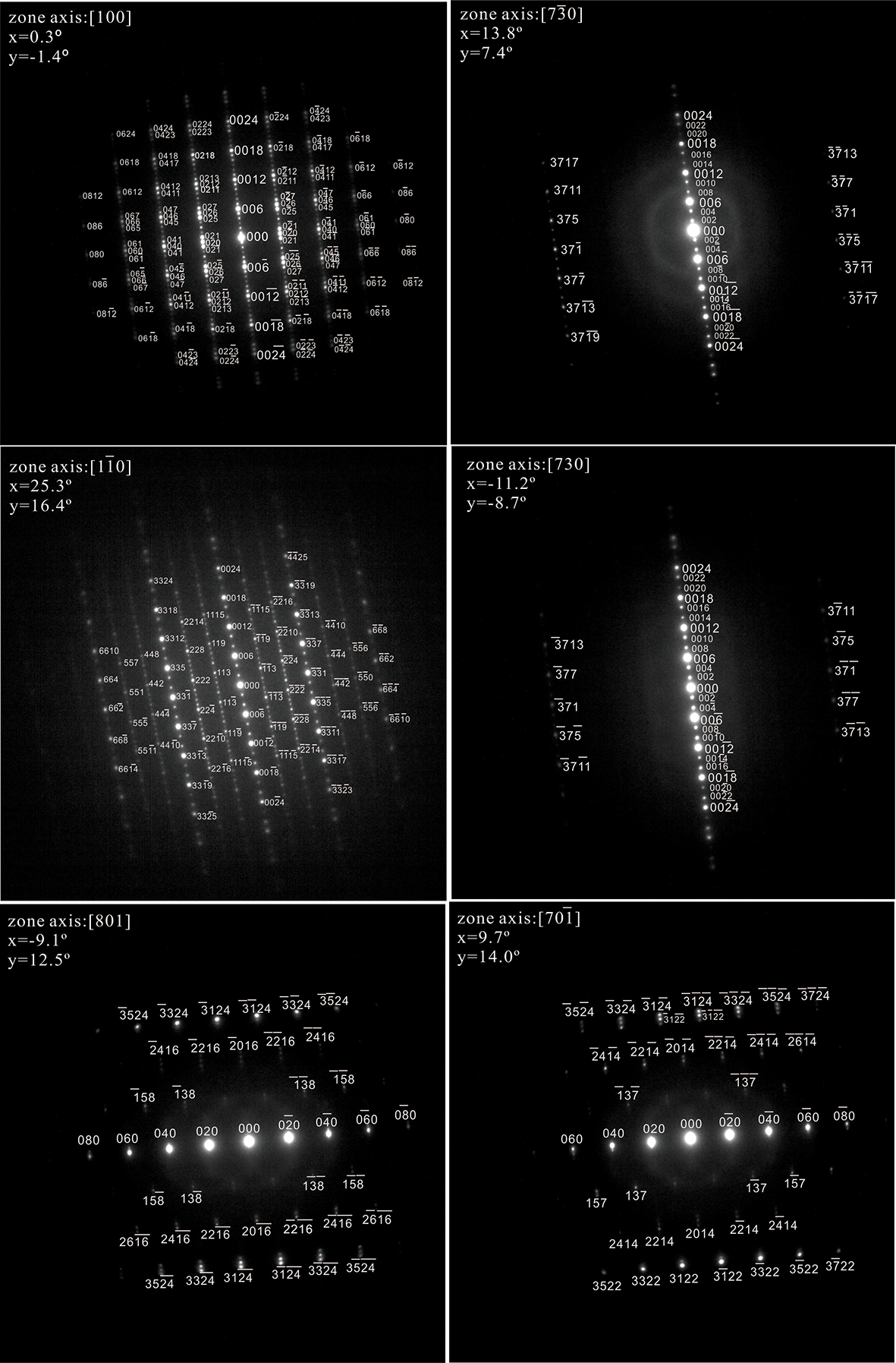
The solid angles calculated from the patterns match with the patterns generated from the structure of parisite-(Ce) (Fig. 9). A series of SAED patterns were taken along two Kikuchi lines. Figure 9 shows the SAED patterns and the indices of the diffraction spots of zones [100], [7 $\bar 3$0], [1
 $\bar 1$0], [730], [801] and [70
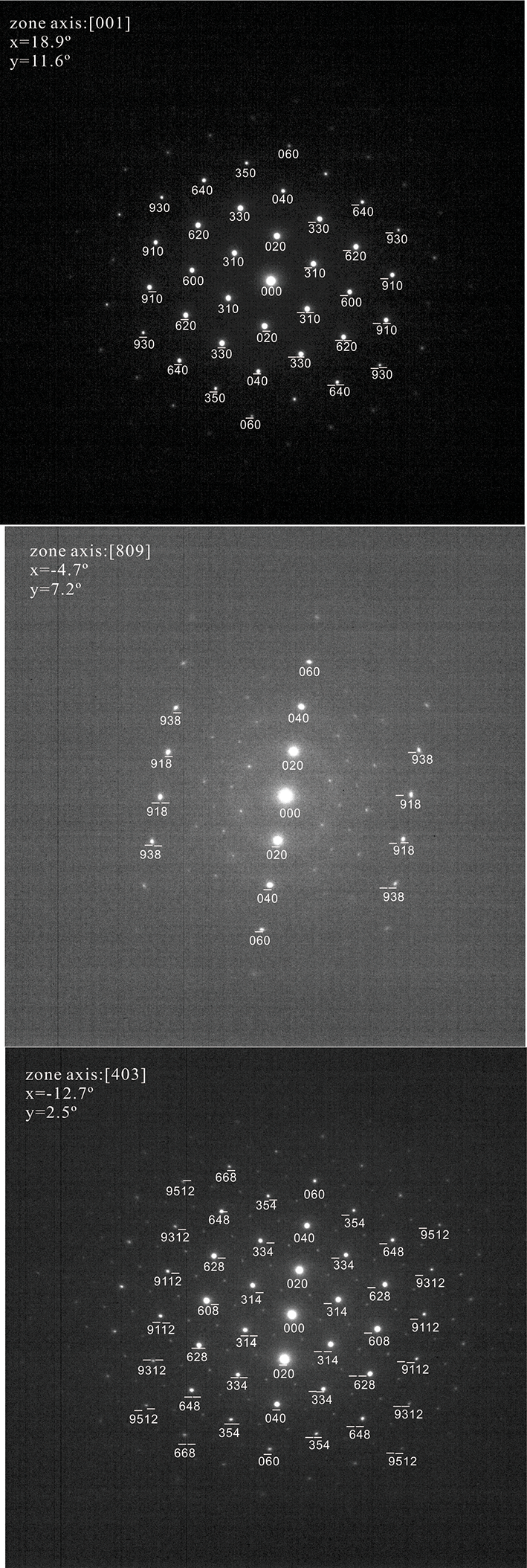
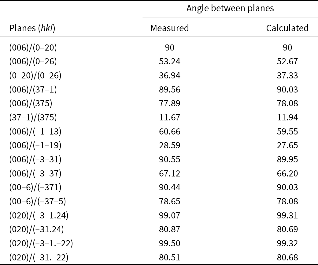
 $\bar 1$], which conform to the monoclinic symmetry of parisite-(Ce). Figure 10 shows the SAED patterns of the diffraction spots of zones [001], [809] and [403]. We also measured the angles between adjacent planes in the SAED patterns of parisite-(Nd). The measured angles of parisite-(Nd) agree well with the calculated angles of parisite-(Ce), with the absolute value of difference <1° (Table 3).
SAED patterns of parisite-(Nd) from 6 different zone axes around the [001] axis with the diffraction spots of zones [100], [7 $\bar 3$0], [1
 $\bar 1$0], [730], [801] and [70
 $\bar 1$].

SAED patterns of parisite-(Nd) of the diffraction spots of zones [001], [809] and [403].

Measured angles of parisite-(Nd) in comparison with calculated angles of parisite-(Ce)

Notes: Measured = measured values based on the SAED patterns of parisite-(Nd) in Fig. 9, Calculated = calculated values based on the cell parameters of parisite-(Ce) (Ni et al., Reference Ni, Post and Hughes2000).
Parisite-(Nd) is the Nd-dominant analogue of parisite-(Ce), as demonstrated by the consistency between their chemical compositions, powder X-ray diffraction data and TEM results. The ionic radius of Nd3+ is 0.983 Å, which is close to the ionic radius of Ce3+(1.01 Å) and La3+ (1.032 Å) (Shannon Reference Shannon1976). The rare elements occupy the same position in the parisite crystal structure. So parisite-(Nd) is believed to have the same structure as parisite-(Ce) (Ni et al., Reference Ni, Post and Hughes2000).
Discussion
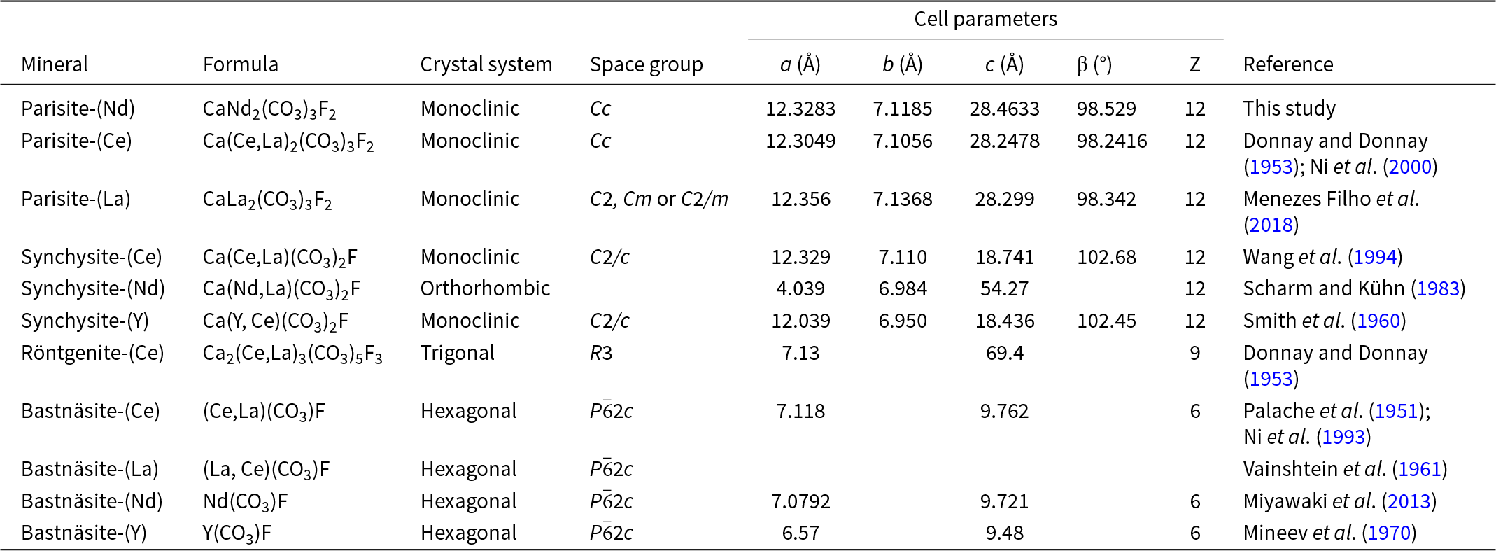
The Ca–REE fluorocarbonate minerals chemically or structurally related to parisite-(Nd) including bastnäsite, synchysite and röntgenite are listed in Table 4 and Table S1. The structure of the Ca–REE fluorocarbonate family of minerals has been described quantitatively in terms of layers parallel to (001) of the hexagonal or monoclinic cell. There are various ways to describe the structure. The most common notation uses two end-members, bastnäsite (B) and synchysite (S), for the characterisation of layer sequences (Van Landuyt and Amelinckx, Reference Van Landuyt and Amelinckx1975; Meng et al., Reference Meng, Wu, Mou and Li2001a, Reference Meng, Wu, Mou and Li2001b). Another method for the interpretation of the stacking sequence can be defined in terms of vaterite (V) and bastnäsite (B) (Ciobanu et al., Reference Ciobanu, Kontonikas-Charos, Slattery, Cook, Ehrig and Wade2017; Capitani, Reference Capitani2019; Capitani, Reference Capitani2020). The structure can also be described in terms of four layers parallel to (001): a REEF structural layer (d), a layer of CO3 groups between two REEF layers (e), a layer of Ca (f), and a layer of CO3 groups between calcium and REEF layers (g) (Donnay and Donnay, Reference Donnay and Donnay1953).
Minerals and phases chemically/structurally related to parisite-(Nd)

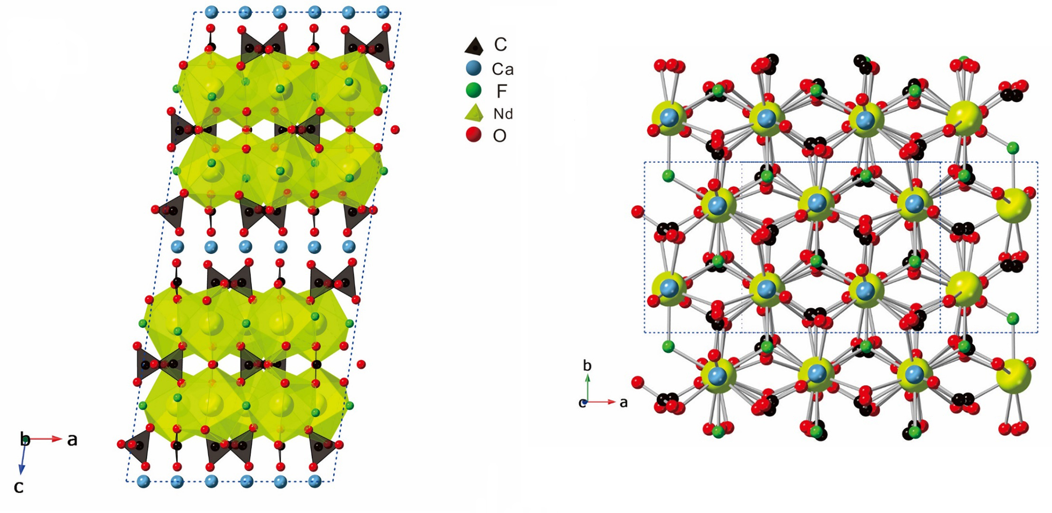
The structure of parisite-(Nd) is regarded as an intermediate product composed of 1:1 ratio of B and S or 2:1 ratio of B and V or dedgfg structure along the (00l) plane (Fig. 11) (Donnay and Donnay, Reference Donnay and Donnay1953). In the structure of parisite-(Ce), Nd atoms coordinate to three F atoms in the same (001) layer and six O atoms, three from the neighbouring (CO3) groups on each side. Ca atoms coordinate with eight O atoms, four from each adjoining (CO3) layer; the six shorter Ca–O bonds form a regular octahedron (Fig. 11). The parisite-(Nd) structure consists of stacked (001) layers of the Nd polyhedra and Ca polyhedra. The polyhedra within the layers share edges, and the polyhedra between the layers are connected by apical oxygen atoms and by vertically orientated carbonate groups (Ni et al., Reference Ni, Post and Hughes2000).
The crystal structure of parisite-(Nd) modified from parisite-(Ce) by Ni et al. (Reference Ni, Post and Hughes2000) using CrystalMaker software (Palmer, Reference Palmer2015).

Not only the nature of the nanometric crystal aggregate of the parisite-(Nd) sample, but also structural complexity makes its crystal structure very difficult to determine. Because the X-ray diffraction power of F, O and C atoms is very weak, it is difficult to calculate the structure factor. In addition, a variety of syntactic intergrowth, polytypes, polysomatic faults, polytypic disorder and twinning, known to occur for parisite-(Ce), also occur in our observations of parisite-(Nd), which brought great difficulties in analysis of the X-ray studies (Wu and Meng, Reference Wu and Meng2000; Meng et al., Reference Meng, Wu, Mou and Li2001a, Reference Meng, Wu, Mou and Li2001b; Ciobanu et al., Reference Ciobanu, Kontonikas-Charos, Slattery, Cook, Ehrig and Wade2017; Capitani, Reference Capitani2019). Taking parisite-(Ce) as an example, its structure was defined with space group R3, a = 7.18 Å and c = 8.41 Å in an early study (Donnay and Donnay, Reference Donnay and Donnay1953). Eventually Ni et al. (Reference Ni, Post and Hughes2000) refined the crystal structure to space group C2/c or Cc with a unit cell of a = 12.305(2), b = 7.1053(5), c = 28.250(5) Å and β = 98.257(14)°. In that study, it was proposed for the first time that the common 6R polytype of parisite was not present, and it may be a 2M polytype. Capitani et al. (Reference Capitani2019) had reported the discovery of a new polytype with C1 space group that may be 2M2, and believed that rhombohedral symmetry in parisite was wrong. The structure of parisite-(La) was proved using a simulation calculation with Ni et al. (Reference Ni, Post and Hughes2000)’s work. However, due to inclusions of synchysite, röntgenite or bastnäsite microblocks, no new structure could be determined for parisite-(La) (Hålenius et al., Reference Hålenius, Hatert, Pasero and Mills2016; Menezes Filho et al., Reference Menezes Filho, Chaves, Chukanov, Atencio, Scholz, Pekov, da Costa, Morrison, Andrade, Freitas, Downs and Belakovskiy2018). In our study, we tried to analyse several particles of parisite-(Nd) many times on a Rigaku Xtalab Synergy-DS 4-circle diffractometer with CuKα radiation but failed. Powder X-ray diffraction and selected-area electron diffraction were, instead, carried out for the crystallographic characterisation to prove the structure of parisite-(Nd) the neodymium analogue of parisite-(Ce).
Supplementary material
The supplementary material for this article can be found at https://doi.org/10.1180/mgm.2024.90.
Acknowledgements
We acknowledge all members of the IMA Commission on New Minerals, Nomenclature and Classification, Professor Ferdinando Bosi, and three anonymous reviewers for their helpful suggestions and comments. Dr Li Ting and Dr Chen Zhi are greatly acknowledged for the help of single crystal X-ray diffraction analysis.
Competing interests
The authors declare none.