In addition to primary congenital leaflet malformation, paediatric aortic regurgitation is often secondary to ventricular septal defects and their surgical repair, infective endocarditis and aortic stenosis and its surgical repair. Reference Tretter and Langsner1 Aortic valve repair in children is still a challenge, and somatic growth makes re-operation nearly inevitable. Other methods include the Ross procedure, artificial valve replacement, and homograft replacement. Reference Buratto and Konstantinov2
The Ozaki procedure, or aortic valve neocuspidisation, involves the complete excision of diseased native leaflets and construction of newly templated neocusps using the pericardium. It has yielded promising results in adult Reference Ozaki, Kawase and Yamashita3,Reference Ozaki, Kawase, Yamashita, Uchida, Takatoh and Kiyohara4 and paediatric Reference Baird, Cooney, Chávez, Sleeper, Marx and Nido5 populations. However, the reported studies have mainly focused on total leaflet reconstruction; single leaflet reconstruction using the Ozaki procedure has been seldom reported. Thus, the present study retrospectively examines single leaflet reconstruction using the Ozaki procedure in nine children with aortic regurgitation performed at our medical centre and describes the outcomes of this alternative surgical treatment for paediatric aortic regurgitation.
Materials and methods
This study was approved by the Ethics Committee of Fuwai Hospital, Chinese Academy of Medical Sciences. During hospitalisation, all parents of the children signed the informed consent for the use of medical records. Clinical data were collected from the hospital database and extracted from the medical records as well as from the outpatient records of the last available follow-up.
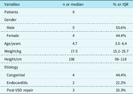
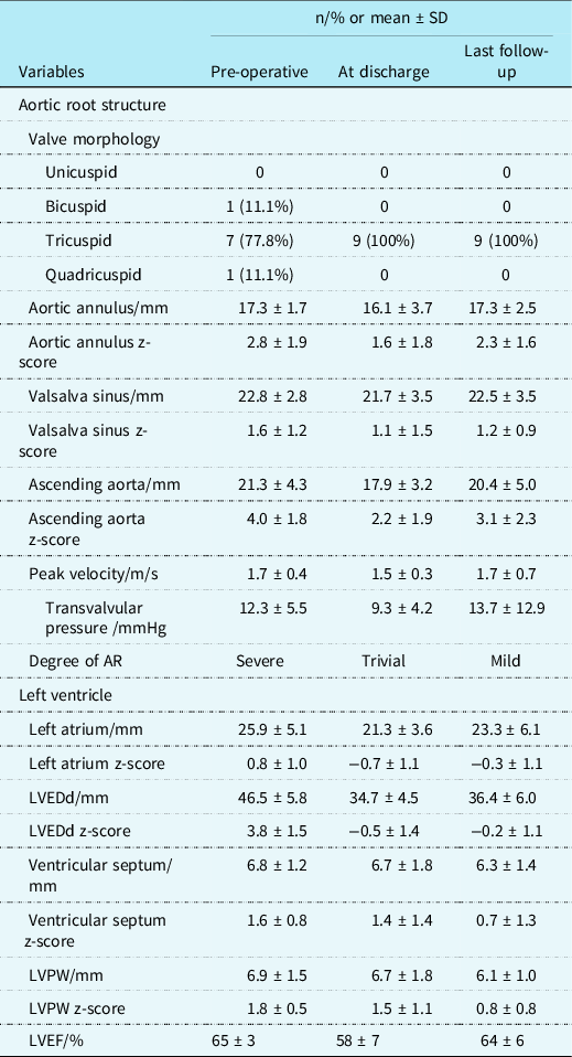
A total of nine children with aortic regurgitation who underwent single leaflet reconstruction using the Ozaki procedure in our centre from May 2017 to September 2019 were retrospectively analysed. No concomitant complex cardiac anomalies were observed. There were four female patients and five male patients. The median surgical age was 4.7 (interquartile range 3.5, 6.4) years. The median weight was 17.5 (15.2, 25.7) kg. The median height was 108 (96, 118) cm. The pre-operative data are shown in Table 1. Eight patients had severe aortic regurgitation, while one had moderate aortic regurgitation. The mean peak forward blood flow velocity of aortic valve was 1.7 m/s, which was in normal range. The left ventricle was significantly enlarged with a mean z-score of 3.8. Pre-operative echocardiography data are shown in Table 2.
Pre-operative data

IQR = interquartile range; SD = standard deviation; VSD = ventricular septal defect.
Indexes evaluated by echocardiography at different time points

AR = aortic regurgitation; LVEDd = left ventricular end-diastolic dimension; LVEF = left ventricular ejection fraction; LVPW = left ventricular posterior wall; SD = standard deviation.
The standard Ozaki procedure for total aortic leaflet reconstruction is described in detail in a previous article. Reference Ozaki, Kawase and Yamashita3 In our centre, surgery was performed through median sternotomy. The pattern of cardiopulmonary bypass was determined by concomitant operations, such as ventricular septal defect repair, under moderate hypothermia at a minimum of 31.0°C. The fat and other redundant tissue on the outer surface of the autologous pericardium was carefully cleansed. The excised pericardium was treated with 0.6% glutaraldehyde solution for 5 minutes and was then rinsed three times for 6 minutes using physiological saline solution. A transverse incision was made through the aortic root to expose the aortic valve. The damaged single leaflet was meticulously excised. Then, the distance between the commissures of the damaged leaflet was measured with a sizing apparatus originally invented by Ozaki and colleagues. A new leaflet of the corresponding size was trimmed from the glutaraldehyde-treated autologous pericardium using an original template. Finally, the annular margin of the pericardial leaflet was running sutured with 5–0 Prolene® to the annulus. Commissural coaptation was secured with additional 5–0 Prolene®. An epicardial temporary pacemaker was placed as per routine.
The evaluation of the surgical efficacy was mainly evaluated based on the symptoms of the patients as well as the morphology and functional status of the aortic valve and left ventricle estimated by echocardiography. The adjusted z-scores of each evaluated index (Table 2) were calculated according to the body surface area because of the somatic growth of children. The severity of aortic stenosis was determined by peak velocity in m/s and mean transvalvular pressure. Aortic regurgitation was graded as none, trivial, mild, moderate, or severe based on a combination of echocardiographic indicators such as area of reflux orifice and others. The patients were evaluated pre-operatively, at discharge, and at the last follow-up. All patients were routinely required to return for outpatient follow-up visits at 3, 6, and 12 months after surgery and once each year afterwards. If the patients visited local hospitals for routine examinations, formal echocardiography reports were requested and sent to our centre. The median follow-up was 22 (14, 33) months. One patient failed to follow up. There was no mortality or re-intervention within the follow-up period. All patients were asymptomatic and could handle daily activities at the last follow-up. One patient had moderate or more regurgitation.
Statistical analysis
Data were collected and analysed in SPSS (version 25, IBM SPSS, Armonk, New York). Normally distributed and skewed continuous variables were described in terms of the mean ±standard deviation and median (interquartile range) values. Paired t-tests and Wilcoxon signed rank tests were used to compare the differences between the groups at different time points. Descriptive statistics were reported for the categorical variables as frequencies or percentages and were compared using the Pearson χ2 or Fisher’s exact test. Values of p < 0.05 were considered statistically significant.
Results
The median surgical age was 4.7 years, with the youngest being the age of 2.3 years. Four patients had aortic valve malformation, which resulted in primary congenital aortic regurgitation. The other five patients had secondary aortic regurgitation, with two having infective endocarditis and three having undergone previous ventricular septal defect repair. Pre-operative data are shown in Table 1.
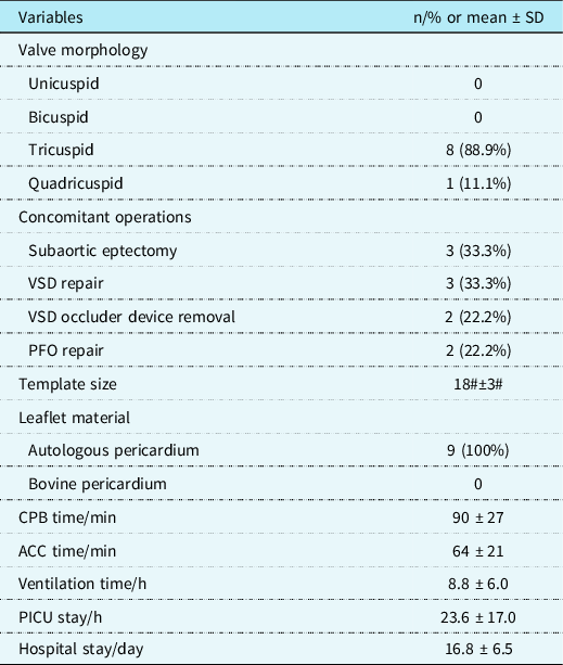
Tricuspid was observed in eight patients, and quadricuspid was confirmed in one patient during surgery. In regard to leaflet lesion, leaflet dysplasia (n = 3), leaflet contracture after compression (n = 2), and leaflet perforation (n = 3) were observed. In the case of quadricuspid, the right and the left coronary leaflets were smaller than normal, while the two non-coronary leaflets were of normal size. The original left coronary leaflet and its adjacent non-coronary leaflet were replaced by a new individual leaflet, constructing a neo-tricuspid aortic valve.
Concomitant surgical procedures were carried out to repair simple cardiac anomalies, such as ventricular septal defect repair and subaortic septectomy. There were no perioperative complications. Perioperative data are shown in Table 3.
Perioperative data

ACC = aortic cross-clamping; CPB = cardiopulmonary bypass; PFO = patent foramen ovale; PICU = paediatric ICU; SD = standard deviation; VSD = ventricular septal defect.
Aortic root structure
One patient was pre-operatively diagnosed as bicuspid by echocardiography but then confirmed as tricuspid during surgery with a dysplastic right coronary leaflet. Other patients had consistent results from echocardiography and surgery. All aortic valves were reconstructed as tricuspid.
In children with aortic regurgitation in our study, the aortic root structure is characterised by a slightly enlarged aortic annulus, a Valsalva sinus of normal size and a significantly enlarged ascending aorta. Herein, these had corresponding z-scores of 2.8, 1.6, and 4.0, respectively. These changes are consistent with the typical pathophysiological changes observed in aortic regurgitation. Because the aortic annulus and Valsalva sinus were not significantly enlarged, the operation mainly focused on the single leaflet reconstruction, and no intervention was simultaneously made to the aortic root. Immediately after the surgery, the above-mentioned indexes were smaller than pre-operatively. The z-score of the ascending aortic diameter decreased to 2.2 (p = 0.016). At the last follow-up, the z-scores were consistent with those immediately after the surgery and smaller than those pre-operatively. The z-score of the diameter of Valsalva sinus and ascending aorta decreased to 1.2 (p = 0.046) and 3.1 (p = 0.006), respectively. The trend of variation in the aortic root structure was roughly consistent with the changes related to the degree of aortic regurgitation.
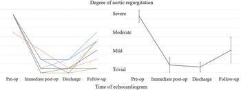
The mean peak forward blood flow velocity of aortic valve did not change significantly pre-operatively and post-operatively. The degree of aortic regurgitation was recorded at four time points: pre-operatively, immediately after surgery, at discharge, and at the last follow-up. The individual trends of variation in the degree of aortic regurgitation of the nine patients are shown in Fig 1 (left). Eight patients had severe aortic regurgitation pre-operatively, while one had moderate aortic regurgitation. After single leaflet reconstruction, the aortic regurgitation degree decreased to less than mild in most patients. At the last follow-up, one patient had moderate regurgitation, three had mild to moderate regurgitation, two had mild regurgitation, one had trivial to mild regurgitation, and two had trivial regurgitation. The rate of freedom from moderate or more aortic regurgitation was 88.9%. The average trend of variation in the aortic regurgitation degree is also shown in Fig 1 (right). At the last follow-up, the average aortic regurgitation degree was mild and significantly lower than the severe degree encountered pre-operatively (p = 0.000).
Degree of aortic regurgitation. Left: The individual trends of variation. Right: The average trend of variation.

Left ventricle
The diameter of the left atrium changed pre-operatively and post-operatively but remained within the normal range. The pre-operative left ventricular end-diastolic dimension showed significant enlargement, with a z-score of 3.8. There was also mild thickening of the left ventricular wall which was roughly consistent with the typical pathophysiological changes in aortic regurgitation. After the single leaflet reconstruction and diuretic treatment, the z-score of the left ventricular end-diastolic dimension decreased to −0.45 at discharge (p = 0.000). The ventricular septum and the left ventricular posterior wall became thinner post-operatively. At the last follow-up, the z-score of the left ventricular end-diastolic dimension was −0.2, significantly lower than the pre-operative value (p = 0.000). The thickness of the ventricular septum and the left ventricular posterior wall continued to decrease, while the z-score of the left ventricular posterior wall thickness was significantly smaller than the pre-operative value (p = 0.022). The decrease in the left ventricular end-diastolic dimension, as well as the thinning of the left ventricular wall, was related to the correction of aortic regurgitation. The data are shown in Table 2.
The pre-operative left ventricular ejection fraction was 65%. It decreased to 58% at discharge (p = 0.017), which might be related to the decrease in cardiac preload. At the last follow-up, the left ventricular ejection fraction was 64%, indicating that cardiac function gradually recovered.
Discussion
Ozaki procedure
The Ozaki procedure is a relatively new method for the treatment of aortic valve disease that has been applied since 2007. Reference Ozaki, Kawase and Yamashita3 The diseased aortic valves are replaced by three new leaflets of equal size made of glutaraldehyde-treated autologous pericardium. A custom-made sizing apparatus is used to measure the distance between each commissure. An original template is made to create a new leaflet of corresponding size trimmed from the glutaraldehyde-treated autologous pericardium. Thus, the process is standardised, Reference Ozaki, Kawase and Yamashita6 and it has been greatly promoted since its original proposal. It is distinct from other valvuloplasty techniques: Reference Prêtre, Kadner, Dave, Bettex and Genoni7–Reference Tao, Zeng and Lim9 The new leaflets made from the pericardium in the Ozaki procedure are deeply coapted up to the same horizontal plane as the top of the commissures. Also, the commissures are sutured with a 5-mm-wide wing extension. With the commissural wing sewn facing the aortic wall, the commissural part of each leaflet can have greater strength and better capacity to tolerate maximal stress.
Ozaki et al Reference Ozaki, Kawase, Yamashita, Uchida, Takatoh and Kiyohara4 retrospectively analysed 850 patients with aortic valve diseases treated by the Ozaki procedure in their centre. They obtained satisfactory mid-term results at the mean follow-up at 53.7 months. Two hundred and fifty-four patients had severe aortic regurgitation pre-operatively. The median surgical age was 71 years. The overall survival rate was 85.9%, and the rate of moderate or more regurgitation post-operatively was 7.3%. Although studies are scarce, the Ozaki procedure has also been applied in children. Christopher et al Reference Baird, Cooney, Chávez, Sleeper, Marx and Nido5 retrospectively analysed a group of 57 children with a median surgical age of 12.43 years and obtained satisfactory short-term results. The rate of post-operative moderate or more regurgitation was 4.0%.
Single leaflet reconstruction
One of the challenges of paediatric aortic valve surgery is that the growth capacity of the new leaflet cannot match the somatic growth of children. Re-intervention is almost inevitable. Reference Buratto and Konstantinov2 In several studies, the new leaflets were made according to physiological size Reference Prêtre, Kadner, Dave, Bettex and Genoni7,Reference Tao, Zeng and Lim9 ; however, the new leaflets in the Ozaki procedure are larger and higher with deep coaptation, as described above.
Several retrospective studies of single leaflet reconstruction to treat paediatric aortic regurgitation have shown satisfactory short-term results, Reference Prêtre, Kadner, Dave, Bettex and Genoni7–Reference Tao, Zeng and Lim9 although the real challenge is in the long term. In a multicentre study, Luke et al Reference Wiggins, Mimic and Issitt10 retrospectively analysed 58 children with aortic valve disease with a median surgical age of 14.8 years. Twenty-three patients had aortic regurgitation. Forty patients with lesions on all leaflets received neo-tricuspidalisation using the Ozaki procedure, while another 18 patients with a single leaflet lesion were treated by single leaflet reconstruction using the method described by Rene et al. Reference Prêtre, Kadner, Dave, Bettex and Genoni7 No significant differences were observed between the two groups in regard to the rate of re-intervention and moderate or more regurgitation at 3 years post-operative.
As previously mentioned, single leaflet reconstruction using the Ozaki procedure has been seldom reported. For cases of aortic regurgitation with a single leaflet lesion, single leaflet reconstruction may not be taking full advantage of the Ozaki procedure. The new leaflet does not have deep coaptation with the normal leaflets preserved. However, if the normal leaflets are preserved, they can match the somatic growth of children and possibly partially compensate for the lack of growth of the large new leaflet inserted by the Ozaki procedure, thus prolonging, in theory, the period of freedom from regurgitation.
Single-centre experience
There were nine children with aortic regurgitation in our study. The median surgical age was 4.7 years. The aortic regurgitation was of severe degree, and the left ventricular end-diastolic dimension significantly enlarged (z-score = 3.8). After single leaflet reconstruction by the Ozaki procedure, there was no mortality or re-intervention during the 22-month follow-up period. One patient had moderate or more regurgitation during follow-up. The short-term results were satisfactory.
The patient with moderate or more regurgitation during follow-up was diagnosed with congenital aortic regurgitation. The surgical age was 2.3 years. The pre-operative aortic annulus (z-score = 4.6), Valsalva sinus (z-score = 3.0), and ascending aorta (z-score = 5.5) were significantly enlarged. The left ventricular end-diastolic dimension significantly increased (z-score = 5.4), with severe regurgitation of the aortic valve. The right coronary leaflet was dysplastic, and severe adhesions were found between the commissures. The adhesions of the commissures were carefully incised, and the right coronary leaflet was replaced by a new leaflet made from glutaraldehyde-treated autologous pericardium using the Ozaki procedure. The z-score of the aortic annulus decreased to 3.3 at discharge, with trivial to mild regurgitation. The z-score of the aortic annulus increased to 4.9, with moderate regurgitation 2 months post-operative. One of the possible reasons was that pure valvuloplasty without concomitant aortic root intervention might be not suitable for aortic regurgitation with an obviously enlarged aortic annulus. Chawki et al Reference ElZein, Roberson, Hammad and Ilbawi11 pointed out that the outcome after valvuloplasty is dependent on the comprehensive repair of all of the involved components of the aortic root.
Individualised surgical interventions should be followed in children with aortic regurgitation. For only leaflet lesions, the Ozaki procedure is an alternative method. Modifications such as making leaflets larger than the standard template and banding the Valsalva sinus to limit its expansion are worth further exploring. For those with a significantly enlarged aortic annulus, concomitant aortic root intervention is necessary.
Leaflet material
In addition to the surgical method, the leaflet material is also an important factor for the prognosis. Glutaraldehyde-treated autologous pericardium and commercial bovine pericardium are widely used at present. In the standardised Ozaki procedure, the excised autologous pericardium was treated with 0.6% glutaraldehyde solution for 5 minutes and then rinsed three times for 6 minutes using physiological saline solution. Previously, the rate of re-intervention for this method was found to be 4.2% during a 4.5-year follow-up. Reference Ozaki, Kawase, Yamashita, Uchida, Takatoh and Kiyohara4 Duran et al Reference Halees, Shahid, Sanei, Sallehuddin and Duran12 retrospectively analysed 92 patients with aortic valve reconstruction surgery. The mean surgical age was 30 years. Bovine pericardium was used in 27 patients, while glutaraldehyde-treated autologous pericardium was used in another 65 patients. There was no significant difference in the re-intervention rate between the two groups after a 16-year follow-up. Similar results were obtained in other studies of paediatric aortic valve repair surgery. Reference Baird, Cooney, Chávez, Sleeper, Marx and Nido5,Reference Wiggins, Mimic and Issitt10 However, most of these studies are retrospective. Prospective, randomised trials are required to reach more definite conclusions. In addition, new materials such as polytetrafluoroethylene have been applied with satisfactory short-term results. Reference Nosál’, Poruban, Valentík, Šagát, Nagi and Kántorová13
Limitations
This study was subject to the usual limitations of a retrospective, non-randomised study design. The sample size was relatively small.
Single leaflet reconstruction using the Ozaki procedure was an effective surgical method for treating children with aortic regurgitation in our centre with satisfactory short-term results. Studies of larger sample size and longer follow-up are needed to draw more definite conclusions.
Acknowledgements
None.
Financial support
This study was supported by the National Key R&D Program of China [2017YFC1308100].
Conflicts of interest
None.
Ethical standards
This study had been approved by the Ethics Committee of Fuwai Hospital, Chinese Academy of Medical Sciences (Ethics approval number: 2017–977).