Introduction
After fertilization, the zygote enters the early embryonic development process, consisting of 2-cell, 4-cell, 8-cell, morula and blastocyst stages. When division reaches the 8-cell stage, the zygote begins to bind tightly and further develops into a morula, from which it develops into a blastocyst. At the blastocyst stage, two cell lineages, the trophectoderm (TE) and the inner cell mass (ICM), can be clearly seen. Early embryonic development is critical for the subsequent implantation and pregnancy process (Calix et al., Reference Calix, Ornaghi, Wilson, Fernandez, Vialard, Barnea and Paidas2017). Abnormal early embryonic development may lead to a miscarriage. In the preimplantation phase, mitochondrial dysfunction can interfere with embryonic development (Babayev and Seli, Reference Babayev and Seli2015; Harvey, Reference Harvey2019). Studies have found that mitochondrial dysfunction may be caused by oxidative stress (Van Blerkom, Reference Van Blerkom2011; Agarwal et al., Reference Agarwal, Durairajanayagam and du Plessis2014; Kauppila et al., Reference Kauppila, Kauppila and Larsson2017). Oxidative stress can cause early embryonic cell apoptosis and autophagy (An et al., Reference An, Peng, Cheng, Lu, Zhou, Zhang and Su2019; Xu et al., Reference Xu, Zhang, Sun, Lan, Wan, Zhang and Sun2019). To maintain normal embryonic development, a certain concentration of reactive oxygen species (ROS) is necessary (Terao et al., Reference Terao, Wada-Hiraike, Nagumo, Kunitomi, Azhary, Harada, Hirata, Hirota, Koga, Fujii and Osuga2019). However, excessive ROS levels may be one of the main reasons for the delay or stagnation of early embryonic development (Goodale et al., Reference Goodale, Hayrabedyan, Todorova, Roussev, Ramu, Stamatkin, Coulam, Barnea and Gilbert2017). In addition, the accumulation of ROS may affect early embryonic development by reducing the mitochondrial membrane potential (ΔΨm) (Wang et al., Reference Wang, Wang, Zou and Xu2009; Yuan et al., Reference Yuan, Liang, Jin, Zhang, Zhang and Kim2017).
Methomyl (S-methyl N-(methyl-carbamoyl); MET) is a carbamate insecticide, also known as environmental oestrogen (EE) (Meng et al., Reference Meng, Qiu, Hu, Fan, Song, Zheng, Wu, Qu, Li, Chen and Xu2016). It is widely used because of its broad-spectrum insecticidal properties. Residues of MET are harmful to the environment and can also cause liver, kidney, nervous system and reproductive system diseases in humans and animals (Hoizey et al., Reference Hoizey, Canas, Binet, Kaltenbach, Jeunehomme, Bernard and Lamiable2008; Shalaby et al., Reference Shalaby, El Zorba and Ziada2010; El-Demerdash et al., Reference El-Demerdash, Attia and Elmazoudy2012; Sakr et al., Reference Sakr, Hassanien, Bester, Arbi, Sobhy, El Negris and Steenkamp2018; Seleem, Reference Seleem2019). Recent studies on the reproductive toxicity of methomyl have mainly focused on male mammals (Farag et al., Reference Farag, Eweidah and El-Okazy2000; Mahgoub and El-Medany, Reference Mahgoub and El-Medany2001; Shalaby et al., Reference Shalaby, El Zorba and Ziada2010;), and there have been few studies on the female reproductive system. In the reproductive system, MET can affect the structure of the testis, epididymis and vas deferens, resulting in a decrease in the number of spermatogenic cells and sperm, the weight of the gonads and the quality of semen (Mahgoub and El-Medany, Reference Mahgoub and El-Medany2001). Shanthalatha and colleagues orally administered 5 mg/kg of MET to female mice for 3 months consecutively and found that the relative weight of the fallopian tubes, uteri and ovaries decreased significantly (Shanthalatha et al., Reference Shanthalatha, Madhuranath and Yajurvedi2012).
Although the toxicity of MET to the reproductive system has been extensively studied, there have been few reports on the effects of MET on the early embryonic development of female mammals. Therefore, in this study, we explored the effect of MET on the in vitro development of early mouse embryos and its possible mechanisms. We exposed early mouse embryos to MET and evaluated the effect of MET on early embryo development by detecting the blastocyst rates, apoptosis, ROS and glutathione (GSH) levels, ΔΨm, autophagy and the expression of oxidation-related gene mRNA.
Materials and Methods
All chemicals and reagents used in this study were purchased from Sigma-Aldrich (St. Louis, MO, USA) unless otherwise stated.
Mice
All mouse experiments were conducted in accordance with the guidelines of the Animal Research Committee of Yanbian University, China. Female Kunming mice aged 6–8 weeks were purchased from the Experimental Animal Production Center of Yanbian University, China. Male Kunming mice (8 weeks old) were housed in a single cage. Mice were kept in temperature-controlled rooms and provided with sufficient food and water at a constant temperature (22 ± 2°C). Female mice were reared for 7 days to adjust their physiological cycle to adapt to the environment for experimental use.
Early embryo collection and in vitro culture (IVC)
In the experiment, female mice were intraperitoneally injected with 10 IU of PMSG (Ningbo Shu Sheng Pharmaceutical Co., Ltd., China) at 15:00 in the afternoon and 10 IU of hCG (Ningbo Shu Sheng Pharmaceutical Co., Ltd, China) were injected intraperitoneally after 48 h for superovulation treatment in female mice. After the female mice were injected with hCG, the female and male mice were caged immediately (1:1) and the female mice were checked for vaginal suppositories the next morning. A vaginal suppository indicates successful fertilization. At 20 h later, the female mice were killed by cervical dislocation. The fallopian tubes were removed by aseptic surgery and placed in a pre-equilibrated 37.5°C, 5% CO2 M2 medium. The enlarged ampullae of the fallopian tubes were cut under a stereomicroscope to collect cumulus–oocyte complexes (COCs). The COCs were transferred to M2 medium containing 0.5% hyaluronidase (1 mg/ml) for 2 min and washed three times until the cumulus cells were removed and the fertilized eggs containing double pronuclei were collected. MET was dissolved in deionized water to prepare a concentrated stock solution. The culture medium was divided into five groups: the control group, 10 μM MET group, 20 μM MET group, 30 μM MET group and 35 μM MET group. The previously collected double-pronuclear fertilized eggs were placed in an overnight pre-equilibrated control group and KSOM embryo medium with different concentrations of MET groups and they were cultured into blastocysts in a 37°C, 5% CO2 in air incubator. The early embryo blastocysts were observed under a microscope (Olympus).
Detection of total cell number and apoptosis in blastocysts
First, blastocysts cultured for 96 h were washed three times with 1% BSA–PBS. The blastocysts were then immersed in 3.7% paraformaldehyde (PFA) and fixed at room temperature for 30 min. Under the same conditions, the blastocysts were incubated in 0.5% Triton X-100 for 30 min to penetrate the membrane and then blocked with 1% BSA–PBS for 30 min. Intracellular apoptosis rates of blastocysts were measured by terminal deoxynucleotidyl transferase dUTP nick end labelling (TUNEL) assay using the In Situ Cell Death Assay Kit (cat. no. 11684795910, Roche, fluorescein-conjugated dUTP:terminal deoxynucleotidyl transferase enzyme; 9:1). Blastocysts were incubated at 37°C in the dark for 1 h and then washed three times with 1% BSA–PBS. The fluorescent dye nuclear stain Hoechst 33342 was used to measure the total cell number in the blastocysts. They were then stained with Hoechst 33342 (10 µg/ml in PBS) for 15 min, washed three times with 1% BSA–PBS and then mounted onto glass slides. DNA was labelled with a blue fluorescence filter and apoptosis was labelled with a green fluorescence filter to obtain images and observed under an apoptosis fluorescence microscopy (Nikon Corp. Tokyo, Japan.). Apoptosis index = positive nuclei/total number of nuclei.
ROS and GSH level assays in early embryos
The early mouse embryos were washed three times with PBS containing 1% BSA and then placed in 10 μmol/l DCFH-DA (2,7-dichlorodihydro-fluorescein diacetate probe, Thermo Fischer Scientific, Waltham, MA, USA) and incubated at 37°C for 20 min in the dark. Then, the embryos were washed three times with 1% BSA–PBS and placed in PBS. The samples were imaged using a fluorescence microscope and the data were analyzed using ImageJ software. The early embryos were washed three times with PBS containing 1% BSA, then placed in 10 μmol/l CMF2HC (CellTracker™ Blue dye 4-chloromethyl-6, 8-difluoro-7-hydroxycoumarin, Invitrogen) and incubated at 37°C in the dark for 20 min. The early embryos were then washed three times with 1% BSA–PBS. The samples were imaged using a fluorescence microscope (Nikon Corp.) and the data were analyzed using ImageJ software.
Assay of ΔΨm in early embryos
The early embryos were washed three times in PBS containing 1% BSA and 2 μM JC-1 (5,5,6,6-tetrachloro-1,1,3,3-tetraethyl-imidacarbocyanine iodide, Invitrogen, Carlsbad, CA, USA) was diluted with 1% BSA–PBS at a ratio of 1:1000, covered with mineral oil and incubated for 30 min. The embryos were placed in JC-1, incubated at 37°C for 30 min in the dark, washed three times and placed in PBS to observe the fluorescence under a fluorescence microscope (Nikon Corp., Tokyo, Japan). The mitochondrial membrane potential in the cells was measured by the change in JC-1 emission fluorescence. The change in the relative ratio of red/green fluorescence reflects the intensity of the membrane potential and the data were analyzed using ImageJ software.
Immunofluorescence
The early embryos were washed three times with 1% BSA–PBS, fixed with 3.7% paraformaldehyde (PFA) for 20 min, permeabilized with 0.5% Triton X-100 for 30 min and incubated with 1% BSA in PBS at 37°C for 1 h. Early embryos were placed in the primary antibody (with 1% BSA–PBS) and sealed away from light at 4°C overnight (anti-LC3). They were incubated with 1% BSA three times and then the embryos were incubated with Alexa Fluor 488-conjugated secondary antibody (1:200, 1% BSA–PBS dilution) at 37°C for 2 h. The early embryos were then washed three times with 1% BSA–PBS (5 min/time), stained with Hoechst 33342 (10 μg/ml in 1% BSA–PBS dilution) for 15 min and then washed three times with PBS. The primary antibody was not added to the negative control group. After the embryos were directly incubated with secondary antibody, they were washed three times with 1% BSA–PBS and then incubated with Hoechst 33342 at 37°C for 15 min in the dark. Finally, the early embryos were sealed on glass slides and the immunofluorescence intensity was analyzed.
Real-time quantitative polymerase chain reaction (RT-qPCR)
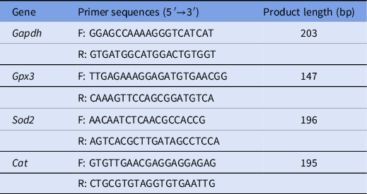
Blastocysts were collected and used with the DynaBeads® mRNA DIRECTTM Kit (Invitrogen Dynal AS, Oslo, Norway) according to the manufacturer’s instructions. Next, the QuantiNova® Reverse Transcription Kit (50) (QIAGEN GmbH, Germany) was used according to the manufacturer’s instructions to reverse transcribe the extracted mRNA to obtain cDNA. A 20-μl reaction system was selected from the KAPA SYBR® FAST kit instructions. The reaction system included: 2 µl cDNA, 1 µl primer (including upstream and downstream primers), 10 µl SYBR Green, 6.6 µl ddH2O, 0.2 µl KSF ROX High and 0.2 µl KSF ROX Low. The PCR reaction program was as follows: pre-denaturation at 95°C for 3 min, amplification of the target gene for 40 cycles, 95°C for 3 min, 60°C for 30 s and 72°C for 20 s. Using Gapdh as the internal control, the 2−ΔΔCT method was used to determine the relative expression level (Livak and Schmittgen, Reference Livak and Schmittgen2001). This study tested the mRNA levels of Gpx3, Sod2 and Cat. The primer sequences used are shown in Table 1.
Sequences of primers used for RT-qPCR

The annealing temperature was maintained at 60°C. F: forward primer; R: reverse primer.
Statistical analysis
Each test was repeated at least three times and the significance of the test data was statistically analyzed using SPSS 19.0 (Inc., Chicago, IL, USA) software for one-way analysis of variance (ANOVA) or independent samples t-test analysis. Differences between treatment groups were assessed using least significant difference (LSD) multiple comparisons. We used ImageJ software to detect and analyze the fluorescence intensity and GraphPad Prism 6.01 software to make a histogram. The test results were expressed as mean ± standard deviation (mean ± SD). The total number of embryos (n) in each group is shown in the histogram and the number of replicates (R) in each experiment is shown in the note. Additionally, the differences between the histograms marked with the letter a or b were statistically significant (P < 0.05) and there was no significant difference between the histograms without a letter (P > 0.05).
Results
Effect of MET on early embryonic development in mice
To explore the effect of MET on the in vitro development of early mouse embryos, this study tested the development rate of blastocysts treated with different concentrations of MET (10, 20, 30 and 35 μM), as shown in Fig. 1A. Compared with the control group, the blastocyst rate was reduced after treatment with 30 and 35 μM MET (47.92 ± 3.61% vs. 31.10 ± 6.57% vs. 23.00 ± 3.36%, P < 0.05; Fig. 1B). We used the lowest concentration with a significant difference in the blastocyst rate for subsequent experiments. This study also counted the total number of cells in the blastocyst and the apoptosis index, as shown in Fig. 1C. The number of cells in the blastocyst in the MET group was not different from that in the control group (71.00 ± 12.69 vs. 59.64 ± 14.75, P < 0.05; Fig. 1D). In this study, a TUNEL assay was used to determine the apoptosis index. The results showed that the apoptosis rate of the MET group was higher than that of the control group (5.25 ± 1.92% vs. 2.01 ± 1.52%, P < 0.05; Fig. 1E). The results showed that MET reduced the development rate of mouse blastocysts.
Effect of MET on the in vitro development of early embryos. (A) Representative images of blastocysts treated with different concentrations of MET (10, 20, 30 and 35 μM). Scale bar: 100 μm. (B) Quantitative relationship between the blastocyst rate of the control group and the different concentrations MET group, R = 5. (C) After 30 μM MET treatment, the number of cells in the blastocyst decreased and the apoptosis rate increased. Scale bar: 200 μm. (D) Statistical analysis of the number of cells in the blastocyst, R = 3. (E) Statistical analysis of the apoptosis rate in blastocysts, R = 3.

Effect of MET on the levels of ROS and GSH in mouse embryos
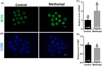
To determine whether MET induces oxidative stress damage in early mouse embryos, the H2DCFDA fluorescent probe was used to determine the level of ROS in blastocysts, as shown in Fig. 2A. The ROS fluorescence intensity of the MET group was higher than that of the control group (1.00 ± 0.35 vs. 2.02 ± 0.71, P < 0.05; Fig. 2C). To further explore the antioxidant level of MET in early mouse embryos, the CMF2HC fluorescent probe was used to determine the GSH level in blastocysts, as shown in Fig. 2B. Compared with that of the control group, the GSH level of the MET group was reduced (1.00 ± 0.05 vs. 0.84 ± 0.11, P < 0.05; Fig. 2D). The results showed that MET induced oxidative stress in early embryos.
Effect of MET on oxidative stress in early embryonic development in vitro. (A) Representative images of ROS levels in early embryos after MET treatment. ROS: green. Scale bar: 200 μm. (B) Representative images of GSH levels in early embryos after MET treatment. GSH: blue. Scale bar: 200 μm. (C) Fluorescence intensity analysis of ROS levels in early embryos in the control group and MET group, R = 3. (D) Fluorescence intensity analysis of GSH level in early embryos of the control group and MET group, R = 3.

The effect of MET on the level of ΔΨm in mouse embryos
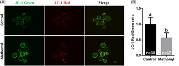
Mitochondrial dysfunction is the main cause of increased ROS levels and affects early embryonic development. Therefore, this study used the JC-1 kit to measure changes in the mitochondrial membrane potential in blastocysts. The higher the ratio of red to green fluorescence, the higher the polarity of the mitochondrial membrane potential and vice versa. As shown in Fig. 3A, compared with the control group, the MET group showed increased green fluorescence and decreased red fluorescence. The results of the immunofluorescence intensity ratio (red/green) analysis showed that the mitochondrial membrane potential level of the MET group was lower than that of the control group (1.00 ± 0.23 vs. 0.56 ± 0.17, P < 0.05; Fig. 3B). Therefore, MET causes mitochondrial dysfunction in early embryos.
Influence of MET on the mitochondria of early mouse embryos. (A) Representative images of mitochondrial membrane potential intensity after MET treatment in early mouse embryos. JC-1 green, JC-1 red. Scale bar: 200 μm. (B) Fluorescence intensity analysis of mitochondrial membrane potential in early embryos after MET treatment, R = 3.

Effect of MET on the autophagy of mouse embryos
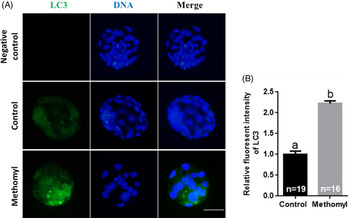
LC3 is widely used as a marker of autophagy. Therefore, to explore whether MET can affect early embryonic autophagy in mice, the expression level of LC3 in the early embryos of each group was determined, as shown in Fig. 4A. The results of the immunofluorescence intensity analysis showed that the blastocyst LC3 level in the MET group was higher than that in the control group (1.00 ± 0.08 vs. 2.22 ± 0.07, P < 0.05; Fig. 4B). The results show that MET induced early embryonic autophagy.
Influence of MET on early embryonic autophagy. (A) Representative images of autophagy in early mouse embryos, stained with LC3. LC3: green, DNA: blue. Scale bar: 500 μm. (B) Statistical analysis of fluorescence intensity of early embryos in the control group and MET group, R = 3.

Effect of MET on antioxidant-related genes in mouse embryos
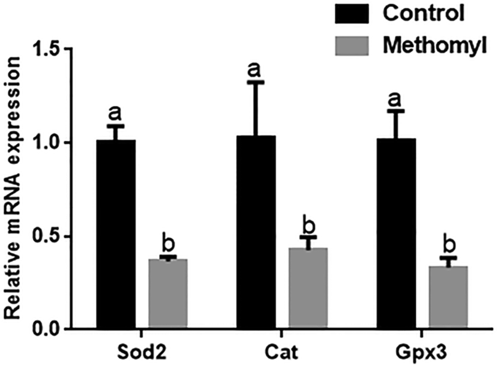
To further explore the effects of MET on oxidative stress damage in early embryos in mice, this study explored the mRNA transcription levels of antioxidant-related genes in early embryos. As shown in Fig. 5, the mRNA transcription levels of the antioxidant-related genes Sod2, Cat and Gpx3 in the control group were higher than those in the MET group (P < 0.05).
The influence of MET on antioxidant-related genes (Sod2, Cat and Gpx3) in early embryos. The mRNA levels of Sod2, Cat and Gpx3 in the early embryo of 30 μM MET group were significantly lower than those of the control group in 96 h in vitro culture, R = 3.

Discussion
The pollution of vegetables, animal feed and water sources by MET is an increasingly serious problem worldwide (Chowdhury et al., Reference Chowdhury, Banik, Uddin, Moniruzzaman, Karim and Gan2012; Van Scoy et al., Reference Van Scoy, Yue, Deng and Tjeerdema2013; Badawy and El-Aswad, Reference Badawy and El-Aswad2014; Derbalah et al., Reference Derbalah, Sunday, Kato, Takeda and Sakugawa2020). Food and feed contaminated by MET have serious consequences for animal and human health. In recent years, many studies have shown that MET causes a series of harmful effects on the reproductive system, leading to reduced animal reproduction and development (Mahgoub and El-Medany, Reference Mahgoub and El-Medany2001; Shalaby et al., Reference Shalaby, El Zorba and Ziada2010; Shanthalatha et al., Reference Shanthalatha, Madhuranath and Yajurvedi2012). In this experiment, the effect of MET on the in vitro development of early mouse embryos was explored. The test results showed that MET can significantly reduce the rate of blastocyst formation, the total number of blastocysts and apoptosis, causing oxidative stress and mitochondrial dysfunction, leading to autophagy, which may be the cause of the decline in early embryonic development in mice.
It is well known that the developmental ability of early embryos affects subsequent embryo implantation. First, the effect of MET on the mouse embryonic development rate was studied. A dose experiment was conducted using doses from 10 μM to 35 μM and it was found that 30 μM MET had obvious toxic effects on early mouse embryos. Previous studies have shown that other environmental oestrogens, zearalenone (ZEA) and bisphenol A (BPA), can cause abnormal early embryonic development in pigs (Alm et al., Reference Alm, Greising, Brüssow, Torner and Tiemann2002; Guo et al., Reference Guo, Zhao, Shin, Niu, Ahn, Kim and Cui2017). Regarding the oestrogen-like effects of MET, oestrogen plays an important role in the early stages of embryonic development (Ying et al., Reference Ying, Hsu, Hong, Cheng and Yang2000). The results of this study showed that after MET treatment, the rate of mouse early embryo blastocyst development was also significantly reduced. In this study, the number of cells in the blastocysts was determined. The results show that compared with that of the control group, the total number of cells in the blastocysts in the MET treatment group was significantly reduced. In addition, the results of the TUNEL assay showed that the apoptosis rate of blastocysts after MET exposure was significantly higher than that of the control group. In summary, MET reduces the developmental ability of early embryos, thereby affecting the quality of blastocysts, which further indicated the toxic effects of MET on early embryonic development.
When exposed to environmental pollutants, oxidative stress is the first reaction in mammals and ROS production is one of the necessary conditions for oxidative stress and the consumption of antioxidants. Oxidative stress during in vitro culture can also lead to reproductive defects in embryos (Dennery, Reference Dennery2007). Oxidative stress plays an important role in reproductive dysfunction mediated by environmental endocrine disruptors and ROS are considered an early marker for toxicity assessment (Valko et al., Reference Valko, Leibfritz, Moncol, Cronin, Mazur and Telser2007). In previous studies of embryonic development, low levels of ROS and high levels of GSH have been shown to be very important for promoting early embryonic development (Zhao et al., Reference Zhao, Xu, Li, Jin, Sun and Gao2020). However, the effect of MET on ROS and GSH content in early mouse embryos is not fully understood. In this study, the addition of 30 μM MET to the KSOM medium significantly reduced the GSH level in early embryos and increased the intracellular ROS level. These results further indicate that MET plays an important role in maintaining high levels of ROS and low levels of GSH. This shows that MET causes oxidative stress in early embryos and reduces the quality of embryonic development.
Glutathione peroxidase (GPX), superoxide dismutase (SOD) and catalase (CAT) are antioxidant enzymes that play key roles in oxidative stress. Antioxidant enzymes act as free radical scavengers by limiting the effect of ROS on tissues, which can protect cells from damage (Kyle et al., Reference Kyle, Miccadei, Nakae and Farber1987). These enzymes work together to eliminate any changes between ROS and physiological concentrations. SOD catalyzes the conversion of superoxide radicals into H2O2, whereas CAT converts H2O2 into water. Therefore, these enzymes can reduce ROS damage (Mansour et al., Reference Mansour, Abbassy and Shaldam2017). SOD is the main defensive enzyme that converts superoxide radicals into H2O2 (Rodríguez-Fuentes et al., Reference Rodríguez-Fuentes, Rubio-Escalante, Noreña-Barroso, Escalante-Herrera and Schlenk2015). In this study, the Sod2 mRNA transcription level of early embryos treated with MET was significantly reduced compared with that in the control group. This result is consistent with those of the Sameeh study, in which the SOD activity in MET-induced rat red blood cells was reduced. (Mansour et al., Reference Mansour, Mossa and Heikal2009). CAT is located in the peroxisome and can efficiently catalyze H2O2 into water and oxygen (Wu et al., Reference Wu, Xu, Shen, Qiu and Yang2011). The mRNA transcription level of Cat was significantly reduced in early embryos exposed to 30 μM MET. Correspondingly, decreased CAT activity was also observed in zebrafish embryos treated with BPA, nonylphenol (NP) and a mixture of the two (Wu et al., Reference Wu, Xu, Shen, Qiu and Yang2011). GPx is a major defence system against oxidative damage of essential intracellular compounds such as proteins and polyunsaturated fatty acids, particularly by reducing hydroperoxides to water. After exposure to MET, the transcription level of Gpx3 mRNA in early embryos decreased significantly at a concentration of 30 μM. In addition, Sameeh and colleagues reported a significant reduction in GPx activity in the plasma of male MET-induced rats (Mansour et al., Reference Mansour, Mossa and Heikal2009). In general, the ability of the biological antioxidant defence mechanism after MET treatment is reduced. In this study, the mRNA transcription levels of antioxidant genes (Sod2, Cat, Gpx3) was significantly reduced, indicating that the antioxidant capacity of MET-treated mice was damaged, which further verified that MET induced oxidative stress in early embryos.
To explore the reasons for the low rate of early embryonic development in mice caused by MET, this study investigated the mitochondrial membrane potential of MET in early mouse embryos and found that compared with that of the control group, the early embryos treated with MET showed lower mitochondrial membrane potential. Mitochondria are the main energy source for ATP production through oxidative phosphorylation (Dumollard et al., Reference Dumollard, Duchen and Carroll2007). ATP is considered the most critical factor in early embryonic development (Van Blerkom, Reference Van Blerkom2011). &x2206;Ψm reflects the activity of early embryo oxidative phosphorylation (Liu et al., Reference Liu, Trimarchi and Keefe2000; Wilding et al., Reference Wilding, Dale, Marino, di Matteo, Alviggi, Pisaturo, Lombardi and De Placido2001). Therefore, &x2206;Ψm is crucial for early embryonic development. Some studies have found that mitochondrial dysfunction can lead to a decline in oocyte quality and interfere with subsequent embryonic development (Babayev and Seli, Reference Babayev and Seli2015; Mastrorocco et al., Reference Mastrorocco, Martino, Marzano, Lacalandra, Ciani, Roelen, Dell’Aquila and Minervini2019). The results of this study indicated that MET exposure can cause mitochondrial dysfunction, which may affect early embryonic development.
Autophagy is a catabolic process that controls cell homeostasis. The basal level of autophagy acts as a cytoprotective mechanism for dysregulated cytoplasmic components through orderly clearance and recycling functions. However, when intracellular components are excessively degraded, autophagy may eventually lead to cell death (Tsujimoto and Shimizu, Reference Tsujimoto and Shimizu2005; Levine and Kroemer, Reference Levine and Kroemer2008). Many studies have shown that ROS are also involved in the induction of autophagy in many cell types (Lee et al., Reference Lee, Cho and Koh2009; Zhang et al., Reference Zhang, Kong, Kang, Su, Li, Zhong and Sun2009; Xu et al., Reference Xu, Cui, Sun, Lee, Li, Kwon, Lee, Hwang and Kim2011). To study whether autophagy was induced in early mouse embryos after MET treatment, the expression of LC3, a molecule involved in the internal regulation of autophagy in the molecular signalling pathway, was detected. The results showed that after MET treatment, the positive signal of autophagy increased; that is, under the action of autophagy, MET reduced the developmental ability of early embryos in mice.
In conclusion, our results indicated that MET-induced oxidative stress leads to mitochondrial dysfunction and autophagy in mouse embryos. Our results revealed the negative effects of MET exposure on early embryo development in mice. This finding provides further evidence for the toxic effects of MET on the reproductive system.
Financial support
This study was supported by the Conservation and Development and Utilization of Yanbian Yellow Cattle Germplasm Resources by Jilin Provincial Development and Reform Commission (2020C037–4) and supported by Research Fund of Engineering Research Center of North-East Cold Region Beef Cattle Science & Technology Innovation, Ministry of Education, China and the 111 Project (D20034).
Conflict of interest
None.
Ethical standards
All mouse care and protocols used were in accordance with the guidelines of the Animal Research Committee of Yanbian University, China.