Introduction
Alzheimer's disease (AD) is the most common neurodegenerative disorder in old age and characterized by two hallmark pathologies: accumulation of β-amyloid peptide (Aβ) derived from the amyloid-precursor protein (AβPP) and neurofibrillary tangles. In the past years, the concept of AD has undergone major changes. Especially, the increasing evidence that the pathophysiological process of Aβ accumulation begins many years before the typical clinical signs of dementia-stage emerge opens up new perspectives to create therapeutic agents targeting Aβ production or dispersing of amyloid deposits. Over the past three decades, research focused on determining the chemical structure of Aβ peptides and their diverse physiological, functional, and pathological roles in the brain. However, it should not be neglected, that Aβ peptides are generated outside of the central nervous system in considerable quantities e.g. by the skeletal muscle, platelets, and in vascular walls. Outside the brain blood platelets are an important source of both APP and Aβ, which can be released after platelet activation (Bush et al., Reference Bush1990).
AD has a widely reported inflammatory component comprising a microglial response to fibrillary Aβ by activation of immune mediators, chemokines, and inflammatory cytokines. In brain tissue, CD (cluster of differentiation) 14, is a critical regulator of the microglial inflammatory response to Aβ deposits (Lucin and Wyss-Coray, Reference Lucin and Wyss-Coray2009) and preferentially expressed on monocytes/macrophages in plasma. Previous research has shown, that e.g. peripheral monocytes can cross the blood–brain barrier and differentiate into microglia cells in the parenchyma of the brain, thus supporting the interconnection between cerebral and peripheral inflammation. Accordingly, a bidirectional connections between Aβ induced plaque formation, neuroinflammation, neurodegeneration, and cerebrovascular disease emerged as so called “vascular hypothesis” of AD first proposed in 1993 (de la Torre et al., Reference de la Torre and Mussivand1993). Numerous studies provide evidence for a central role of vascular dysfunction e.g. presenting as cerebral amyloid angiopathy, microbleeds, or microinfarctions as well as cardiovascular disease and arteriosclerosis in the pathogenesis and disease presentation of AD. Platelet activation is a key component of different types of thrombotic disorders, such as peripheral and cerebral vascular disease, and arteriosclerosis. The formation of platelet–monocyte aggregates can be used to quantify platelet activation, which is mediated by platelet surface P-selectin (CD62P) (Jung et al., Reference Jung, Cho, Moon, Sung Hur, Yoon and Yoon2014). Therefore, the detection and quantification of platelet–monocyte aggregates could be a sensitive and reliable marker for platelet activation in AD (Jung et al., Reference Jung, Cho, Moon, Sung Hur, Yoon and Yoon2014).
In this project, we aimed to investigate whether circulating levels of platelet–monocyte aggregates in EDTA blood are different between cognitively healthy subjects and AD patients. Furthermore, we examined whether there is a differential impact of Aβ –40, –42, and pyroglutamate–Aβ peptides on blood of healthy controls and AD patients. Thus, this study will reveal, whether there is a pathological platelet–monocyte interaction in AD.
Methods
Cognitively healthy controls (n = 46) and patients suffering from AD (n = 40) were recruited at the Landeskrankenhaus Hall/Tirol and the Memory Clinic Innsbruck, Austria. Diagnosis was performed as reported previously by us (Marksteiner et al., Reference Marksteiner, Imarhiagbe, Defrancesco, Deisenhammer, Kemmler and Humpel2013). Briefly, diagnosis of AD was established by a structured routine process, including clinical assessment, neuropsychological tests, and neuroimaging (magnetic resonance imaging). Probable AD was diagnosed according to National Institute of Neurological and Communicative Disorders and Stroke and the Alzheimer's disease and Related Disorders Association criteria. The geriatric depression scale 30-item version (GDS) was applied for all participants. Exclusion criteria for healthy subjects and AD patients included (1) another primary neurological or mental disorder (except depression), (2) any kind of metabolic decompensation or had any signs of peripheral inflammation (e.g. rheumatic disease), (3) long-term alcohol or drug abuse, (4) or any current, clinically significant cardiovascular disease. The study was approved by the ethics committee of Innsbruck Medical University, Austria.
Blood was processed within 3 h after collection. Fluorescence-activated cell scanning (FACS) analysis on whole blood was performed with a BD FACS (FACScan; Becton Dickinson). A total of 50 µl whole EDTA blood was incubated with (A) CD62P-FITC (BD555523) and CD14-PercP (Miltenyi 130-094-969) or (B) IgG1kappa-FITC control (BD555748) and IgG2a-PercP control (Miletnyo 130-094-967) or (C) CD42a-FITC (BD 348083) and CD14-PercP for 15 minute at room temperature (RT). Then, 1 ml erythrocyte lysis buffer (Miltenyi 130-094-183) was added, vortex and incubated 15 minute at RT. After centrifugation (300g 5 minute), the pellet was dissolved in 200 µl FACS flow and immediately measured in the FACS. In order to analyze the effects of Aβ peptides in platelet–monocyte aggregates, 2 µl of 1 mg/ml stock of (a) human Aβ40 (SP-BA40-1 Innovagen, Sweden), (b) human Aβ42 (SP-BA42-1 Innovagen, Sweden), or (c) Pyroglutamate3-(Pyr3) Aβ (3-42) (SP-5046-5-1 Innovagen, Sweden) was added to 200 µl whole blood, and incubated for 1 h at 37 °C and then 50 µl was used for the whole blood FACs analysis as described above.
Statistical analysis was performed by Students T-test where p < 0.05 was significant. Possible confounding variables such as Apo E genotype and cardiovascular comorbidities were added as covariates.
Results
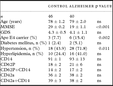
Table 1 shows the characteristics of 46 cognitively healthy controls and 40 age-matched AD patients. The Mini-Mental State Examination (MMSE) of the AD patients was significantly lower corresponding to a mild to moderate dementia stage. Neither controls nor AD patients suffered from depression. Regarding cardiovascular risk factors, AD patients showed a higher prevalence of hypertension. Further, the presence of the Apo E4 genotype was higher in AD patients compared to healthy controls.
Demographic data, somatic comorbidities, and results of monocyte–platelet interaction between healthy controls and AD patients

Whole EDTA blood of healthy controls or Alzheimer patients (n gives the number of patients) was analyzed by FACS for CD14, CD62P, and CD42a. Values are given as mean±SEM in % of the total cell population. Statistical analysis was performed by Students-T test (*** p < 0.001, ns not significant). MMSE, mini-mental state examination; GDS, geriatric depression scale.
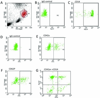
FACS analysis was used to analyze platelet–monocyte aggregates. Monocytes were identified using CD14-PercP antibodies versus a respective PercP-IgG control (Figure 1A–C).
Results of FACS analysis of platelet–monocyte aggregates. The cell populations in whole blood are shown, and the monocytes (M) are circled and differentiate from lymphocytes (L) and neutrophils (N) (A). Figure C shows specific labelling of monoyctes with CD14, while background is defined with an IgG control (B). Platelets bound to monocytes were identified by CD42a (D) and CD62P (F) labelling. Again an IgG control revealed background staining (D). Figure G shows the co-staining of CD42a and CD14 (upper right quadrant) defining the Platelet-Monocyte-Aggregates. Note that single platelets (P) are very small at the very left/below (A).

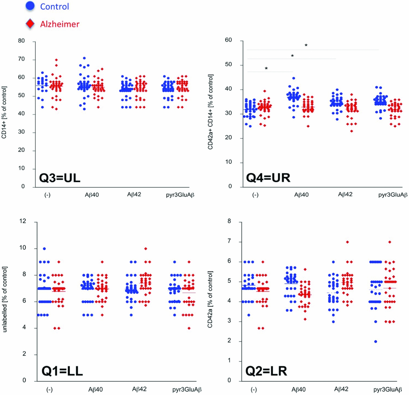
Platelets co-localized with monocytes were identified by CD42a-FITC (Figure 1E) and CD62-FITC (Figure 1F) versus a respective FITC-IgG control (Figure 1D). Figure 1G shows the co-localization of monocyte–platelets in the upper right quadrant. Levels of CD14, CD62P, and CD42a were not different between healthy controls and AD patients (Table 1). When whole blood of healthy controls was incubated with Aβ-40 or -42 or Pyr3 peptides all three slightly increased the monocyte–platelet aggregates (Figure 2). However, when blood of AD patients was incubated none of these three Aβ peptides had an effect on the monocyte–platelet aggregates.
Effects of beta-amyloid peptides on the platelet–monocyte-aggregates (CD42a-CD14) in whole blood. Whole EDTA blood (200 µl) from healthy controls (n=14–32) or AD patients (n=15–33) was incubated with or without 2 µl (1 mg/ml) beta-amyloid (Aβ) peptides for 1hr at 37°C and then analyzed by FACS as shown in Figure 1. The quadrant Q1 (lower left) represents unlabeled cells, Q2 (lower right) CD42a+, Q3 (upper left) CD14+ and Q4 (upper right) CD42a+/CD14+. Values are given as mean±SEM as percent of control. Statistical analysis was performed by Students-T test (* p < 0.05, ns not significant).

Discussion
In this study, we examined monocyte–platelet interaction in patients with AD compared to healthy controls in plasma before and after incubation with different beta-amyloid peptides. Monocytes and monocyte–platelet aggregates were identified by CD14, CD62P, and CD42a antibodies and analyzed by FACS analysis. Our results provide evidence that monocyte–platelet interaction in peripheral plasma of AD-patients differ from healthy controls.
The concentration of peripheral plasma monocyte was determined by CD14 antibodies between AD patients and age-matched healthy controls. We found neither difference for CD14 nor CD62P and CD42a in peripheral plasma of healthy controls compared to AD patients before beta-amyloid peptide incubation. Other studies on the role of CD14 in brain tissue have documented that CD14 deletion result in decreased plaque burden in transgenic murine model of AD (Reed-Geaghan et al., Reference Reed-Geaghan, Reed, Cramer and Landreth2010). Our results suggest that there might be critical differences in immune cells and their response to beta-amyloid peptide on the brain and peripheral side of the blood–brain barrier. In this respect it has to be noted that prior analyzes of Aβ40 and Aβ42 plasma levels in AD patients compared to healthy controls found no group differences (Roher et al., Reference Roher2009).
Furthermore, we found that solely healthy controls but not AD patients show an increase of monocyte–platelet aggregates followed by beta-amyloid peptide incubation. These results are somewhat surprising, since there is a well-known inflammatory component mediated by microglial response to fibrillary forms of β-amyloid in the AD-brain (Lucin and Wyss-Coray, Reference Lucin and Wyss-Coray2009). In the CNS, Aβ can elicit an aggressive neurotoxic response by binding to Toll-like receptor (TLR) 4 co-receptor CD14 (Liu et al., Reference Liu2005) and triggering microglia to secrete numerous different neurotoxic factors. One the other hand, our findings may support the previously reported impaired peripheral clearance of β-amyloid by autophagy of activated peripheral immune cells, such as macrophages (Pickford et al., Reference Pickford2008). Earlier work suggests, that on the one hand the peripheral system is potent in clearing brain Aβ and preventing AD pathogenesis (Xiang et al., Reference Xiang2015), on the other hand failure in the clearance of Aβ peptides may therefore result in inflammation and neurotoxicity (Giri et al., Reference Giri2000). In healthy individuals, there is a steady-state level of cerebral Aβ production and clearance via microglia phagocytosis and proteolytic degradation by enzymes (Wang et al., Reference Wang, Zhou and Zhou2006). Our results may indicate an impaired immune response to Aβ peptides and consequently their clearance in peripheral blood in AD.
Further, our results of enhanced monocyte–platelet aggregation measured by CD62P and CD42a values before and after incubation with beta-amyloid peptides are somewhat in line with prior findings. Outside the brain, platelets are a source of β-amyloid and β-amyloid precursor protein (Fernandez et al., Reference Fernandez1995). However, previous work showed that the vast majority of platelets are less activated in AD. Järemo et al. (Reference Järemo, Milovanovic, Buller, Nilsson and Winblad2013) found, that platelet reactivity estimated from membrane-attached P-selectin was lower in AD patients compared to healthy controls after provocation with thrombin receptor activation peptide. In contrast, they found that membrane-attached P-selectin without agonist stimulation was not different between the AD and control groups described as an apparently paradoxical P-selectin reaction. Further, they found that soluble P-selectin was higher in the AD patients but membrane-attached P-selectin without agonist stimulation was not different between the disease and control groups, which was linked to lower platelet reactivity. Together with our results these findings encourage further research on the P-selectin paradox and its relevance as a biomarker for AD.
To sum up our findings may underline the earlier reported interplay of circulating monocytes, with can infiltrate the brain and act as amyloid phagozytes as described in murine models of AD (Reed-Geaghan et al., Reference Reed-Geaghan, Reed, Cramer and Landreth2010). Especially, the better understanding of changes in monocyte–platelet interaction after beta-amyloid incubation in the periphery could provide an easy applicable biomarker in clinical routine. Besides diagnostic applications, interventions targeting the peripheral monocyte–platelet interaction with beta-amyloid peptides may open additional therapeutic approaches in AD.
Our study has certain limitations most notably the small sample size of the sample. This pilot study should therefore be followed by future analysis on larger samples.
Further, the analysis of peripheral blood may not correspond to the situation in cerebrospinal fluid (CSF) or with the other side of the blood–brain barrier. Even though, a lumbar puncture is difficult and stressful for patients, our results underpin the possible relevance of the monocyte–platelet interaction before and after incubation with different beta-amyloid peptides and therefore should be studies additionally in CSF of AD patients.
Conflict of interest
None
Description of authors’ roles
Ch. Humpel designed the study, carrying out the statistical analysis, supervised the data collection, and assisted with writing the paper. M. Defrancesco supervised the data collection and wrote the paper. J. Marksteiner assisted with designing the study and writing the paper.
Acknowledgment
We thank Monika Greil for blood processing and FACS analysis. This study has been supported by the Austrian Nationalbank (Nr 15887).