It might seem that the answer to the question posed in our title is remarkably simple – namely, any chamber identified within the ventricular mass. Had this been the answer accepted by paediatric cardiologists and morphologists during the development of the speciality, then we would have been spared the several polemics of the latter half of the last century concerning the nature of the univentricular heart or single ventricle.Reference Van Praagh, Ongley and Swan1–Reference Van Praagh, Plett and Van Praagh4 In the light of these controversies, some of them continuing today, the question is not so much “what is a ventricle”, but rather “what is the morphologic nature of the second chamber found in patients with double inlet left ventricle”? By answering this question, we can also provide an answer to the more general question. In the process, we also provide convincing morphological and developmental evidence that the small chamber in question is truly an incomplete right ventricle.Reference Anderson and Ho5, Reference Jacobs and Anderson6
The anatomic boundaries of the ventricular mass
If we are to provide an accurate definition for chambers within the ventricular mass, we must first offer sound anatomic definitions for the extent of the ventricular mass itself. This is because some accounts of ventricular structure start with the premise that it is possible to recognise so-called “segments” within the congenitally malformed heart over and above the generally accepted atrial, ventricular, and arterial components. These additional segments are described as the atrioventricular canal and the conus.Reference Van Praagh7 There is no question but that a discrete atrioventricular canal can clearly be defined in the developing heart, as can a discrete muscular outflow tract.Reference Sizarov, Ya, de Boer, Lamers, Christoffels and Moorman8 The myocardium of the atrioventricular canal, however, consists of non-working myocardium, or primary myocardium,Reference Moorman and Christoffels9 which eventually contributes to the entire left ventricular free wall, the atrial vestibular myocardium, as well the atrioventricular nodal myocardium, the latter persisting as the compact atrioventricular node and the right and left atrioventricular nodal rings.Reference Aanhanen, Mommersteeg and Norden10 The myocardium of the outflow tract subsequently differentiates into, and becomes incorporated within, the right ventricular mass, persisting for the larger part as the infundibulum of the morphologically right ventricle.Reference Anderson, Webb, Brown, Lamers and Moorman11 During the process of development, as we will describe, it is also possible to distinguish between the primary myocardium of the linear cardiac tube and the ballooning components that eventually become the apical parts of the ventricular chambers. All this knowledge is important as we establish the mechanisms of cardiac development. Such knowledge is now achieving increasing importance in validating the presumed morphogenesis of cardiac malformations, as had been predicted very many years ago.Reference Abbott12 It is inappropriate, however, to use terms specifically derived from the developing heart when describing the postnatal organ, be it normal or congenitally malformed.
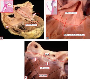
In the postnatal heart, it is an easy matter to recognise the boundaries of the ventricular mass, as obvious anatomical landmarks delimit the borders at its inlet and outlet. The atrioventricular junctions form the boundaries at the inlet. These fibro-fatty tissue planes provide the insulation ensuring that, at all points apart from the penetration of the specialised atrioventricular conduction axis, there is an anatomic separation between the atrial and ventricular myocardial masses (Fig 1a). The hinge lines of the leaflets of the atrioventricular valves co-localise with the atrioventricular junctions. The margin of the ventricular muscle mass at its outlet is also a discrete anatomic entity, being formed by the boundary between the muscular walls of the ventricles and the non-myocardial walls of the arterial trunks. These anatomic ventriculo-arterial junctions (Fig 1b and c) are crossed by the hinge lines of the arterial valves, but remain recognisable as discrete anatomical boundaries, particularly in the right ventricle, where all three leaflets of the pulmonary valve are supported by infundibular muscle (Fig 1b). In the left ventricle, the muscular walls support only two of the leaflets of the aortic valve. This is because it is the fibrous continuity almost always found in the normal heart between the leaflets of the aortic and mitral valves that supports the larger part of the non-coronary aortic valvar leaflet (Fig 1c).
The images show the boundaries of the ventricular mass. Panel (a) is a four-chamber section across the atrioventricular junctions, with the arrows showing the plane of insulation between the atrial and ventricular chambers. The inset shows how, at some places in the left atrioventricular junction, the hinge of the mitral valve coincides with the fibrous plane of insulation. Panels (b) and (c) show the opened orifices of the pulmonary (b) and aortic (c) valves, respectively, having removed the valvar leaflets. The inset in panel (b) shows how, at the nadir of attachment of the leaflets, the hinge is well below the anatomic ventriculo-arterial junction, which is marked by the dashed line, and the arrow in the inset. As seen in panel (c), in the aortic valve only two of the valvar leaflets are supported by ventricular musculature (arrows).

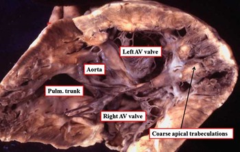
Within the ventricular mass as thus described, it is the rule to find two discrete anatomic chambers, even when the hearts themselves are congenitally malformed. Only very rarely are hearts encountered with a solitary chamber making up the ventricular mass, and such solitary chambers possess exceedingly coarse apical trabeculations (Fig 2). This chamber, of necessity, is the ventricle. In hearts with two anatomic chambers, the question remains as to how best to describe them when one is malformed and incomplete. In the past, one of us was party to creating hugely complex definitions so as to justify describing those hearts with one big left ventricle in the presence of a smaller chamber as “univentricular hearts”.Reference Anderson, Tynan and Freedom13 The deficiency of this approach became apparent when it was pointed out that anatomic structures are best defined on the basis of their own intrinsic morphology, using the most constant components of the structure in question for arbitration, rather than relying on other features that themselves might be variable.Reference van Praagh, David, Wright and van Praagh14 If this principle, dubbed the morphological method, is to be used in the context of the ventricular mass, it then becomes essential to determine how best to describe the individual components to be found within each of the two normal ventricles, because only in this manner will we be able to analyse those ventricles that are incomplete.
This is a solitary ventricle of indeterminate morphology. It receives both atrioventricular (AV) valves, and gives rise to the pulmonary (Pulm.) trunk and the aorta. Note the particularly coarse apical trabeculations. Despite taking sections antero-superiorly and postero-inferiorly through the ventricular walls, it was not possible to find a remnant of a second ventricular chamber.

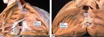
Ventricular subdivision, however, has itself previously been a bone of contention. It had been popular, and remains popular for some,Reference Van Praagh7 to divide the ventricles into two parts, namely the sinus and the conus. Examination of the normal ventricles, however, provides no obvious landmarks to support this simplistic division (Fig 3). Indeed, it had been description of abnormal ventricles on such a bipartite approach that had produced most of the problems surrounding so-called “single ventricles”, as the small chamber seen in the setting of double inlet left ventricle was deemed to lack its “sinus”, and hence be no more than the right ventricular conus.Reference Van Praagh, Wise, Dahl and Van Praagh15 Analysis of normal ventricles in a tripartite manner, as suggested by Goor and Lillehei,Reference Goor and Lillehei16 resolves all these problems, as the three functional components of both normal ventricles are readily identified (Fig 3). The inlet components extend from the atrioventricular junctions to the distal attachments of the tension apparatus of the atrioventricular valves. The outlet components support the leaflets of the arterial valves, being abbreviated in the left ventricle because of the fibrous continuity between the atrioventricular and arterial valves (Fig 1c). The third ventricular component is then the most constant when either ventricle is congenitally malformed. Owing to the nature of its trabeculations, this third component also serves as the best discriminator between the morphologically right and left ventricles, as in the postnatal situation the trabeculations are coarse in the morphologically right ventricle, but fine and criss-crossing in the morphologically left ventricle (compare Fig 3a and b). As we will show, when using this tripartite approach for description, it becomes an easy matter to describe abnormal ventricles on the basis of their size, and the manner in which the inlet and outlet components are shared, or not shared, between the two apical trabecular components.Reference Anderson and Ho5, Reference Jacobs and Anderson6
The images show the opened morphologically right (a) and left (b) ventricles. There are no obvious landmarks that permit distinction of a so-called sinus and conus within these normal ventricles. In contrast, it is an easy matter to recognise the ventricular inlet, outlet, and apical trabecular components.

Normal development of the ventricular mass
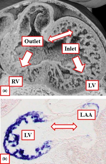
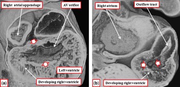
Before proceeding to describe the anatomic nature of incomplete ventricles as seen in congenitally malformed hearts, it is helpful to consider the way the right and left ventricles normally develop, because this information provides valuable evidence to support our hypothesis that the apical trabecular components provide the basis for descriptions of abnormal chambers. Over the past two decades, the old notion that the linear cardiac tube contains all the so-called cardiac segments has been unequivocally disproved. We now know that, during the process of cardiac development, new cardiomyocytes are gradually added to the forming cardiac plate at both its arterial and venous poles. When rediscovered, this gradual addition was proposed to be derived from a so-called “second cardiac field”, although consensus is beginning to accrue to show that the new material is added consecutively from a single cardiac field.Reference Moorman, Christoffels, Anderson and van den Hoff17 We also know that the myocardium of the initial cardiac tube, even subsequent to the process of looping, has particular molecular characteristics that enable it to be recognised as primary myocardium. Subsequent to looping of the tube, the components of the definitive chambers that permit their anatomic distinction develop by a process of ballooning from the cavity of the tube.Reference Moorman and Christoffels9 The atrial appendages balloon in parallel from the atrial component of the primary tube. The apical ventricular components, in contrast, balloon in series from the inlet and outlet components of the ventricular loop, respectively (Fig 4a). Concomitant with initiation of the process of ballooning, it becomes possible to distinguish the cardiomyocytes of the ballooning components from those making up the primary cardiac tube. The cardiomyocytes forming the walls of the primary tube express neither Connexin40 nor atrial natriuretic peptide. Both these gene products, in contrast, are expressed in the cardiomyocytes forming the walls of the atrial appendages and the developing ventricular apical components (Fig 4b). This permits description of the apical ventricular myocardium, and the myocardium of the appendages, as chamber myocardium. These considerations help our understanding of the morphology of the abnormal ventricles seen in congenitally malformed hearts. This is because, when the apical trabecular components are first formed, the circumference of the atrioventricular canal, readily recognised as a discrete component of the developing heart, is supported exclusively by the walls of the developing left ventricle, accepting that the proximal parts of these walls adjacent to the atrioventricular canal were initially primary rather than specific chamber myocardium (Fig 5a). This point is of significance because, in the roof of the embryonic interventricular communication, the primary myocardium provides a direct link between the walls of the developing right atrium and right ventricle.
The images show (a) a four-chamber section across the ventricular loop in a human embryo at Carnegie stage 15, and (b) a long-axis section through the developing left atrium and ventricle from a mouse embryo at embryonic day 9.5. The section of the human embryo shows how the apical trabecular components of the developing right (RV) and left (LV) ventricles balloon from the inlet and outlet components of the loop, respectively (arrows). Note that, at this stage of development, the atrioventricular canal (double-headed arrow) opens almost exclusively to the developing LV. The section through the mouse heart has been stained to show atrial natriuretic factor, which is coloured blue. The protein is localised in the apical part of the LV, and the developing left atrial appendage (LAA), but absent from the atrioventricular canal (double-headed arrow), which is a remnant at this stage of the primary myocardium of the initial linear cardiac tube.

The images are from a human embryo at Carnegie stage 14. Panel (a) is a frontal section through the developing ventricles just distal to the atrioventricular (AV) canal. It shows the AV orifice opening to the developing left ventricle, with the interventricular communication (between arrows) providing the entrance to the developing right ventricle. A sagittal section (b) from the same embryo shows how the developing right ventricle at this stage possesses an apical component, and supports the ventricular outflow tract, but has no direct communication with the right atrium other than via the interventricular foramen (between arrow heads).

When viewed from its right ventricular aspect, the developing chamber can be seen to possess an apical component, which supports the entire circumference of the developing outflow tract (Fig 5b). We also know that, at this stage, an antibody to the nodose ganglion fortuitously marks the entire circumference of the interventricular communication, initially no more than a ring within the primary cardiac tube.Reference Lamers, Wessels and Verbeek18 By tracking the location of the cardiomyocytes marked by this antibody, we have previously inferred that the entirety of the definitive right ventricle is derived from the myocardium found distal to the interventricular ring.Reference Lamers, Wessels and Verbeek18 Equally importantly, we showed that the muscular ventricular septum was formed concomitant with the apical growth of the trabecular components, with the atrioventricular bundle forming on the crest of the septum.Reference Lamers, Wessels and Verbeek18 The ventricular conduction pathways are now known to be derived from the dense network of trabeculations that initially pack both the developing apical components (Fig 4a). These trabeculations lose their prominence with maturation of the heart, failing to proliferate at the same rate as the cardiomyocytes in the compact parts of the ventricular walls. It is also the case that, in early stages of development, there is little obvious difference in the coarseness of the trabeculations in the developing ventricles. It is these initial trabeculations that eventually persist in part as the ventricular bundle branches, which are carried on either face of the developing muscular ventricular septum. These developmental facts, therefore, provide strong evidence, first, that the morphologically right ventricle, when initially recognised in the developing heart, possesses only apical trabecular and outlet components. Second, that any septal structure carrying the atrioventricular bundle on its crest, with bundle branches on either side, is the primordium of the definitive muscular ventricular septum.
Complete versus incomplete ventricles
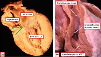
We have shown that, in the normal heart, each ventricle possesses an inlet, an outlet, and an apical trabecular component. We have also seen that, during normal development, the atrioventricular canal is initially supported exclusively by the primary myocardium from which, at its outer curve, will balloon the apical component of the left ventricle, whereas the outflow tract is supported exclusively by the primary myocardium of the outlet component, from which balloons the apical component of the morphologically right ventricle. During normal development, the primary myocardium of the linear cardiac tube undergoes significant remoulding such that the right atrium obtains direct access to the cavity of the right ventricle, while the aorta is re-oriented so as to arise from the cavity of the left ventricle. Failure of these processes of remodelling, or alternatively excessive remodelling of the atrioventricular canal, then provides a rational explanation for the structure of the chambers seen within the ventricular mass of congenitally malformed hearts. Thus, in hearts with classical tricuspid atresia, there is absence of the right atrioventricular connection (Fig 6a). This parallels the situation seen during early cardiac development, when the developing right ventricle has no direct connection with the cavity of the right atrium (Fig 5b). Moreover, as in early cardiac development, the second chamber seen in patients with tricuspid atresia has an apical component of right ventricular morphology (Fig 6a). In most instances, this right ventricle, incomplete compared with the normal heart in that it lacks its inlet component, gives rise to the pulmonary trunk, with the aorta having been transferred to the dominant left ventricle. In a proportion of cases, nonetheless, the right ventricle can give rise to the aorta when the ventriculo-arterial connections are discordant. In rarer circumstances, the right ventricle can give rise to both arterial trunks, then being more akin to the situation encountered during normal development, or even lack a direct arterial exit, when both trunks are transferred to the dominant left ventricle.
The images show the characteristic anatomy of the usual variant of tricuspid atresia. As shown by panel (a), a four-chamber section, there is absence of the right atrioventricular connection (green dotted lines), with the left atrium being the only chamber in communication with a ventricle, in this instance the dominant left ventricle (LV). As seen in panel (b), the anterior second chamber has an apical trabecular component of right ventricular (RV) morphology, and as is usually the case, supported the pulmonary (pulm.) trunk. Blood enters the incomplete RV through the interventricular communication, or ventricular septal defect.

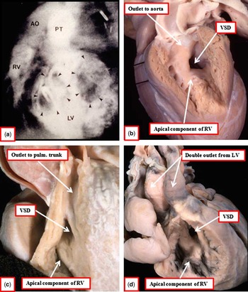
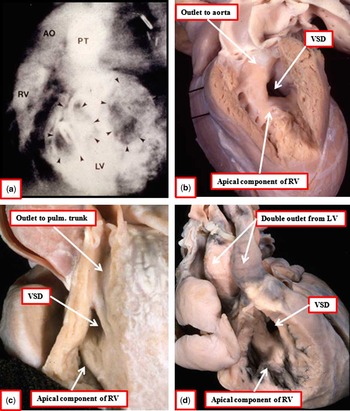
The essence of tricuspid atresia, therefore, is that the left ventricle is dominant, and the right ventricle incomplete, lacking its inlet component. This situation can then be compared with the arrangement of double inlet left ventricle, in which, as in the developing heart, the atrioventricular junctions open exclusively to the cavity of the left ventricle, with a second chamber, if present, supplied with blood through an interventricular communication, or ventricular septal defect (Fig 7a). As we have already discussed, this second chamber was initially considered to be no more than an infundibulum, and there are still some who interpret the chamber to represent an infundibular outlet chamber. When we examine the nature of this chamber, however, we see that it possesses an apical trabecular component with coarse trabeculations (Fig 7b). It is also well established that the atrioventricular conduction axis is carried on the crest of the septum, separating this apical part of the second chamber from the dominant left ventricle. Furthermore, although in most instances it is the aorta that arises from this second chamber in the setting of double inlet left ventricle, in a small proportion of cases the pulmonary trunk can arise from the second chamber. This arrangement is known as the Holmes heart. In this setting, the second chamber is indistinguishable from the incomplete right ventricle found in patients with tricuspid atresia and concordant ventriculo-arterial connections (compare Figs 6b and 7c). In even rarer circumstances, it is also possible, as with tricuspid atresia, to find patients with hearts in which both arterial trunks arise from the dominant left ventricle. If it were true that the second chamber was no more than an infundibulum, then such patients would exhibit solitary ventricles. In fact, the patients have well-formed second chambers with unequivocal right ventricular apical trabeculations (Fig 7d). All of this evidence, therefore, supports the notion that the second chamber seen in patients with double inlet left ventricle is the incomplete right ventricle.
The images show the salient features of the second chamber found in patients with double inlet left ventricle (LV). The angiogram shown in panel (a) is taken to profile the septum. Both atrioventricular orifices (arrowheads) enter the dominant LV. The blood enters the second chamber (right ventricle (RV)) through a ventricular septal defect (VSD). In this heart, as is usually the case, the aorta (AO) arises from the small chamber, and the pulmonary trunk (PT) from the dominant LV. Panel (b) shows a left-sided chamber from a patient with double inlet LV. Note that the chamber has an apical component with coarse trabeculations. In this patient, the AO arose from the small chamber, but when the PT arises from the small chamber, as seen in panel (c), the anatomy is indistinguishable from that seen in tricuspid atresia (compare with Fig 6b). Rarely, both arterial trunks arise from the dominant LV, as in the heart shown in panel (d). The small chamber still possesses apical trabeculations of right ventricular type, and is fed through a VSD.
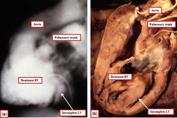
Analysis of malformed hearts on the basis of sharing the inlet and outlet components between the two apical components then provides equally compelling evidence to support the hypothesis that, in patients with dominance of the right ventricle, the second chamber seen within the ventricular mass is an incomplete left ventricle. If we analyse still further the patients with dominant left ventricles, we find that the incomplete right ventricle is always carried on the shoulders of the ventricular mass, either on the right side or the left side. This is analogous to the situation seen during development, when the developing right ventricle itself is carried antero-superiorly on the shoulder of the initially dominant left ventricle. In the patients with dominant right ventricles, in contrast, the incomplete left ventricle is always found postero-inferiorly, usually in the left quadrant of the ventricular mass, but sometimes in right-sided and inferior position. In addition, most usually, patients with double inlet right ventricle also have double outlet from the dominant ventricle, with the incomplete left ventricle exhibiting no more than its apical trabecular component (Fig 8). This morphologic arrangement is consistent with the notion that there was exaggerated displacement of the atrioventricular canal during cardiac development.
The images show (a) an angiogram from a patient with double inlet to a dominant right ventricle (RV) through a common atrioventricular valve, and double outlet from the dominant ventricle. The specimen shown in panel (b) is from a different patient, but has been sectioned to show the same anatomy. Note that the incomplete left ventricle (LV) is positioned postero-inferiorly.

What about the size of the chambers?
Almost without exception, incomplete ventricles, be they of right or left morphology, are also hypoplastic when they lack their inlet component. Ventricles can also be incomplete when lacking their outlet component, as, for example, with double outlet right ventricle, when the left ventricle is incomplete. Such incomplete ventricles lacking an outlet component, however, can still be of sufficient size to drive either the systemic or the pulmonary circulation. It is because of the small size of the ventricles lacking their inlet component, however, that the arrangements with either double inlet ventricle or absence of one atrioventricular connection produce the functionally univentricular arrangement, with the incomplete ventricle being of insufficient size to support either the pulmonary or the systemic circulations. On occasion, incomplete right ventricles can be incorporated as part of the so-called “one-and-a-half” ventricle surgical repair. It does not follow, however, that normally constituted ventricles are always of sufficient size to support biventricular circulatory patterns. Indeed, the small left ventricle seen in the setting of hypoplastic left heart syndrome usually possesses all three of its components, but is rarely, if ever, capable of supporting the systemic circulation. In the setting of pulmonary atresia with intact ventricular septum, the mural hypertrophy produced by this arrangement is often sufficient to squeeze out the cavity of the apical trabecular and outlet parts of the normally constituted right ventricle, again producing the substrate for a functionally univentricular arrangement.
What, then, is a ventricle?
On the basis of both developmental and morphological evidence, we would suggest that a ventricle is best defined as any chamber within the ventricular mass possessing an apical trabecular component. Such ventricles can be of right or left morphology, and always coexist. The ventricles are normally formed when possessing all three of the inlet, apical trabecular, and outlet components, but incomplete when lacking one or both of the inlet and outlet components. Ventricles that are incomplete because of lack of the inlet component are always hypoplastic, with incomplete right ventricles being positioned antero-superiorly within the ventricular mass, and incomplete left ventricles located postero-inferiorly. Patients having such incomplete ventricles because of the lack of the inlet component have functionally univentricular hearts, although the functionally univentricular arrangement can also be produced in the setting of normally constituted but hypertrophied ventricles. Full analysis of ventricular morphology, therefore, requires attention not only to component make-up, but also size.