Introduction
Microphorella Becker is a little known genus of parathalassiine flies that is classified in an expanded concept of Dolichopodidae (Sinclair and Cumming Reference Sinclair and Cumming2006). The genus currently includes 16 described species recorded from the Nearctic, Palearctic, Oriental, and Australasian Regions (Yang et al. Reference Yang, Zhang, Yao and Zhang2007). Like other parathalassiines, they are small (1–2 mm) nonmetallic greyish to dark coloured flies that are generally found along sea coasts and river banks. Parathalassiines are distinguished from other dolichopodids by the following combination of features: antenna with single articled arista-like stylus, wing with cell dm usually present and emitting three veins (i.e., M1, M2, M4), crossvein bm-m nearly complete but not usually joining M1, and male terminalia with the left epandrial lamella bearing a ventral process. Seven extant genera of Parathalassiinae are currently recognized, with Microphorella belonging to a group of four genera that also includes Chimerothalassius Shamshev and Grootaert, Eothalassius Shamshev and Grootaert, and Thalassophorus Saigusa (see Saigusa Reference Saigusa1986; Shamshev and Grootaert Reference Shamshev and Grootaert2002, Reference Shamshev and Grootaert2004, Reference Shamshev and Grootaert2005; Brooks and Cumming Reference Brooks and Cumming2011).
We are actively studying generic limits within the Microphorella genus group and potential paraphyly of some of the genera (Cumming and Brooks Reference Cumming and Brooks2006; Brooks and Cumming Reference Brooks and Cumming2010). Although very weakly defined, we recognize Microphorella on the basis of a wing with a reduced anal lobe and apically convex cua (anal) cell, a very short to absent anal (CuP + CuA) vein, a long R1 and cell dm present, and a head with a weakly developed gena, small short rounded or narrow palpi, and arista-like stylus not lengthened.
Here we describe two new species from western North America that belong to a distinctive species group, which we assign to Microphorella. Specimens of this species group were first collected in 1960 near Terrace, British Columbia by Jim Chillcott, who initiated the Manual of Nearctic Diptera project. Subsequently more specimens from Yukon Territory, Alberta, and Washington were located in the Canadian National Collection of Insects and the United States National Museum of Natural History. In 2008 we collected additional specimens in the Terrace area to complete our study.
Material and methods
Specimens examined in this study are deposited in the Canadian National Collection of Insects, Ottawa, Canada (CNC) and the United States National Museum of Natural History, Washington, District of Columbia, United States of America (USNM).
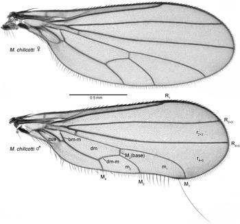
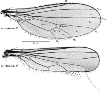
Terms used for adult structures primarily follow McAlpine (Reference McAlpine1981) and Cumming and Wood (Reference Cumming and Wood2009), except for the antenna and wing venation where Stuckenberg (Reference Stuckenberg1999) and Saigusa (Reference Saigusa2006) are followed, respectively. In the system outlined by Saigusa (Reference Saigusa2006), the dipteran wing vein A1 (as used in McAlpine Reference McAlpine1981) is homologized with the mecopteran CuP, and consequently CuA1 (of McAlpine) is termed M4, whereas CuA2 is CuA, the anal cell is cell cua, and the anal vein (A1 + CuA2) is CuP + CuA. The wing vein homologies as they relate to Microphorella are shown in Figures 1 and 2. Homologies of the male terminalia follow Sinclair and Cumming (Reference Sinclair and Cumming2006). Macrotrichia are referred to as bristles, setae, setulae, or hairs depending on relative decreasing size.
Male and female terminalia were macerated in 85% lactic acid heated in a microwave oven. Figures of the male genitalia are oriented with the anatomically dorsal and ventral parts directed towards the top and bottom of the page, respectively, following Sinclair and Cumming (Reference Sinclair and Cumming2006: figs. 347–350). In lateral views of the male genitalia, hatched areas delimit aspects of the medial surface of the opposing side that project beyond the limits of the facing side.
Systematics
Microphorella chillcotti species group
Diagnosis
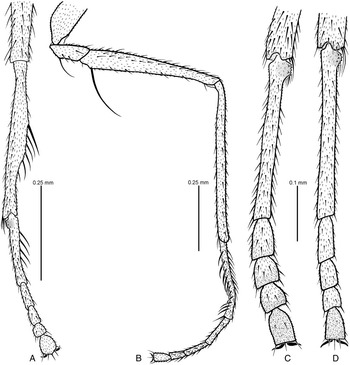
The Microphorella chillcotti species group can be distinguished from other parathalassiines by the following characters: male wing (Figs. 1♂, 2♂, 3) narrowed with costal section below cell m1 bearing fringe of thickened elongate setae and one elongate bristle arising just anterior to M1, cell r4 + 5 expanded apically, cells m1 and m2 compressed; wing of both sexes (Figs. 1, 2) with R1 long extending beyond middle of wing, crossvein bm-m incomplete and anal vein (CuP + CuA) absent; male foreleg (Fig. 4A) with tibia swollen bearing one strong posteroventral bristle and posterior comb-like row of curved setae, with basitarsus bearing posterodorsal comb-like row of setae; male midleg (Fig. 4B) with trochanter and femur each bearing strong basiventral bristle, and with basitarsus bowed and compressed bearing comb-like row of curved setae on each side; foreleg and hindleg of both sexes with spinose tubercle at base of basitarsus (Figs. 4A, 4C, 4D); hypopygium (Figs. 5, 8) with basally articulated ventral epandrial process, ventral surstylar lobe bearing large spatulate prensiseta, and with elongate medial process arising from posterior end of hypandrium; female abdomen with apical segments retracted into segment 6, tergite 10 bearing acanthophorite spine-like setae, and cercus heavily sclerotized narrow and pointed (Fig. 9).
Description
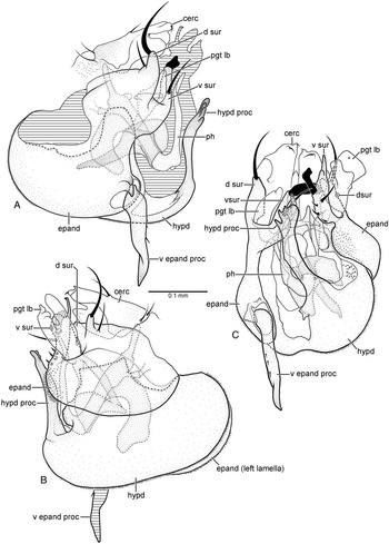
Male:Head: Slightly broader than thorax in dorsal view; ovoid in lateral view; about 1.3× broader than high in anterior view. Neck inserted high on head. Ocellar triangle conspicuous. Occiput concave on upper median part. Eyes dichoptic entirely covered with uniform ommatrichia, medial edge with weak emargination near antenna, ommatidia of uniform size. Frons broadly widening above. Face narrowing towards clypeus, about as broad as distance between inner margins of posterior ocelli at margin with clypeus. Clypeus not separated from face, slightly higher than wide, slightly widening below, apical margin truncate, weakly produced. Bristles of head well differentiated: pair of inclinate fronto-orbitals well separated from base of antennae, pair of lateroclinate anterior ocellars, pair of small proclinate posterior ocellars, pair of inclinate postocellars, pair of lateroclinate inner verticals, pair of lateroclinate outer verticals, single row of short postocular setae; postgena with row of short setae along edge of mouth opening. Antenna inserted above middle of head in profile; scape short, funnel shaped; pedicel longer than scape, spheroidal, with subapical circlet of setulae; postpedicel about 2× longer than wide, drop shaped, clothed in moderately long fine hairs; stylus about 1.8× longer than postpedicel with very short hairs. Palpus narrow, about 3× longer than wide, clothed with minute pile, with one to two lateral setulae and one long preapical seta. Proboscis short, projecting ventrally or slightly anteroventrally; epipharyngeal carina indistinct; epipharyngeal blades narrow; labellum with six geminately sclerotized pseudotracheae. Gena narrow. Thorax: Mesoscutum moderately arched, prescutellar depression usually apparent. Prosternum fused with proepisternum forming precoxal bridge. Antepronotum narrow with two setulae per side. Postpronotal lobe distinct with two to three small setulae. Mesonotum shield shaped in dorsal view, slightly longer than wide, bristles well differentiated. Acrostichal setae absent; five dorsocentral bristles with weak anterior accessory setula, one presutural supra-alar bristle, zero to two postsutural supra-alar bristles, two notopleural bristles, and one postalar bristle per side; scutellum broadly subtriangular with one pair erect inclinate bristles near apex. Mesopleuron bare. Legs:Foreleg (Fig. 4A): Coxa with fine setae on anterior surface and stronger setae along apical margin; femur subequal in length to tibia, devoid of well-developed bristles; tibia swollen in middle portion bearing one strong posteroventral bristle at basal third and comb-like posterior row of about four curved setae; tarsus slightly shorter than tibia; basitarsus nearly as long as tarsomeres 2–5 combined, slightly compressed near middle, with spinose anterior tubercle at base and comb-like row of about four posterodorsal setae; tarsomeres 2–4 decreasing in length apically; tarsomere 5 slightly widened, subequal in length to tarsomere 2. Midleg (Fig. 4B): Coxa with a few weak anterior setae along apical margin and one fine hair-like seta laterally near middle; trochanter with strong basiventral bristle; femur shorter than tibia, with one long basiventral bristle nearly half femur length; tibia devoid of well-developed bristles; tarsus subequal in length to tibia; basitarsus as long as tarsomeres 2–5 combined, bowed and compressed with ventral surface flat, pale and nude, anteroventral and posteroventral margins each with comb-like row of curved setae, one strong apicoventral seta; tarsomeres 2–4 decreasing in length apically; tarsomere 5 subequal in length to tarsomere 4. Hindleg: Coxa with two setae on outer surface; femur subequal in length to tibia, devoid of well-developed bristles; tibia devoid of well-developed setae; tarsus slightly shorter than tibia; basitarsus slightly longer than tarsomeres 2–5 combined, with spinose posteroventral tubercle at base; tarsomeres 2–4 decreasing in length apically; tarsomere 5 distinctly longer than tarsomere 4 (Figs. 4C, 4D). Wing (Figs. 1♂, 2♂, 3): Narrowed compared to female. Pterostigma absent, membrane entirely covered with minute microtrichia, anal lobe weakly developed, alula absent. Costa circumambient, with a series of three well-developed setae at extreme anterior base. Anterior section bearing two rows of short spine-like setae from Sc to R2 + 3, setae finer and longer from R2 + 3 to M1. Posterior section of costa weakly sinuous between M4 and M1, base and anal section with fringe of rather elongate setae; costal section below cell m1 with fringe of about 10 thickened elongate setae, increasing in length distad, and one long bristle arising just anterior to M1, bristle about as long as wing height. All longitudinal veins complete, reaching wing margin, Sc faint apically. R1 reaching costa well beyond middle of wing. Cell r4 + 5 expanded apically with M1 curving posteriorly in apical portion and strongly diverging from R4 + 5. Cells m1 and m2 compressed, cell m2 wider than high. M2 and M4 short beyond dm-m crossvein. Base of Rs originating opposite humeral crossvein. Short r-m crossvein present in basal fifth of wing. Crossvein bm-m incomplete. Cell dm present, extending to middle of wing. Cells br, bm and cua in basal fourth of wing. Cell br slender, longer than bm and cua. Cells bm and cua subequal in length and width, both slightly broader than br. Cell cua closed with distal end rounded. Anal vein (CuP + CuA) absent. Calypter with fine setae. Abdomen: Abdominal muscle plaques present. Tergites 1–6 with short posteromarginal setae; sternites 2–6 with scattered setulae; tergite 7 with two setulae posterolaterally, otherwise bare; sternite 7 bare. Segments 1–4 symmetrical with simple tergites and sternites; at least segments 6–7 narrowed and laterally compressed to form cavity on right side for hypopygium. Sternites 6–7 simple, not contorted, lacking pregenitalic processes. Sternite 8 quadrate, with several setae posteriorly, tergite 8 atrophied. Hypopygium (Figs. 5, 8): Lateroflexed to right; inverted with posterior end directed anteriorly; small and compact, less than half length of abdomen; asymmetrical; foramen unformed. Epandrium divided into left and right lamellae. Left epandrial lamella largely overlapping left side of hypandrium, hourglass shaped in lateral view (Figs. 5A, 8A), ventral portion broadly rounded and fused with hypandrium but epandrial margin distinct; ventral epandrial process present (Figs. 5A, 5C, 8A, 8E), articulated at base, bearing three short setulae, with pointed apex. Left surstylus (Figs. 5A, 5C, 8A, 8E) bilobed, dorsal and ventral lobes separated by deep U-shaped cleft through which left postgonite lobe protrudes. Dorsal lobe of left surstylus with apical tubercle bearing large sickle-like bristle and one seta near base of tubercle. Ventral lobe of left surstylus digitiform with one long seta near mid-length and large spatulate apical seta (prensiseta). Right epandrial lamella (Figs. 5B, 8B) ovoid in lateral view, smaller than left lamella, not fused with hypandrium, lacking ventral process. Right surstylus (Figs. 5B, 5C, 8B, 8E) bilobed, dorsal and ventral lobes separated by deep U-shaped cleft through which right postgonite lobe protrudes. Dorsal lobe of right surstylus with preapical tubercle bearing large sickle-like bristle and one seta near base of tubercle. Ventral lobe of right surstylus flap-like with three short basal setae, one long seta, and large curved spatulate seta (prensiseta) along middle portion, apex with narrow finger-like process. Hypandrium reniform in lateral view (Figs. 5B, 8B), with elongate medial hypandrial process arising from posterior end (Figs. 5A, 5C, 8A, 8C, 8D, 8E). Postgonite with basal internal portion cradling base of phallus and ejaculatory apodeme, V-shaped in dorsal view with lateral apodeme-like processes flanking ejaculatory apodeme; left and right postgonite lobes protruding out from between dorsal and ventral lobes of surstylus. Left postgonite lobe short, globular (Figs. 5A, 8A). Right postgonite lobe large, with expanded bilobate apex (Figs. 5B, 8B). Phallus tubular, bent upwards (Figs. 5A, 8A). Ejaculatory apodeme long, curved downwards, keel-like, laterally flattened. Cercus short (Figs. 5C, 8E).
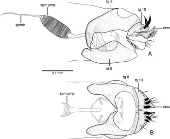
Female: Similar to male except face slightly broader; palpus with short preapical seta; foreleg with tibia unmodified, lacking posteroventral swelling in basal half and devoid of well-developed bristles; fore basitarsus without row of posterodorsal setae, spinose anterior tubercle at base slightly less produced; midleg with trochanter and femur each lacking strong basiventral bristle; mid basitarsus unmodified, not bowed and without comb-like rows of setae; hind basitarsus with spinose anterior tubercle at base slightly less produced; wing broader with M1 straighter and cell m2 longer, posterior margin slightly convex without lengthened costal setae and elongate costal bristle (Figs. 1♀, 2♀); apical abdominal segments retracted into segment 5 (segments 6–8 mainly glabrous); terminalia (Fig. 9) with tergite 8 medially divided, narrowly fused with sternite 8 anterolaterally; tergite 10 medially divided with three acanthophorite spine-like setae on each side; cercus heavily sclerotized, narrow, pointed; spermatheca unsclerotized unpigmented tube with sperm pump at base.
Remarks
The Microphorella chillcotti species group is a distinctive lineage that is easily separated from other species of Microphorella. The two new species described here are based on relatively few specimens collected from a small number of localities; further collecting in western North America and perhaps eastern Asia may uncover additional new species that belong to this lineage. In addition, continued phylogenetic analysis of the Microphorella genus group and the remainder of Parathalassiinae may warrant a higher taxonomic ranking (i.e., genus or subgenus) for this species group in the future.
Microphorella chillcotti sp. nov.
(Figs. 1, 4A, 4B, 4C, 5, 6, 7)
Microphorella chillcotti sp. nov., wings of female and male. Abbreviations: bm-m, basal medial crossvein; cua, anterior cubital (=anal) cell; dm, discal medial cell; dm-m, discal medial crossvein; M1, 1st medial vein; M2, 2nd medial vein; M4, 4th medial vein; m1, 1st medial cell; m2, 2nd medial cell; R1, 1st radial vein; R2 + 3, 2nd + 3rd radial vein; r2 + 3, 2nd + 3rd radial cell; R4 + 5, 4th + 5th radial vein; r4 + 5, 4th + 5th radial cell.

Type material
Holotype ♂ labelled: “CANADA: BC: Exchamsiks/ River, 58 km SW of Terrace/ 54°20.00′N, 129°17.81′W/ 17–18.VIII.2008, MT/ S.E. Brooks & J.M. Cumming”; “HOLOTYPE/ Microphorella chillcotti/ Brooks & Cumming” [red label] (CNC). Paratypes:CANADA:British Columbia: 2♀, same data as holotype (CNC); 1♀, same data as holotype, except 15–17.VIII.2008 (CNC); 2♂, 1♀, same data as holotype except, 18.VIII.2008, S.E. Brooks (CNC); 1♂, 32 miles SW of Terrace, 9.VII.1960, J.G. Chillcott, cutover swampland (CNC); 1♀, same data except, 13.VII.1960, open scrubby cutover area (CNC); 1♂, Buckinghorse Provincial Campground, Alaska Highway DC-175 (281.6 km), 27.VI.1978, Paul H. Arnaud Jr. (USNM). Alberta: 1♂, mile 121/122 Kananaskis Road, 14.VII.1973, J.A. Downes, J.A.D. 909/4/122 (CNC); UNITED STATES OF AMERICA: Washington: 5♂♂, 8♀♀, Olympic Nat. Park, Hoh River Rain Forest, 7.VII.1968, W.W. Wirth (USNM).
Diagnosis
Males of Microphorella chillcotti are most easily distinguished from those of M. vockerothi by the wing which is distinctly expanded apically as opposed to being strap-like, the hind tarsus with tarsomeres 2–5 widened, and the hypopygium with a straight ventral process of the left epandrial lamella, a hypandrial process with an expanded toothed apex, and cerci that are broad apically. Females of M. chillcotti appear to be indistinguishable from those of M. vockerothi.
Description
Male: Body length 1.2–1.5 mm, wing length 1.7–1.9 mm. Dark brown in ground colour with blue-green and rusty brown pruinosity, legs slightly paler. Head: Largely dark, dull blue-green pruinose, with dark brown ground colour; head slightly broader than thorax in dorsal view; setae black. Frons dull rusty-brown pruinose. Face dark brown with dull green-blue pruinosity. Clypeus concolorous with face. Antenna entirely dark brown. Palpus dark brown. Thorax: Dark brown ground colour with blue-green and rusty brown tinged pruinosity. Scutum densely pruinose, blue-green with prominent rusty area between dorsocentral bristles and dorsal margin of notopleuron. Mesonotum usually with one postsutural supra-alar bristle (sometimes lacking). Halter with light brown stalk, knob pale brownish-white. Legs (Figs. 4A–C): Dark brown, paler at knees, tarsi lighter on ventral surface; femora, tibiae, and tarsi mostly covered with fine hairs. Hindleg with tarsomeres 2–5 distinctly widened (Fig. 4C). Wing (Fig. 1♂): Hyaline, about 2.6× longer than wide, widening towards apex, dorsal and ventral margins divergent, apex expanded and semicircular, veins dark brown; R2 + 3 and R4 + 5 parallel in distal half; cell r4 + 5 expanded apically with M1 curved posteriorly near apex and strongly diverging from R4 + 5; calypteral setae brown. Abdomen: Dark brown ground colour with blue-green tinged pruinosity. Hypopygium (Fig. 5) dark brown with weak blue-green tinge, projecting apical components paler; epandrium with left epandrial lamella hourglass-shaped in lateral view with middle constriction pronounced (Fig. 5A); ventral epandrial process straight (Figs. 5A, 5C); dorsal lobe of left and right surstylus with moderately long seta near base of tubercle (Figs. 5A, 5B); hypandrium reniform; hypandrial process with expanded apex bearing teeth (Figs. 5A, 5C); phallus U-shaped in lateral view (Fig. 5A); cercus with broad apex (Fig. 5C).
Female: Body length 1.2–1.3 mm, wing length 1.7–1.9 mm. Similar to male except as noted under Microphorella chillcotti species group description.
Distribution
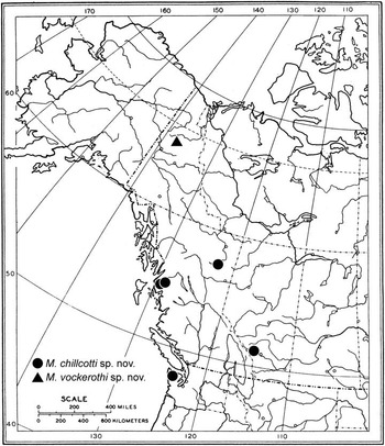
This new species is known from northern British Columbia south to the Olympic Peninsula, Washington and east to Kananaskis, Alberta (Fig. 6).
Remarks
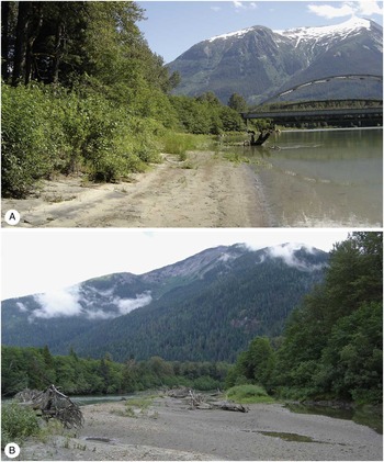
Collection records indicate this species occurs in open scrubby cut-over areas including swampland and flood plains with willows, patches of flowers, grasses, and sedges. We have collected this species from sandy-gravel river banks along the Exchamsiks River near Terrace, British Columbia (Fig. 7).
Etymology
Microphorella chillcotti is named in honour of CNC dipterist Jim Chillcott, who collected the first known specimens of this species from the area of Terrace, British Columbia in 1960.
Microphorella vockerothisp. nov.
Microphorella vockerothi sp. nov., wings of female and male. Abbreviations: bm-m, basal medial crossvein; cua, anterior cubital (=anal) cell; dm, discal medial cell; dm-m, discal medial crossvein; M1, 1st medial vein; M2, 2nd medial vein; M4, 4th medial vein; m1, 1st medial cell; m2, 2nd medial cell; R1, 1st radial vein; R2 + 3, 2nd + 3rd radial vein; r2 + 3, 2nd + 3rd radial cell; R4 + 5, 4th + 5th radial vein; r4 + 5, 4th + 5th radial cell.

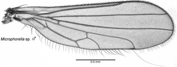
Microphorella sp., wing of male.

Male legs of Microphorella chillcotti species group. (A) fore tibia and tarsus of M. chillcotti sp. nov., dorsal view; (B) midleg of M. chillcotti sp. nov., anterior view; (C) hind tarsus of M. chillcotti sp. nov., dorsal view; (D) hind tarsus of M. vockerothi sp. nov., dorsal view.

Type material
Holotype ♂ labelled: “YUKON: Dempster/ Hwy 135–145 km/ 14.VI.84 S&J Peck/ Spruce-willow for.”; “collected with/ car net”; “HOLOTYPE/ Microphorella vockerothi/ Brooks & Cumming” [red label] (CNC). Paratypes: CANADA: Yukon Territory: 7♂, 8♀, same data as holotype (CNC).
Additional material
CANADA:Yukon Territory: 1♂, Dempster Hwy 135–145 km, 14.VI.1984, S. & J. Peck, Spruce-willow forest, collected with car net (CNC) (see “Remarks” below).
Diagnosis
Males of Microphorella vockerothi are most easily distinguished from those of M. chillcotti by the wing which is strap-like and not distinctly expanded apically, the hind tarsus with tarsomeres 2–5 slender, and the hypopygium with an arched ventral process of the left epandrial lamella, an apically pointed hypandrial process, and cerci that are narrow apically. A single male specimen has been examined that has a bifid apex on the hypandrial process (see “Remarks” below). Females of M. vockerothi appear to be indistinguishable from those of M. chillcotti.
Description
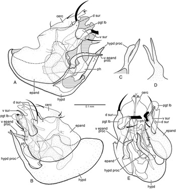
Male: Body length 1.2–1.4 mm, wing length 1.6–1.7 mm. Dark brown in ground colour with blue-green and rusty brown pruinosity, legs slightly paler. Similar to M. chillcotti except as follows: Mesonotum lacking postsutural supra-alar bristle. Hind tarsus with tarsomeres 2–5 slender (Fig. 4D). Wing not expanded towards apex, strap-like, about 3.5× longer than wide (Fig. 2♂); R2 + 3 and R4 + 5 weakly converging beyond distal half, becoming parallel apically; cell r4 + 5 expanded apically with M1 angled posteriorly near apex and strongly diverging from R4 + 5. Hypopygium (Fig. 8) with left epandrial lamella hourglass-shaped in lateral view with middle constriction indistinct (Fig. 8A); ventral epandrial process arched (Figs. 8A, 8E); dorsal lobe of left and right surstylus with short seta near base of tubercle (Figs. 8A, 8B); hypandrium shallowly reniform; hypandrial process with pointed apex, lacking teeth (Figs. 8A, 8B, 8E); phallus S-shaped in lateral view (Fig. 8A); cercus with narrow apex (Fig. 8E).
Female: Body length 1.0–1.4 mm, wing length 1.6–1.7 mm. Similar to male except as noted under Microphorella chillcotti species group description.
Distribution
Microphorella vockerothi is currently only known from the type locality along a 10 km stretch of the Dempster Highway in the Ogilvie Mountains north of Dawson City, Yukon Territory. The specimens were collected along this 10 km stretch with a large cone-shaped net attached to the front of a car.
Remarks
The type series of Microphorella vockerothi was collected along with a single male specimen that differs in the following features: wing with veins R2 + 3 and R4 + 5 slightly divergent apically, anal area more reduced, cell m2 narrower and with setae along posterior margin of cell slightly longer (Fig. 3); hypopygium with apical process of the hypandrium bifid (Figs. 8C, 8D). It is possible that this single male is an aberrant specimen of M. vockerothi, but these differences could also indicate that this specimen belongs to a separate undescribed species. For this reason we have excluded the specimen from the type series of M. vockerothi. Further collecting efforts are required around the type locality of M. vockerothi to resolve the status of this specimen.
Etymology
This species is named in honour of our mentor Dr. J. Richard (Dick) Vockeroth. Dick was instrumental in the development, growth, and curation of the CNC dolichopodid collection and it is our great pleasure to name this species in his honour. Dick is one of the great characters of dipterology, totally brilliant and completely eccentric. Anyone who has had the opportunity to spend time with him has a story (or many) to tell. Unfortunately, ill-health has prevented Dick from working since 2009 and the Diptera Unit is not the same without him.

Microphorella chillcotti sp. nov., male. (A) hypopygium, left lateral view; (B) hypopygium, right lateral view; (C) hypopygium, posterior view. Abbreviations: cerc, cercus; d sur, dorsal lobe of surstylus; epand, epandrium; hypd, hypandrium; hypd proc, hypandrial process; pgt lb, postgonite lobe; ph, phallus; v epand proc, ventral epandrial process; v sur, ventral lobe of surstylus.

Known distribution of Microphorella chillcotti sp. nov. (dots); Microphorella vockerothi sp. nov. and male specimen of Microphorella sp. (triangle).

Exchamsiks River locality near Terrace, British Columbia, Canada, where specimens of Microphorella chillcotti sp. nov. were collected by a Malaise trap placed along sandy portion of the river bank (A), and by sweeping over sandy gravel habitat near the river's edge (B).

Microphorella vockerothi sp. nov. and Microphorella sp., male. (A) hypopygium of M. vockerothi, left lateral view; (B) hypopygium of M. vockerothi, right lateral view; (C) hypandrial process of Microphorella sp., left lateral view; (D) hypandrial process of Microphorella sp., posterior view; (E) hypopygium of M. vockerothi, posterior view. Abbreviations: cerc, cercus; d sur, dorsal lobe of surstylus; epand, epandrium; hypd, hypandrium; pgt lb, postgonite lobe; ph, phallus; v epand proc, ventral epandrial process; v sur, ventral lobe of surstylus.

Female terminalia of Microphorella vockerothi sp. nov. (A) left lateral view; (B) dorsal view. Abbreviations: cerc, cercus; spm pmp, sperm pump; spmth, spermatheca; st, sternite; tg, tergite.

Discussion
The phylogenetic relationships of the Microphorella chillcotti species group to other species of Microphorella or to other members of the Microphorella genus group are unclear. Various modifications of the male wing are relatively widespread throughout the Microphorella genus group, for example in the highly modified apex of the male wing of Thalassophorus spinipennis Saigusa (see Figs. 2C, 2D in Brooks and Cumming Reference Brooks and Cumming2011). We also know of at least three undescribed species of Microphorella from eastern Asia that have somewhat lengthened costal setae along the hind margin of the mid portion of the male wing. However, unlike males of the M. chillcotti species group, these species don't have the single long costal seta near the wing apex. Given that such modified setae probably function as male secondary sexual features and that various male wing modifications are widely distributed throughout this genus group, a more detailed morphological analysis will be required to determine the phylogenetic significance of these features.
Problems also persist with the definition of Microphorella, which appears to be paraphyletic with respect to at least Thalassophorus and Eothalassius (Cumming and Brooks Reference Cumming and Brooks2006; Brooks and Cumming Reference Brooks and Cumming2010, Reference Brooks and Cumming2011). Synonymizing these genera with Microphorella at this time would result in a very weak generic concept. Subdividing Microphorella into several genera might be a more reasonable alternative. However, a detailed phylogenetic analysis of Parathalassiinae is needed to determine the number and limits of genera to be included in the subfamily. Until this is accomplished, Microphorella will continue to be a problematic genus in the Parathalassiinae.
Acknowledgements
We are indebted to Norm Woodley (USNM) for the loan of specimens. Toyohei Saigusa (Fukuoka) kindly provided us with information on undescribed species of Microphorella from eastern Asia, including a photograph of a male wing. We thank Bradley Sinclair (CNC) for useful discussions on homology and character state distributions within the Microphorinae and Parathalassiinae.