It is commonly understood that hearts have either a single or two separate arterial trunks. Concordant ventriculoarterial connections are defined as the aorta arising from the left ventricle and pulmonary trunk arising from the right ventricle. In 1982, Diaz-GongoraReference Diaz-Gongora, Quero-Jimenez, Espino-Vela, Arteaga and Bargeron 1 published a report describing a heart with three separate arterial trunks. In the pathologic description, the pulmonary trunk arose from the right ventricle and connected to the left pulmonary artery and arterial duct. The aorta arose from the left ventricle posterior to the pulmonary trunk and had a normal position and orientation. A third arterial trunk – deemed the “intermediate trunk” – was discovered anterior to the aorta and posterior to the pulmonary trunk, arising directly from the left ventricle between the two vessels. The intermediate trunk connected to the right pulmonary artery. The anterior wall of the intermediate trunk and the posterior wall of the pulmonary trunk were in direct connection but had completely separate lumens. The origin of the intermediate trunk was anterior and slightly to the left of the aortic valve and had a rudimentary valve structure. No other cardiac malformations were present. Pelletier et alReference Pelletier, Sokoloski and Kardon 2 published a similar case of an infant diagnosed with three arterial trunks. Imaging showed the pulmonary trunk arising appropriately from the right ventricle. The aorta arose normally from the left ventricle. A third smaller arterial trunk originated from the left ventricle anterior and slightly leftward of the aorta connecting to the right pulmonary artery. Similarly, the anterior wall of the third trunk and the posterior wall of the pulmonary trunk were in continuity, but the lumens were separate. They also described a rudimentary semilunar valve.
In these reports, the occurrence of an intermediate trunk was an isolated anomaly. We present the first case of a “tritruncal” heart with all outlets arising from the right ventricle, in combination with additional outflow tract derangements.
Report of case
Our patient was diagnosed prenatally with double-outlet right ventricle, large malalignment ventricular septal defect, and interrupted aortic arch type B. Postnatal transthoracic echocardiogram confirmed the prenatal diagnosis, but pulmonary artery anatomy could not be clearly delineated.
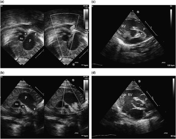
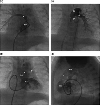
Cardiac catheterisation revealed three arterial trunks arising from the right ventricle. The complete anatomic diagnosis included the usual atrial arrangement, concordant atrioventricular connections, ventricular right-hand topology, and a tritruncal outlet with all three outlets arising from the right ventricle – “triple outlet right ventricle”. The aortic arch anatomy was an interrupted left aortic arch type B. The systemic and pulmonary venous anatomy was normal. An interventricular communication was present with posterior deviation of the outlet septum crowding the subaortic space. The right ventricle outlets include the pulmonary trunk arising anteriorly, the aorta arising posterior and rightward of the pulmonary trunk, and a third “intermediate” trunk arising from the right ventricle. The intermediate trunk was positioned between the pulmonary trunk and aorta, posterior to, and slightly rightward of the pulmonary trunk. It was located leftward and adjacent to but slightly anterior to the aorta. The pulmonary valve connects to the pulmonary trunk, left pulmonary artery, and arterial duct. The aortic valve connects to the ascending aorta, right common brachiocephalic artery, and left common carotid artery. The intermediate trunk had a rudimentary valve from the right ventricle that guards the entrance to an isolated right pulmonary artery. The pulmonary trunk and right pulmonary artery valve leaflets are in fibrous continuity with one another. The anterior wall of the intermediate trunk was directly adjacent to the posterior wall of the pulmonary trunk, but they had separate lumens. The outlet septum was below the pulmonary trunk and the aortic valve (Figs 1, 2).
( a , b ) Echocardiogram depicting the pulmonary trunk giving rise to the left pulmonary artery and discontinuity with the right pulmonary artery. ( c ) Modified parasternal view showing the right ventricular outflow tract and pulmonary trunk with the right pulmonary artery arising separately from the right ventricle with distinct valvar tissue. ( d ) Sweeping rightward and slightly anteriorly the aorta is then seen in long axis with posterior deviation of the outlet septum crowding the subaortic space. *=right pulmonary artery; #=left pulmonary artery; %=outlet septum; Ao=aorta; D=patent arterial duct; P=pulmonary trunk; RV=right ventricle.

( a ) Angiogram of the right pulmonary artery with the catheter in the right ventricle immediately below the rudimentary valve. ( b ) A catheter is positioned retrograde from the aorta across the arterial duct into the pulmonary trunk. The left pulmonary artery is seen arising from the pulmonary trunk with the patent arterial duct. ( c , d ) A right ventriculogram demonstrates three distinct great arteries arising from the right ventricle in anterior and lateral views. The pulmonary trunk is superimposed and not well seen on the anterior view but is well seen on the lateral. *=right pulmonary artery; #=left pulmonary artery; Ao=aorta; D=patent arterial duct; P=pulmonary trunk; RV=right ventricle.

The patient underwent aortic arch repair, right pulmonary artery unifocalisation, arterial duct ligation, and pulmonary artery band on the fourth day of life. At surgery, the right pulmonary artery was transected close to the aorta and probe placement showed a direct communication into the right ventricle. The right pulmonary artery was anastomosed to the distal pulmonary trunk (end to side). Postoperative course was complicated by supravalvar aortic stenosis and right pulmonary artery stenosis requiring reoperation and subsequent stent implantation. The patient died at 6 months of age secondary to infection.
Discussion
The two previously published cases discussed the presence of an intermediate trunk as an isolated anomaly. Our case is unique in that it is the first to describe additional outflow tract derangements. The segmental classification of the previously published cases is a challenge. All three cases have concordant atrioventricular connections; however, in the two previously published cases, the ventriculoarterial connections are neither concordant nor discordant, given a concordant right ventricular to pulmonary trunk connection but a “double-outlet left ventricle”. With the difficulties in segmental classification, the terminology tritruncal heart appropriately describes these two cases. Unique to our case, all three outlets – the pulmonary trunk, the aorta, and the intermediate trunk – arise from the right ventricle making the term triple outlet right ventricle a clear anatomic description.
As identified in mouse models, development of the ventricular outflow tracts involves the separation of an intrapericardial arterial trunk into two distinct aortic and pulmonary components. The outflow tract has a subvalvar ventricular outflow region, the valve and valve sinuses, and the distal outflow tract. Protruding cushions form on opposing sides of the distal outflow tract dividing the lumen into two parts and neural crest cells then complete the “septation” into the aortic and pulmonary channels. Simultaneously, the caudal extrapericardial sixth arch arteries are directed towards the pulmonary component, whereas the cranial fourth arch arteries are directed towards the aortic component.Reference Anderson, Chaudhry and Mohun 3 , Reference Phillips, Mahendran, Singh, Anderson, Chaudhry and Henderson 4 With this understanding, one cannot fully explain the formation of the tritruncal heart. It seems unlikely that a “septation” of the intrapericardial outflow tracts into three distinct channels with three distinct valves would occur. A more plausible explanation may result from the malposition of the extrapericardial sixth arch arteries during the interaction with the intrapericardial distal outflow tract. The embryologic mechanism may be similar to another rare anatomic anomaly, common arterial trunk with “crossed pulmonary arteries”. The ostium of the left pulmonary artery originates superior and rightward of the right pulmonary artery, which arises inferiorly and leftward. The right and left pulmonary arteries cross as they follow their appropriate courses to their respective lungs.Reference Babaoglu, Altun, Binnetoglu, Donmez, Kayabey and Anik 5 Theories suggest that this occurs secondary to faulty differential growth during the partitioning of the outflow tract into the aortic and pulmonary components, as well as an abnormal rotation to the pulmonary component bringing the right pulmonary artery inferiorly.Reference Jue, Lockman and Edwards 6 , Reference Recto, Parness, Gelb, Lopez and Lai 7 Our case may illustrate an extreme version of this entity where the developing right pulmonary artery is left in communication with the subvalvar outflow tract just as it arises from the ventricular mass, but does not explain a separate valve.
This is the first published case of a triple outlet right ventricle and demonstrates the known rare entity of a tritruncal heart in combination with additional outflow tract and aortic arch derangements. The identification and management of this complex cardiac anatomy is fascinating for embryologists and clinicians alike. The tritruncal heart is an unusual congenital anomaly that challenges our understanding of outflow tract development and raises new questions for future researchers.
Acknowledgements
None.
Financial Support
This research received no specific grant from any funding agency, commercial or not-for-profit sectors.
Conflicts of Interest
None.