Introduction
Stress and the consequent release of stress-related neuroendocrine hormones are well recognised to adversely affect various aspects of ruminant health including food intake and rumination. In an effort to better understand the mechanisms by which stress may influence ruminant physiology, we need to examine whether hitherto unexplored areas of interaction within the animal may account for the deleterious effects of stress on ruminant health. One of those areas concerns the ability of stress to modulate the microbial composition of the ruminant intestinal tract. The physiological factors that may influence the microbial composition of the ruminant intestinal tract are incompletely understood. We need to examine whether the production of stress-related neuroendocrine hormones and their release into the intestinal tract can directly influence the resident microbial composition including, but not limited to the continued proliferation of human-associated pathogens such as Escherichia coli O157:H7. The elucidation of such neuroendocrine–bacterial interactions in ruminants can lead to new ways to improve various aspects of animal health and production by creating a favourable environment that ultimately will foster a beneficial microbial composition that also retards the persistence of potential food-borne pathogens.
Stress and its impact on farm animal productivity
Stress is a word used to describe experiences that are challenging psychologically or physiologically. A stressor is the stimulus that causes the stress, and in the context of farmed livestock can be physical, psychological or both. In mammals, stress results in bi-directional communication between the brain and the peripheral organs such that stressful stimuli perceived by the central nervous system (CNS) can directly affect organ functioning, and that physiological changes within the organs of the body can directly affect the CNS. In this context, the many different types of stressors experienced by farm animals can modulate similar hormonal and physiological responses within the stressed subject, and a characteristic chemical hallmark of stress is activation of the sympathetic–adrenal–medullary axis (SAM) and the hypothalamic–pituitary–adrenal (HPA) axis. Activation of the HPA and SAM axes (Mishra et al., Reference Mishra, Segal, Gunter, Kurup, Mishra, Murali, Pierson, Sandovsky-Losica and Stevens1994; Webster et al., Reference Webster, Tonelli and Sternberg2002; Reiche et al., Reference Reiche, Nunes and Morimoto2004) results in systemic elevations of the glucocorticoids (e.g. cortisol) and catecholamine stress hormones (adrenaline and noradrenaline (NA)). These stress hormones are major regulators of behaviour, fuel metabolism, cardiovascular function and thermogenesis, collectively acting to maintain homeostasis during the exposure to stressors (the ‘flight or fight’ response) (Mishra et al., Reference Mishra, Segal, Gunter, Kurup, Mishra, Murali, Pierson, Sandovsky-Losica and Stevens1994; Webster et al., Reference Webster, Tonelli and Sternberg2002; Reiche et al., Reference Reiche, Nunes and Morimoto2004).
Much research in recent years has focussed on examining emotional states in farm animals and assessing their impact on animal welfare and productivity (reviewed in Boissy et al., Reference Boissy, Manteuffel, Jensen, Moe, Sprujit, Keeling, Winckler, Forkamn, Dimitrov, Lagbein, Bakken, Veissier and Aubert2007). Examples of stressors for farm animals principally come from housing environments and management practices. They typically include changes in environmental temperatures, particularly chronic exposure to extremes of heat or cold, restraint such as tethering, isolation from herd members, negative social experiences (with other animals, or with humans during handling procedures), and physical stress resulting from poor diet, dehydration, injury and illness such as infection. Any of these stressors that can be further sub-classified as acute or chronic, avoidable or unavoidable and mild or severe, can have significant negative impacts on feeding, and ultimately livestock productivity.
Farm animal interactions with humans
Cows and cattle are highly sensitive to changes within their living environments (Hemsworth et al., Reference Hemsworth, Barnett, Tilbrook and Hansen1998; Hemsworth, Reference Hemsworth2003). In particular, interactions with other herd members, and with handlers can have significant impact on stress levels in ruminants. For instance, dairy cows that had experienced negative interactions with humans produced less milk, released higher levels of the stress hormone cortisol in their milk, and exhibited higher incidences of avoidance behaviour towards handlers. In contrast, animals treated gently showed less fear towards humans, and were easier to handle (Hemsworth et al., Reference Hemsworth, Barnett, Tilbrook and Hansen1998). Other studies, such as that of Munskgaard et al. also demonstrated the importance of gentle treatment by handlers during procedures such as milking. These workers found that aversive handler contacts such as hits on the head caused avoidance behaviour towards the handler inflicting the aversive treatment (Munskgaard et al., Reference Munskgaard, De Passille, Rushen, Herskin and Kristensen2001). Interestingly, this social learning of the cows was further demonstrated when the animals displayed the same stress-associated behavioural avoidance when they were observers of other herd members receiving aversive treatment. In contrast to the Hemsworth et al. study (Hemsworth et al., Reference Hemsworth, Barnett, Tilbrook and Hansen1998), on this occasion milk production was not reduced in the cows undergoing or observing aversive treatments (Munskgaard et al., Reference Munskgaard, De Passille, Rushen, Herskin and Kristensen2001). A follow-up study by Munksgaard and co-workers showed that acute stress in cows resulted in activation of the adrenocortical axis, and changes in the animal’s response to pain (nociception; Herskin et al., Reference Herskin, Munksgaard and Ladewig2004). The stressors examined were similar to those routinely experienced in animal management such as social isolation in novel surroundings, head fixation by tethering, and introduction of novel neighbours in adjacent stalls. Herskin and co-workers showed that plasma cortisol levels increased following all of these handling type stressors, and that the stressed cows, but not the control cows, demonstrated hypoalgesia in response to a mild pain stimulus. Stressed cows also exhibited greater incidences of avoidance type behaviour, and importantly, exhibited negative changes in their feeding habits, such as feed intake or rumination (Herskin et al., Reference Herskin, Munksgaard and Ladewig2004).
Effects of different stressors on ruminant physiology and microbial flora
Animals respond to changes in their environment by adapting their behaviour. This includes changes in feeding behaviour, particularly feed intake. Upsets in digestion can also occur, which eventually will cause reduced efficiency in utilisation of nutrients and energy for physical maintenance. For instance, when ruminant animals are exposed to outdoor low ambient temperatures they typically shiver; this muscle action, while generating heat, results in more foraging and greater food intake, and represents a diversion of feed material away from biomass-associated increase. In dairy cattle, cold stress can also result in reduced milk secretion, probably due to direct cooling of the mammary glands (Tamminga and Schrama, Reference Tamminga and Schrama1998). Heat stress has also been shown to significantly affect the physiology, hormonal balances and growth performance of diary cows and cattle, in part due to a reduction in feed intake (Tamminga and Schrama, Reference Tamminga and Schrama1998). Heat stressed ruminants can exhibit increased body temperatures, increased rates of respiration, which result in increased energy requirements. They also have greater water intake needs and typically produce less milk. Interestingly, Tajima et al. have reported that the diversity of the rumen microflora in Holstein heifers was altered in response to the changing housing temperatures (Tajima et al., Reference Tajima, Nonaka, Higuchi, Takasuri, Kurihara, Takenaka, Mitsumori, Kajikawa and Aminov2007). Profiling the bacterial species composition of rumen fluid using 16sRNA gene cloning revealed that elevations in environmental temperature and humidity (2 weeks in a climactic chamber maintained at 33°C and 80% humidity v. 20°C and 60% humidity) resulted in consistent changes in microbial diversity, reduced weight gain and an overall lowering of rumen pH. Significant reductions in the levels of short chain fatty acids, the key energy sources of ruminant animals were also found. Later work from the same group (Uyeno et al., Reference Uyeno, Sekiguchi, Tajima, Takenaka and Kurihara2010) identified the temperature responsive rumen species in the heat-stressed heifers as members of the Clostridium coccoides-Eubacterium rectale family of bacteria and the genus Streptococcus, both of which increased in numbers, and the genus Fibrobacter, whose population sizes decreased. The host factors that triggered these changes in rumen species diversity have yet to be elucidated, but it is clear that the rumen microbes were responsive to the changes within the chemical milieu of their host resulting from the heat stress. It can be suggested that a causal relationship might therefore exist between the increased stress hormone levels in the heat-challenged heifers and the increased levels of Streptococcus since previous work has shown that members of the genus Streptococcus demonstrate increased growth in response to catecholamines (Freestone et al., Reference Freestone, Lyte, Haigh and Williams1999; Freestone et al. (unpublished data)). Furthermore, following exposure to catecholamine stress hormones, members of the Streptococcus genus produce a potent acting growth stimulator that has cross species acting activity (Freestone et al. (unpublished data)). The phenomenon of microbes recognising and responding to changes in the environment of their host is termed microbial endocrinology. The role of microbial endocrinology in developing a more holistic understanding of the interaction of ruminant animals with their diverse microbial lodgers during stress is the subject of the remainder of this chapter.
Microbial endocrinology
Microbial endocrinology is a new interdisciplinary research area that represents the intersection of microbiology, endocrinology and neurophysiology. (Lyte, Reference Lyte1993 and Reference Lyte2004a; Freestone et al., Reference Freestone, Sandrini, Haigh and Lyte2008). Its objective is to provide a new paradigm with which to examine and understand the interaction of microorganisms with their host under physical and behavioural conditions present in health and disease. At its foundation is the tenet that microorganisms have evolved specific systems for sensing our hormones, which they then use as an environmental cue that they are inside a suitable host and that is time to initiate growth and pathogenic processes. In the context of the theme of this chapter, microbial endocrinology provides a platform by which to develop a holistic understanding of the factors that shape the interactions between microbes and their bovine host during episodes of stress (Freestone et al., Reference Freestone, Sandrini, Haigh and Lyte2008).
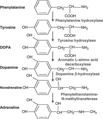
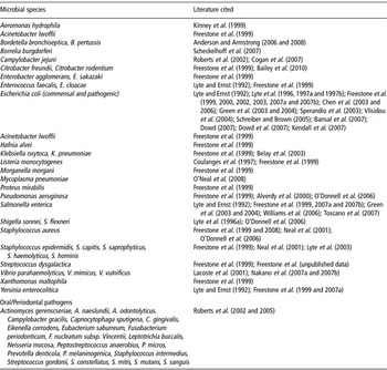
The catecholamine family of stress-elaborated hormones, include the neurotransmitters adrenaline, NA and dopamine, are the most extensively investigated host-derived chemical effectors in microbial endocrinology investigations (Freestone et al., Reference Freestone, Sandrini, Haigh and Lyte2008). Figure 1 shows the structures and biosynthetic pathway of the catecholamine stress hormones, and Table 1 the diversity of catecholamine responsive bacteria. Although the spectrum of bacteria shown is heavily populated by species inhabiting the highly innervated gastrointestinal tract (where NA and dopamine containing nerve terminals are abundant) (Furness, Reference Furness2006), catecholamines are found in secretions throughout the mammalian body and bacteria occupying a wide variety of locations will come into contact with them, and so similarly evolve an ability to sense changes in the stress hormone levels of their host. Several reports have also shown neuroendocrine hormones to be potent growth factors for respiratory pathogens. These include Klebsiella pneumoniae, (Freestone et al., Reference Freestone, Lyte, Haigh and Williams1999) as well as various Bordetella species (Anderson and Armstrong, Reference Anderson and Armstrong2006), such as B. bronchiseptica and B. pertussis. In a serum-based media, it was further shown that norepinephrine stimulated growth of B. bronchiseptica as well as inducing expression of BfeA, a siderophore receptor important for growth in vivo (Anderson and Armstrong, Reference Anderson and Armstrong2006). Mycoplasmas can be potent respiratory pathogens of ruminants, and recently O’Neal et al. used microarray analysis to show that gene expression in Mycoplasma hyponeumoniae underwent substantial changes when exposed to NA, including up-regulation of genes encoding host tissue attachment factors (O’Neal et al., Reference O’Neal, Schafer, Madsen and Minion2008). Stress hormone enhancement of growth and virulence is not solely a feature of pathogens of mammalian hosts, as it has been demonstrated that norepinephrine can increase the infectivity of bacteria causing infections in non-vertebrates species such as oysters (Vibrio species) (Lacoste et al., Reference Lacoste, Jalabert, Malham, Cueff and Poulet2001) and amphibians such as frogs (Aeromonas hydrophila; Kinney et al., Reference Kinney, Austin, Morton and Sonnenfeld1999).
Catecholamine biosynthetic pathway. In mammals, the pathway for the synthesis of catecholamines is from the aromatic amino acid tyrosine (most commonly from food sources; it should be noted that various co-factors needed in the pathway are not shown). Synthesis of catecholamines is to a degree tissue specific, and phenylethanolamine N-methyltransferase, required for adrenaline synthesis, is not expressed in cells of the enteric nervous system. This may explain why a response-specificity for catecholamines present within the gut, NA and dopamine has been demonstrated for several enteric pathogens (Freestone et al., Reference Freestone, Haigh and Lyte2007a; note that this figure was adapted with permission from Freestone et al., Reference Freestone, Sandrini, Haigh and Lyte2008).

Stress hormone responsive bacteria

How do stress hormones stimulate bacterial growth? Most analyses of stress hormone growth induction in vitro have utilised serum-based culture media, which is typically bacteriostatic through iron chelation by serum-transferrin, a glycoprotein with very high ferric iron binding affinity. In such media, bacteria typically grow poorly unless supplemented with the catecholamines in which the growth induction can be >100 times that of the un-supplemented control cultures (Lyte and Ernst, Reference Lyte and Ernst1992; Freestone et al., Reference Freestone, Lyte, Haigh and Williams1999). Insight into the growth induction mechanism may be provided by appreciating that the iron-binding catechol moiety found in siderophores such as enterobactin is also present in the catecholamine family of stress hormones (Freestone et al., Reference Freestone, Lyte, Neal, Maggs, Haigh and Williams2000, Reference Freestone, Williams, Haigh, Maggs, Neal and Lyte2002 and Reference Freestone, Haigh, Williams and Lyte2003; Figure 1). Research from our laboratories and those of others have shown that NA, adrenaline and dopamine and certain of their metabolites (such as dihydroxymandelic acid and dihydroxyphenylglycol; Freestone et al., Reference Freestone, Williams, Haigh, Maggs, Neal and Lyte2002 and Reference Freestone, Haigh and Lyte2007a) all share the ability to stimulate growth in serum or blood growth by enabling bacteria to steal iron from normally inaccessible transferrin or lactoferrin (Freestone et al., Reference Freestone, Lyte, Neal, Maggs, Haigh and Williams2000, Reference Freestone, Williams, Haigh, Maggs, Neal and Lyte2002, Reference Freestone, Haigh, Williams and Lyte2003, Reference Freestone, Haigh and Lyte2007a, Reference Freestone, Walton, Haigh and Lyte2007b and Reference Freestone, Sandrini, Haigh and Lyte2008; Roberts et al., Reference Roberts, Matthews, Socransky, Freestone, Williams and Chapple2002 and Reference Roberts, Matthews, Socransky, Freestone, Williams and Chapple2005; Anderson and Armstrong, Reference Anderson and Armstrong2006 and Reference Anderson and Armstrong2008). Iron is essential for growth of all bacterial pathogens, and its limitation in blood and mucosal secretions via its sequestration by transferrin and lactoferrin is one of the most important innate immune defences against infection (Ratledge and Dover, Reference Ratledge and Dover2000). Catecholamines, therefore enable bacterial pathogens that lack specific acquisition systems for transferrin and lactoferrin-complexed Fe to acquire the iron needed growth in vivo.
Another mechanism by which catecholamines can induce growth of Gram-negative bacteria, particularly enteric species involves induction of a novel growth stimulator (Lyte et al., Reference Lyte, Frank and Green1996a; Freestone et al., Reference Freestone, Lyte, Haigh and Williams1999). Interestingly, this novel growth stimulator that we termed the noradrenaline-induced autoinducer (NA-AI; Lyte et al., Reference Lyte, Frank and Green1996a; Freestone et al., Reference Freestone, Lyte, Haigh and Williams1999), induces its own synthesis and is heat stable, highly cross-species acting activity that stimulates increases in growth of magnitude similar to that achievable with the catecholamines (Freestone et al., Reference Freestone, Lyte, Haigh and Williams1999). The NA-AI induces bacterial growth independently of Tf or Lf (Freestone et al., Reference Freestone, Haigh, Williams and Lyte2003), and is also able to rapidly stimulate the recovery to active growth of viable, but non-culturable E. coli O157:H7 or Salmonella as well as increasing the rate of germination of Bacillus anthrax spores (Reissbrodt et al., Reference Reissbrodt, Rienaecker, Romanova, Freestone, Haigh, Lyte, Tschäpe and Williams2002; Voigt et al., Reference Voigt, Fruth, Tschape, Reissbrodt and Williams2006). The NA-AI is also recognised by periodontal pathogens (Roberts et al., Reference Roberts, Matthews, Socransky, Freestone, Williams and Chapple2005). In terms of its synthesis, induction of the E. coli O157:H7 NA-AI requires only a transient 4 to 6 h exposure to catecholamines (Lyte et al., Reference Lyte, Frank and Green1996a; Freestone et al., Reference Freestone, Lyte, Haigh and Williams1999), after which the NA-AI activity induces its own synthesis. This indicates that the effects of catecholamine released during acute stress could have lasting and wide acting effects on the gut and other tissue microflora long after catecholamine levels in the host animal have returned to normal.
Microbial endocrinology as a lens to holistically understand how stress influences the health of ruminant livestock
It has been recognised for several decades that stress can increase susceptibility to infection in most animals. Until the advent of Microbial Endocrinology, stress influences on infection had been solely attributed to stress hormone modulation of host immune function. The evidence to support this exclusive view is not unreasonable, as sympathetic nerve fibres densely innervate lymphoid organs and terminate in the proximity of immune cell populations, almost all of which possess receptors for the glucorticoids and catecholamines released from stress-induced activation of the HPA and SAM axes (Mishra et al., Reference Mishra, Segal, Gunter, Kurup, Mishra, Murali, Pierson, Sandovsky-Losica and Stevens1994; Webster et al., Reference Webster, Tonelli and Sternberg2002; Reiche et al., Reference Reiche, Nunes and Morimoto2004). The overall effect of stress hormone binding to immune cells is usually a reduction in function. However in any infection, there are always two opposing players – the infectious agent as well as the immune system. Over the last decade, however, it has become clear that bacteria are also able to sense the chemical mediators of their host’s stress response (Lyte, Reference Lyte1993 and Reference Lyte2004a; Freestone et al., Reference Freestone, Sandrini, Haigh and Lyte2008). Viewing stress influences on infection through the lens of microbial endocrinology provides a more holistic understanding of what is happening to all the cells of the stressed animal including its resident microbes.
Before we begin to examine how stress modulates bovine susceptibility to infection through its effects on the endogeneous microflora, it would be informative to examine the literature on models of stress and infection in other animals. In mice, psychological stress appears to be as important as physical stress in its ability to affect the outcome of an infection. Social stress has been shown to have a marked impact on susceptibility to enteropathogen challenge in a mouse social conflict stress model, even when stress enhanced certain aspects of the immune response such as phagocytosis (Dréau et al., Reference Dréau, Sonnenfeld, Fowler, Morton and Lyte1999). In this study, E. coli O157:H7 was inoculated into semi-permeable chambers implanted in the peritoneal cavity of the mice, and incubated for 24 h, after which the chambers were removed, and the final growth levels determined. Note that the implant chambers were fully permeable to proteins and any chemical effectors produced by the mice such as hormones but not to cells. Within the chambers the bacteria grew significantly better in the social conflict stressed mice compared with the control non-stressed mice, despite a fourfold increase in the anti-microbial phagocytic activity in the blood of the stressed animals (Dréau et al., Reference Dréau, Sonnenfeld, Fowler, Morton and Lyte1999). In another rodent stress model, stressors such as social conflict and physical restraint (which have been similarly shown to be significant stressors of bovine species) caused translocation to systemic sites of both the gut and cutaneous microflora (Bailey et al., Reference Bailey, Engler and Sheridan2006 and Reference Bailey, Dowd, Parry, Galley, Schauer and Lyte2010). Significant spilling over of catecholamines from the systemic circulation into the gastrointestinal (GI) tract can occur during acute stress (Eisenhofer et al., Reference Eisenhofer, Aneman, Friberg, Hooper D Fåndriks, Lonroth, Hunyady and Mezey1997). Increased direct release of catecholamines by the enteric nervous system (ENS) under stress has also been demonstrated experimentally by Zhou et al. who showed that in the GI tract of rats expression of tyrosine hydroxylase (the rate limiting enzyme of catecholamine biosynthesis; see Figure 1) became up-regulated in response to physical stress such as surgical perforation of the bowel (Zhou et al., Reference Zhou, Simms and Wang2004). A related study by Alverdy et al. employed a mouse model of surgical stress (a partial hepatectomy v. a sham laparotomy) to show that surgery can cause direct increases in the levels of NA released into the GI tract (Alverdy et al., Reference Alverdy, Holbrook, Rocha, Seiden, Wu, Musch, Chang, Ohman and Suh2000). Alverdy and colleagues also reported that mice subjected to the partial hepatectomy became significantly more susceptible to gut-derived sepsis by the opportunistic pathogen Pseudomonas aeruginosa, possibly because of NA-induced increases in the expression of the PA-I lectin, an essential P. aeruginosa host attachment factor (Alverdy et al., Reference Alverdy, Holbrook, Rocha, Seiden, Wu, Musch, Chang, Ohman and Suh2000).
A neurotoxin model of stress (Lyte and Bailey, Reference Lyte and Bailey1997) showed that acute stress could massively disrupt the balances of the resident gut microflora. Administration of the neurotoxin 6-hydroxydopamine (6-OHDA) ablates noradrenergic nerve terminals, causing a rapid release of systemic stores of NA contained within nerve terminals, which then spill over into the circulation and other tissues (including the GI tract). In only 24 h following the 6-OHDA treatment of mice, analysis of the GI microflora showed a >10-fold increase in the caecal levels of Gram-negative bacteria, principally E. coli. Overgrowth of E. coli in vivo is implicated in gut-derived sepsis (Nieuwenhuijzen et al., Reference Nieuwenhuijzen, Deitch and Goria1996). Significantly, a similar four-log increase in the numbers of coliform bacteria directly attaching to the walls of the caecum was also observed: gut tissue attachment is of course one of the first stages in bacterial translocation and subsequent gut derived sepsis. In animals, the 6-OHDA ablated nerve terminals usually regenerate within 14 days, and indeed examination of the gut microflora of the mice after this time showed the enteric species balance had returned to normal (Lyte and Bailey, Reference Lyte and Bailey1997). That the changes in the microflora were specific to NA release was confirmed by administering desipramine before the 6-OHDA, a drug that sterically hinders binding of NA to noradrenergic nerve terminals. Desipramine-6-OHDA treated mice did not undergo gut microflora changes, suggesting that the systemic release of catecholamines were responsible for the microflora pertubations seen (Lyte and Bailey, Reference Lyte and Bailey1997). Later in vitro work by Freestone et al. (Reference Freestone, Williams, Haigh, Maggs, Neal and Lyte2002) showed that commensal E. coli responded to the GI derived catecholamines NA and dopamine, as well as their metabolites, with growth increases of nearly 100 greater than those of non-supplemented control cultures.
Stress can directly modulate GI function, which as well as affecting livestock productivity may indirectly modulate the behaviour of the microflora. Stressors have been shown to affect secretion of gastric acid, which may in turn influence survival of ingested pathogens. Lenz et al. showed that restraint stress significantly increased or decreased gastric acidity, depending upon temperature (Lenz et al., Reference Lenz, Raedler, Greten, Vale and Rivier1988). Another effect of stress on GI function is a reduction in motility. Stress can reduce gastric emptying, slowing transit in the small intestine through stressor-induced increases in corticotrophin releasing hormone (Nakade et al., Reference Nakade, Tsuchida, Fukuda, Iwa, Pappas and Takahashi2005). The collective message of this and the above research is that natural defences of mammals, such as those provided by the commensal microflora, stomach acid and normal GI motility can be significantly compromised by psychological and physical stress (Bailey et al., Reference Bailey, Engler and Sheridan2006). What might then be the response of an opportunistic enteropathogen to such stress induced changes in the GI tract of their bovine host? A very recent report may shed some light into this question. Work from Bailey et al. (Reference Bailey, Dowd, Parry, Galley, Schauer and Lyte2010) demonstrated that such stress-induced changes can indeed alter the microbial diversity of the gut to such a degree that provides a greater opportunity for a pathogen to infect a host. Bailey et al. stressed mice and showed through the use of a metagenomics-based 454 pyrosequencing techniqure that the microbial diversity of the colon was altered to such a degree as to provide a more favourable environment for infection with the gut pathogen Citrobacter rodentium. Critically, Bailey et al. showed that these changes in microbiota occurred in the absence of any demonstrable change in a number of immune-related parameters (Bailey et al., Reference Bailey, Dowd, Parry, Galley, Schauer and Lyte2010).
Re-examination of Table 1 shows that the spectrum of stress hormone responsive bacteria is weighted towards enteric pathogens. This is not too surprising given that NA- and dopamine-containing sympathetic nerve terminals distributed throughout the body, including the intestinal tract where they make up part of the ENS (Eisenhofer et al., Reference Eisenhofer, Aneman, Friberg, Hooper D Fåndriks, Lonroth, Hunyady and Mezey1997). Indeed, around half of all the NA present within the body is synthesised and utilised within the ENS (Furness, Reference Furness2006). Thus, an enteric microbe will be well accustomed to the presence of catecholamines, and perhaps might have learned to sense changes in the levels of fitness of their host via monitoring catecholamine levels? Perhaps stressed hosts are more likely to die in the near future and bacteria need to prepare themselves to colonise a new host. Where the old host is falling prey to a carnivore, replication to high numbers and expression of virulence factors might facilitate this process. Where the old host is not eaten, the capacity to cause diarrhoea and vomiting might increase the chances of finding a new host. Support for this hypothesis comes from the observation that stress in food-producing animals such as cows and pigs has been shown to produce changes in the gut microflora relevant to pathogen dissemination. For instance, even a mild physical interaction of humans with piglets, such as a single daily weight measurement involving minimal movement or handling, resulted in increased faecal excretion of E. coli and total coliforms relative to control pigs (Dowd et al., Reference Dowd, Callaway and Morrow-Tesch2007).
An understanding of the communication process that is taking place between the microflora and their host is necessary, not just in the interests of animal welfare, but because the microorganisms respond most strongly to the hormonal changes that take place during host stress are those which can potentially cause life threatening pathogens of humans. The release of even low numbers of E. coli O157:H7 in the faeces of cattle and their entry into the food chain is a primary route to extremely costly human infections (Tarr and Neill, Reference Tarr and Neill2001). Preventing the initial colonisation and proliferation of this fast-growing enteropathogen, which can live asymptomatically within ruminants, is of considerable interest to the livestock and meat production industries. There have therefore been a number of investigations that have focussed on a microbial solution to this problem, such as administration of lactic acid bacteria to cattle. Another factor that must also be considered is perhaps modification of animal husbandry techniques. An extensive review by Terlouw and co-workers investigated the impact of various animal-handling practices employed in the hours/days leading up to slaughter of livestock (Terlouw et al., Reference Terlouw, Arnould, Auperin, Berri, Le Bihan-Duval, Deiss, Lefevre, Lenskink and Mounier2008). The duration of the journey that cattle experience when they are transported to abattoirs, and the length of time undergoing processing before slaughter, inevitably result in multiple episodes of stress (confirmed by enhanced cortisol release), all of which could affect enteropathogen growth and subsequent dissemination onto carcasses and consumer meat products. Some of the stressors experienced by animals before slaughter may not be avoidable, while some of them could be addressed (Terlouw et al., Reference Terlouw, Arnould, Auperin, Berri, Le Bihan-Duval, Deiss, Lefevre, Lenskink and Mounier2008). For instance, in the ruminant gut, enteropathogens such as E. coli O157:H7 are normally greatly outnumbered by resident microbes, and held in check by various anti-microbial factors released by the endogenous microflora (such as organic acids). Nutritional stress, such as the routine withdrawal of feed before animal slaughter can reduce the concentration of these acids in the gut, resulting in pH increases that can positively influence the proliferation of E. coli. Even if meat obtained from stressed animals does not become microbiologically contaminated during the slaughter process, metabolic stress before the animal’s dispatch can significantly affect its quality and flavour (Terlouw et al., Reference Terlouw, Arnould, Auperin, Berri, Le Bihan-Duval, Deiss, Lefevre, Lenskink and Mounier2008).
Stress hormone modulation of bovine enteropathogen virulence
A number of in vitro reports have shown that stress hormones can act as potent stimuli for bacterial attachment to bovine GI tissues. NA was shown to enhance expression of the K99 pilus adhesin of enterotoxigenic E. coli and type 1 fimbriae of commensal E. coli (Hendrickson et al., Reference Hendrickson, Guo, Laughlin, Chen and Alverdy1999). Later work by Vlisidou and co-workers examined the effect of NA on the adherence and enteropathogenicity of E. coli O157:H7 using a bovine ligated ileal loop model of infection (Vlisidou et al., Reference Vlisidou, Lyte, Van Diemen, Hawes, Monaghan, Wallis and Stevens2004). NA was found to enhance E. coli O157:H7-induced intestinal cell inflammatory and secretory responses, and significantly increased adherence of the bacteria to the intestinal mucosa. In the Vlisidou et al. study, NA modulation of enteritis and adherence was dependent on the ability of the E. coli O157:H7 to form attaching and effacing lesions. In another study by Chen et al. NA also promoted caecal adherence of a non-O157:H7 E. coli strain and E. coli O157:H7 eae (intimin, host cell tight attachment protein) and espA (type III translocator protein) mutants incapable of intimate mucosal cell attachment (Chen et al., Reference Chen, Lyte, Stevens, Vulchanova and Brown2006). Later work by Bansal et al. demonstrated that in addition to stress hormones enhancing attachment to host cells, E. coli O157:H7 showed also a positive chemotaxis response to NA and adrenaline (Bansal et al., Reference Bansal, Englert, Lee, Hegde, Wood and Jayaraman2007).
In addition to the catecholamines, glucocorticoid-type hormones are also released during stress, (Mishra et al., Reference Mishra, Segal, Gunter, Kurup, Mishra, Murali, Pierson, Sandovsky-Losica and Stevens1994; Reiche et al., Reference Reiche, Nunes and Morimoto2004), which may be significant as the adrenocorticotropic hormone has been shown to increase attachment of E. coli O157:H7 to colonic mucosa (Schreiber and Brown, Reference Schreiber and Brown2005). As already mentioned, dietary factors such as plant-derived catechol-containing compounds may also influence pathogen behaviour. Catechins, tannic acid, caffeic and chlorogenic acids, all traditionally considered as bacteriostatic agents, were able in host-like environments containing lactoferrin and transferrin (serum or blood) to act as highly potent enteropathogen growth stimulators by enabling bacterial access to the iron within the proteins (Freestone et al., Reference Freestone, Walton, Haigh and Lyte2007b). Tyramine, which is structurally related to the catecholamines (dopamine) and widespread in fermented dairy products due to the abundant tyrosine decarboxylase activity of Enterococcus faecalis starter cultures, significantly enhanced adherence of E. coli O157:H7 to mammalian gut tissues (Lyte, Reference Lyte2004b).
A number of in vitro transcriptional profiling studies have confirmed that exposure to stress hormones leads up to regulate expression of enteropathogen virulence genes relevant to host cell attachment. For instance, Dowd carried out microarray analyses of E. coli O157:H7 virulence gene expression in the presence of NA (Dowd, Reference Dowd2007). He reported that E. coli O157:H7 grown in serum-containing culture medium containing NA showed differential regulation of 101 genes compared with similarly grown non-supplemented controls. Genes showing a higher level of expression included genes for host cell attachment (eae, espB and espA) and the stx1 and stx2 shiga toxin genes. This latter result confirms an earlier report that NA increased E. coli O157:H7 shiga toxin production (Lyte et al., Reference Lyte, Arulanandam and Frank1996b). Work by Kendall et al. (Reference Kendall, Rasko and Sperandio2007) showed that exposure to the adrenaline-induced expression of the locus of enterocyte effacement (LEE) operon in an E. coli O157:H7 luxS mutant. Although adrenaline is a non-gut catecholamine, this is an interesting result, as the LEE locus contains 41 open reading frames coding for genes required for E. coli O157:H7 intimate attachment to host mucosa. Kendall et al. also noted adrenaline-induction of flagellar genes in their luxS mutant, which may be significant as motility is important in the pathogenesis E. coli O157:H7 (Kendall et al., Reference Kendall, Rasko and Sperandio2007).
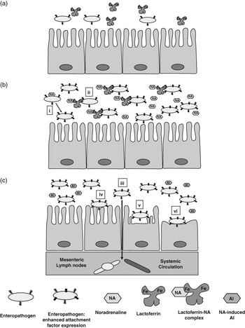
Figure 2 is a cartoon showing what might happen to a few enteropathogens in the gut of a cow experiencing an episode of acute stress. The three panels combine what is known about how catecholamine stress hormones modulate enteropathogen growth and virulence, and demonstrate that catecholamine release during acute stress could have lasting and wide acting effects on the gut microflora long after catecholamine levels have returned to normal.
Model showing how enteropathogenic bacteria might behave in the gastrointestinal (GI) tract of a stressed bovine host. This cartoon shows how an initial few cells of an invading enteropathogen, such as E. coli O157:H7 or Salmonelle enterica, might behave when their bovine host becomes acutely stressed. Sequence of events: (a) Bovine gut during calm times. Lactoferrin maintains iron limitation in mucosal secretions, which are therefore bacteriostatic. The number of pathogens during this time of calm is therefore low and controlled by endogenous factors. (b) Bovine gut during an acute stress. Enteric nervous system activity increases with the consequent release of catecholamines (noradrenaline (NA) and dopamine) within the gut. Work from in vitro and in vivo reports shows that the encounter of the pathogen with the stress hormone could result in two major events. The first (i) is that NA acts as a cue to prime virulence factor expression – in this case production of attachment factors such as adhesins. The second (ii) is that the catecholamine interacts with the lactoferrin (and any transferrin that might also be present), converting a normally bacteriostatic set of proteins in to a useful nutritional iron source and providing support for enhanced bacterial growth in the gut (Freestone et al., Reference Freestone, Lyte, Neal, Maggs, Haigh and Williams2000, Reference Freestone, Williams, Haigh, Maggs, Neal and Lyte2002 and Reference Freestone, Haigh and Lyte2007a). (c) Several hours after the acute stress have passed and the levels of catecholamines in the gut have returned to normal. For enteric pathogens such as E. coli O157:H7, only a transient 4-h exposure to catecholamines is sufficient to induce production of a novel autoinducer of growth (NA-AI; Lyte et al., Reference Lyte, Frank and Green1996a; Freestone et al., Reference Freestone, Lyte, Haigh and Williams1999). The NA-AI not only stimulates growth, but can also enhance shiga toxin production (Lyte et al., Reference Lyte, Arulanandam and Frank1996b). Any pathogens present might also respond to other signals produced by the intestinal microbial flora, such as the LuxS-dependent AI-3, which also activates transcription of genes involved in attaching and effacing lesion formation (Sperandio et al., Reference Sperandio, Torres, Jarvis, Nataro and Kaper2003; Kendall et al., Reference Kendall, Rasko and Sperandio2007). The increasing numbers of pathogens, and possibly also endogenous gut bacteria (Lyte and Bailey, Reference Lyte and Bailey1997; Bailey et al., Reference Bailey, Engler and Sheridan2006 andReference Bailey, Dowd, Parry, Galley, Schauer and Lyte2010), could affect gut integrity leading to bacterial translocation (iii) either to the mesenteric lymphatic tissue or, in a worst-case scenario, directly into the systemic circulation, where even commensal bacteria could lead to sepsis and multiple organ failure. Previous exposure to NA might also cause increased expression of E. coli virulence genes, leading to increased attachment of the pathogen to the gut mucosa, either through non-intimate attachments (Chen et al., Reference Chen, Lyte, Stevens, Vulchanova and Brown2006) (iv) or through espA- or locus of enterocyte effacement (LEE)-dependent intimate attachment (Vlisidou et al., Reference Vlisidou, Lyte, Van Diemen, Hawes, Monaghan, Wallis and Stevens2004; Kendall et al., Reference Kendall, Rasko and Sperandio2007) (v and vi), causing the attaching and effacing lesions characteristic of enteropathogenic and enterohaemorrhagic E. coli (note that this figure was adapted with permission from Freestone et al. Reference Freestone, Sandrini, Haigh and Lyte2008).

Conclusion: less stressed cows may be better nourished and microbiologically safer
Stress is a natural part of life, for ruminants and man alike. However, this review has shown that stressors of farm animals can affect not only the animal experiencing the stress, they can also directly cause changes in the microflora of the animals, including rumen microbes, such that the productivity of the animals is reduced, which clearly indicates that stress has economic consequences. Murine models have shown that stressors can directly modulate the composition of the gut microflora, and evidence is increasing that the microbes within ruminant livestock may also be sensitive to the stress experienced by their host. In the infection context, if future studies show that bovine stress influences enteropathogen carriage and shedding, then reducing stress by adopting better animal husbandry techniques, thereby decreasing the potential for stress hormone-induced promotion of undesirable bacterial species such as E. coli O157:H7 may make meat microbiologically safer, and possibly of better quality.