Various devices are used for transcatheter closure of ventricular septal defects.Reference Yang, Tai, Khin and Quek 1 Although specific devices have been developed for transcatheter ventricular septal defect closure, some occluder devices are reportedly used in an off-label manner.Reference Santhanam, Yang, Chen, Tai, Rajgor and Quek 2 The Occlutech Duct Occluder (Occlutech, Helsingborg, Sweden) is a newly introduced device for transcatheter closure of patent ductus arteriosus.Reference Ríos-Méndez 3 In a previous article, we presented the first report of closure of a ventricular septal defect using the Occlutech Duct Occluder.Reference Atik-Ugan and Saltik 4 We herein describe a patient in whom two different devices (Occlutech Duct Occluder and Amplatzer Duct Occluder II; St. Jude Medical, Saint Paul, Minnesota, United States of America) were successfully used to close a perimembranous ventricular septal defect with a ventricular septal aneurysm. To the best of our knowledge, this is the first report of transcatheter closure of a ventricular septal defect using both an Occlutech Duct Occluder and Amplatzer Duct Occluder II.
Case
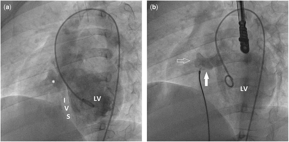
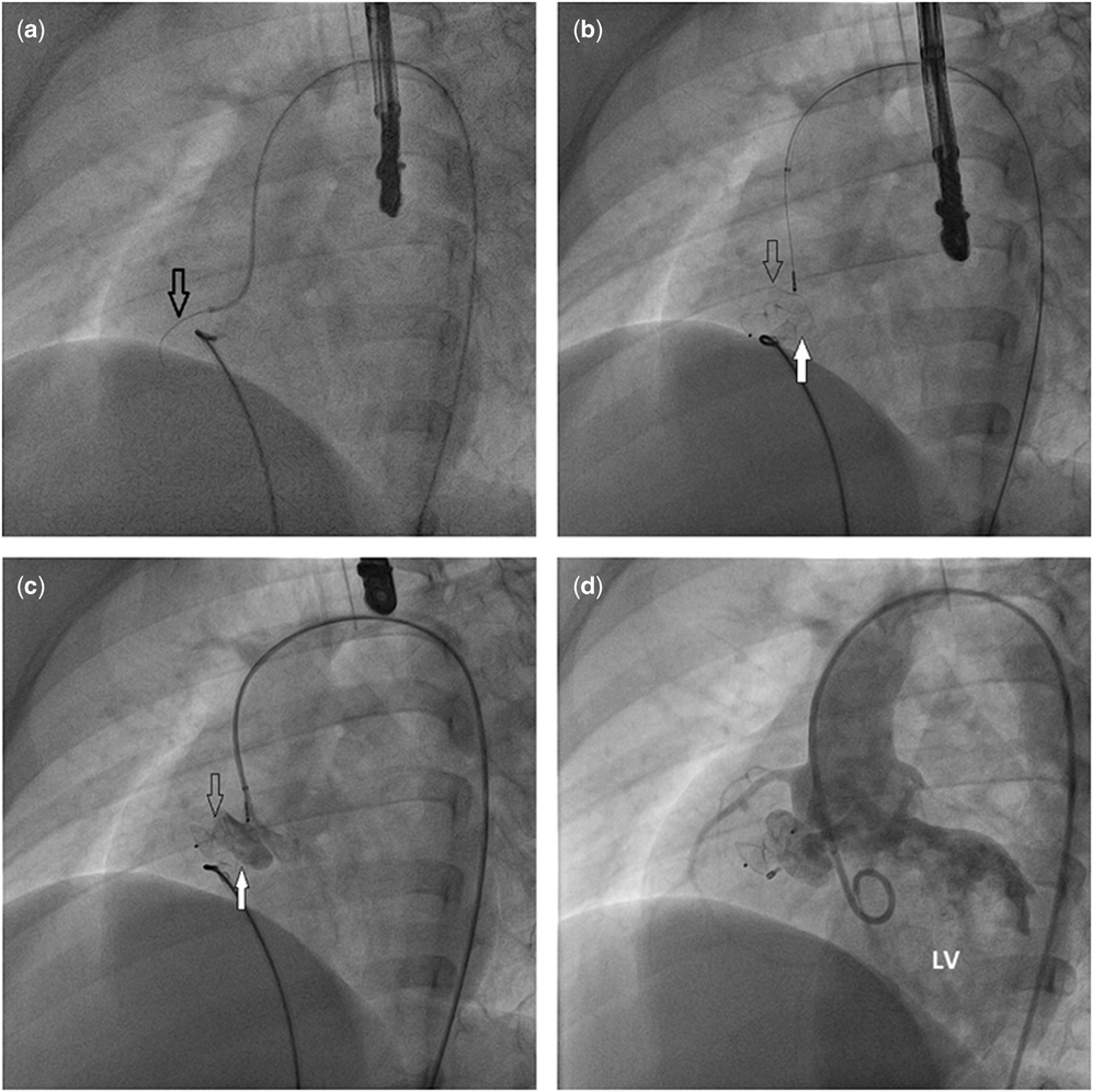
A 12-year-old boy with a ventricular septal defect was admitted to our outpatient clinic. Transthoracic echocardiography revealed a perimembranous ventricular septal defect that was partially restricted by a septal aneurysm and enlarged left cardiac chambers. After obtaining informed consent from his parents, cardiac catheterisation was performed under general anaesthesia. A left ventricular angiogram showed a 14-mm ventricular septal defect restricted by a ventricular septal aneurysm on the right ventricular side with two exits (Fig 1a). The larger exit measured 5.1 mm. Oximetric data revealed a shunt ratio of 1.85. We decided to close the defect with an Occlutech Duct Occluder. The defect was crossed from the arterial side, and an arteriovenous loop established. A 6-Fr, 45° Occlutech Delivery Sheath (Occlutech, Helsingborg, Sweden) was introduced from the femoral vein and passed across the defect into the left ventricle. A 6/8 short shank Occlutech Duct Occluder was then advanced through the sheath. The disc was delivered fully, the device was pulled back to place the disc at the left ventricular end of the defect, and the shank was delivered. Before releasing, left ventricular angiography and transthoracic echocardiography showed relatively large residual flow through the second orifice of the ventricular septal aneurysm (Fig 1b). Because the second orifice was far from the first one and the aneurysm was large enough to anchor two devices, we decided to use an additional device. The residual defect was crossed from the arterial side (Fig 2), and a 5-Fr TorqVue LP sheath (St. Jude Medical) was introduced from the arterial side and passed across the defect into the right ventricle. A 4/4 Amplatzer Duct Occluder II was then advanced through the sheath. The distal disc was opened on the right side of the defect, and the rest of the device was placed on the left side of the aneurysm. Hand-injection of contrast through the arterial long sheath demonstrated appropriate positioning of the two devices. After confirming the positions of the devices with echocardiography, the Occlutech Duct Occluder was released, followed by the Amplatzer Duct Occluder II. Control angiography showed no residual shunt through the ventricular septal defect. An electrocardiogram revealed sinus rhythm with normal atrioventricular conduction. After 24 hours, transthoracic echocardiography showed good positioning of both devices with no residual shunt or aortic or tricuspid regurgitation. The patient was discharged from the hospital the following day with no complications. The patient was doing well at the 11-month follow-up.
( a ) Left ventricular angiogram showing the ventricular septal defect restricted by a large ventricular septal aneurysm. LV=left ventricle; IVS=interventricular septum.*Large ventricular septal aneurysm. ( b ) Left ventricular angiogram showing the Occlutech Duct Occluder within the aneurysm at an appropriate position (filled arrow) and relatively large residual flow through the second orifice of the ventricular septal aneurysm (arrow).

( a ) Crossing of the residual defect from the arterial site with a hydrophilic guidewire (arrow). ( b ) Amplatzer Duct Occluder II (arrow) placed at the second orifice of the ventricular septal aneurysm. ( c ) Hand-injection of contrast through the arterial sheath demonstrating appropriate positioning of the two devices: Occlutech Duct Occluder (filled arrow) and Amplatzer Duct Occluder II (arrow). ( d ) Control angiography after release of the devices demonstrating total occlusion of the defect. LV=left ventricle.

Discussion
Transcatheter closure of a perimembranous ventricular septal defect with an aneurysm remains challenging. Two methods may be used for positioning the device: closing the inlet or closing the outlet of the aneurysm.Reference Bian, Ma and Wang 5 Most aneurysms thereby allow implantation of an occluder with the device retention skirt entirely within the aneurysm and the other portion of the device secured in an opening of the aneurysm on the right ventricle side.Reference El Said, Bratincsak, Gordon and Moore 6 Sometimes the aneurysmal defect has multiple holes in the right side of the septum and only one opening inlet in the left side.Reference Wu, Qin and Zhao 7 Closure of multi-hole ventricular septal defects in the perimembranous location using a large single device is an option. However, if the defects are far apart, this is not always possible. In these circumstances, using two different devices is another alternative. Devendran et alReference Devendran, Koneti and Jesudian 8 reported the first case of transcatheter closure of two perimembranous ventricular septal defects with aneurysms using two Amplatzer Duct Occluder II devices. In 2017, Zhao et alReference Zhao, Han and Zhang 9 described four patients whose ventricular septal defects were closed with Amplatzer Duct Occluder II devices and perimembranous ventricular septal defect occluder devices. To our knowledge, this is the first case in which both an Occlutech Duct Occluder and Amplatzer Duct Occluder II were used for closure of a perimembranous aneurysmal ventricular septal defect with two separate openings in the right side of the septum.
In our patient, the ventricular septal aneurysm had two main orifices located on the upper and lower sides. We believe that the Occlutech Duct Occluder has some structural advantages such as its wider proximal part, which may help the device to hold onto the interventricular septum by acting as a retention disc. We initially attempted to close the defect with a 6/8 Occlutech Duct Occluder. Left ventricular angiography and transthoracic echocardiography showed that although the retention skirt was entirely within the aneurysm the retention disc did not close the ventricular septal defect and a relatively large residual flow through the second orifice of the ventricular septal aneurysm was detected. In such cases, a generally accepted option is to change the device to a larger one. After detailed evaluation of the images, we concluded that a single occluder may have failed to close all defects completely and that the septal aneurysm was long enough to anchor two devices. Therefore, we decided to use two occluders instead of one large device.
In previously published cases, the operators implanted the second device after releasing the first device.Reference Devendran, Koneti and Jesudian 8 , Reference Zhao, Han and Zhang 9 In our case, we did not release the first device until the second device had settled. During the manipulations of the second device implantation, we were able to monitor the patient’s heart rhythm, and we continued this rhythm monitoring even after both devices were in place. After observation of normal sinus rhythm, both devices were released simultaneously. This approach may provide early prediction of rhythm disorders and atrioventricular block, which is the most serious complication of transcatheter ventricular septal defect closure.
In conclusion, transcatheter closure of a multi-hole perimembranous ventricular septal defect with an aneurysm using two different devices is feasible and safe in selected patients. However, the indications for the technique should be strictly controlled.
Acknowledgements
The authors thank Angela Morben, DVM, ELS, and those from Edanz Group (www.edanzediting.com/ac) for editing a draft of this manuscript.
Financial Support
The clinicians received no grants from any funding agency and no support from any commercial or not-for-profit sector.
Conflicts of Interest
None.
Ethical Standards
The authors assert that this work complies with the ethical standards of the relevant national guidelines on human experimentation and with the Helsinki Declaration of 1975, as revised in 2008. This case was approved by the patient’s family.