INTRODUCTION
Serine protease inhibitors (serpins) are members of a superfamily of proteins found in a wide variety of organisms including viruses, microbes, plants and animals. All serpins have a common conserved motif known as the reactive centre loop (RCL) (Irving et al. Reference Irving, Pike, Lesk and Whisstock2000; Gettins, Reference Gettins2002a , Reference Gettins b ; Gettins and Olson, Reference Gettins and Olson2009) which is cleaved by target protease at a specific peptide bond between residues designated P1 and P1′, leading to the production of a stable conformational structure (Carrell and Owen, Reference Carrell and Owen1985). Serpins play key regulatory roles in a number of physiological processes including immune responses (Drouet et al. Reference Drouet, Bouillet, Csopaki and Colomb1999; Biro et al. Reference Biro, Herincs, Fellinger, Szilagyi, Barad, Gergely, Graf and Sarmay2003; Zou et al. Reference Zou, Picheng, Weng, Mita and Jiang2009), blood coagulation (Debrock and Declerck, Reference Debrock and Declerck1997; Sugino et al. Reference Sugino, Imamura, Mulenga, Nakajima, Tsuda, Ohashi and Onuma2003), signalling cascades (O'Donnell and Blackman, Reference O'Donnell and Blackman2005), fibrinolysis (Mak et al. Reference Mak, Enghild and Dubin1996), inflammation (Congote, Reference Congote2007; Thorgersen et al. Reference Thorgersen, Ghebremariam, Thurman, Fung, Nielsen, Holers, Kotwal and Mollnes2007), and tumour suppression (Irving et al. Reference Irving, Pike, Lesk and Whisstock2000; Liu et al. Reference Liu, Li, Wang, Dong, Zhang and Xiang2009). In addition, serpins have been shown to play central roles in host immune modulation and/or evasion by pathogens, leading to the suggestion that pathogen serpins may have evolved specifically to limit host immune activation by mediating the inhibition of host immunomodulatory signals (Chopin et al. Reference Chopin, Matias, Stefano and Salzet1998; Fenwick and Webster, Reference Fenwick and Webster2006).
With respect to parasitic helminths, it has been reported that serpins play important roles in the host-parasite relationship and that this family of proteins might also be involved in host immune modulation and/or evasion (Zang et al. Reference Zang, Yazdanbakhsh, Jiang, Kanost and Maizels1999; Zang and Maizels, Reference Zang and Maizels2001; Yan et al. Reference Yan, Liu, Song, Xu and Dissous2005; Merckelbach and Ruppel, Reference Merckelbach and Ruppel2007; Yi et al. Reference Yi, Xu, Yan and Li2010). A detailed review of serpins from the parasitic helminths and their roles in the host-parasite interaction is available (Molehin et al. Reference Molehin, Gobert and McManus2012). Despite significant research regarding the biology and immunology of schistosomiasis, much is still unknown regarding the mechanisms employed by schistosomes in evading the host immune response or the involvement of serpins in these processes. In particular, there are no reports on the functional characterization of serpins in the Asiatic Schistosoma japonicum or their possible role in the mechanism of resistance against schistosomiasis.
The transcriptional changes occurring during the S. japonicum life cycle, as explored by microarray analysis, included the differential expression of a putative serpin annotated (Contig00668; Gene ID: SJCHGC01857) (Gobert et al. Reference Gobert, Moertel, Brindley and McManus2009). The gene was then cross-referenced with the S. japonicum genome database using BLASTn (Schistosoma japonicum Genome Sequencing and Functional Analysis Consortium, 2009 ), which enabled us to identify a full-length cDNA sequence (termed SjB10; accession number FN320630.1) putatively encoding an intracellular S. japonicum serpin. We also present structure-to-function bioinformatics analysis of SjB10, its production as a recombinant protein and its subsequent biochemical and functional characterization.
MATERIALS AND METHODS
Ethics statement
All work was conducted with the approval of the QIMR Berghofer Medical Research Institute Animal Ethics Committee (Project number 288).
Identification of a S. japonicum serpin
Source sequence, encoding S. japonicum SjB10 (GenBank accession FN320630.1), was obtained using BLAST (Basic Local Alignment and Search Tool) (Altschul et al. Reference Altschul, Gish, Miller, Myers and Lipman1990) with the BLASTp, BLASTn and BLASTx algorithms against the S. japonicum Gene Index (SjGI) available at DFCI (http://compbio.dfci.harvard.edu/tgi/) and a S. japonicum gene expression database (Gobert et al. Reference Gobert, Moertel, Brindley and McManus2009). Validation of sequence accuracy was carried out by inspecting and confirming the presence of start and stop codons, the expected amino acid length range for serpins (350–450 amino acids of translated protein sequence) (Gettins, Reference Gettins2002b ; Huntington, Reference Huntington2006), and the presence of two amino acid motifs which are highly conserved for serpins: NAVYFKG and DVNEEG (Miura et al. Reference Miura, Kawabata, Wakamiya, Nakamura and Iwanaga1995; Han et al. Reference Han, Zhang, Min, Kemler and Hashimoto2000).
Bioinformatics analysis
The SjB10 coding sequence was compared with known serpins in GenBank using the BLASTp program. In order to gain insight on probable functionality, the deduced amino acid sequence for SjB10 was scanned against amino acid motif entries such as THMMM, PROSITE, SignalP servers, ScanProsite (ExPASY Bioinformatics Resource Portal; http://www.expasy.org/proteomics). The RCL of SjB10 was determined based on the consensus 20/21 residue peptide ‘p17 [E]-p16 [E/K/R]-p15 [G]-p14 [T/S]-p13 [X]-P12-9 [AGS]-p8-1 [X]-p1′ – 4′’ (Hopkins et al. Reference Hopkins, Carrell and Stone1993; Irving et al. Reference Irving, Pike, Lesk and Whisstock2000). For instance, residues in position 12–9 could either of alanine (A), glycine (G) or serine (S). The putative scissile bond (P1–P1′) and the P1 residue was predicted based on the fact that there are generally 17 amino residues (P17 to P1) between the start of the hinge region of the RCL and the scissile bond (Gettins, Reference Gettins2002b ). P1, P2 etc. represent the designation of residues in the RCL as already published (Schechter and Berger, Reference Schechter and Berger1967) in which the scissile bond is between residues P1 and P1′, residues N-terminal to this are designated P2, P3, etc. and those C-terminal P2′, P3′. Multiple sequence alignment was carried out using the MUSCLE program (Edgar, Reference Edgar2004a , Reference Edgar b ) in the MEGA 6.0 program (Tamura et al. Reference Tamura, Peterson, Peterson, Stecher, Nei and Kumar2011). SignalP 4.0 server (Petersen et al. Reference Petersen, Brunak, von Heijne and Nielsen2011) was employed to predict the presence or absence of secretory signal on the polypeptide. Putative N-glycosylation sites were found using the NetNGly1.0 server (Gupta et al. unpublished, http://cbs.dtu.dk/services/NetNGlyc/). Theoretical molecular mass and isoelectric points of the mature SjB10 protein was calculated using the Compute pI/Mw tool (Bjellqvist et al. Reference Bjellqvist, Hughes, Pasquali, Paquet, Ravier, Sanchez, Frutiger and Hochstrasser1993).
Structural based sequence alignment and comparative modelling
The SjB10 amino acid sequence was subjected to structural based alignment using STRAP (Gille and Frommel, Reference Gille and Frommel2001) and the MUSCLE algorithm (Edgar, Reference Edgar2004a ). The secondary structures were superimposed on the structurally aligned sequences using the human α1-antitrypsin tertiary structure (PDB 1HP7) as the archetypal template (Kim et al. Reference Kim, Woo, Seo, Yu and Ryu2001). The aligned structures were subsequently viewed using DeepView-SWISS PdbViewer v4.1 (Guex and Peitsch, Reference Guex and Peitsch1997) and GeneDoc software (Nicholas et al. Reference Nicholas, Nicholas and Deerfield1997). The SjB10 polypeptide was subjected to molecular modelling using the SWISS-MODEL workspace in the automated mode (Arnold et al. Reference Arnold, Bordoli, Kopp and Schwede2006). The best 3D structure template was predicted using the alignment of the SjB10 sequence against structures of Protein Data Bank (PDB) proteins using BLASTP (Altschul et al. Reference Altschul, Gish, Miller, Myers and Lipman1990). QMEAN was used to estimate model reliability and predict quality (Benkert et al. Reference Benkert, Kunzli and Schwede2009a , Reference Benkert, Schwede and Tosatto b ). Illustrations of the 3D structures were generated using DeepView-SWISS PdbViewer.
Phylogenetic analysis
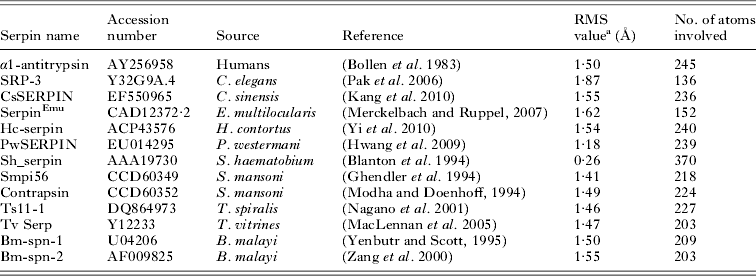
Multiple amino acid sequence alignments of SjB10 and other parasite serpins retrieved from GenBank (Accession Numbers in Table 1) were created using the MUSCLE algorithm (Edgar, Reference Edgar2004a , Reference Edgar b ) in an open source Phylogeny.fr program (Dereeper et al. Reference Dereeper, Guignon, Blanc, Audic, Buffet, Chevenet, Dufayard, Guindon, Lefort, Lescot, Claverie and Gascuel2008). Phylogenetic analyses were performed using the default setting of the ‘One Click mode’ in the Phylogeny.fr program (Dereeper et al. Reference Dereeper, Guignon, Blanc, Audic, Buffet, Chevenet, Dufayard, Guindon, Lefort, Lescot, Claverie and Gascuel2008) with PhyML for tree building (Guindon and Gascuel, Reference Guindon and Gascuel2003), TreeDyn for tree drawing (Chevenet et al. Reference Chevenet, Brun, Banuls, Jacq and Christen2006) and Gblocks for automatic curation (Castresana, Reference Castresana2000).
Structural alignment of SjB10 with human α1-antitrypsin, Caenorhabditis elegans and other parasitic helminth serpins

a Abbreviation: RMS: root mean squared.
Parasite materials
A Chinese field isolate (Anhui strain) of S. japonicum was maintained in Oncomelania hupensis hupensis snails and in BALB/c mice (Animal Resource Centre, Western Australia) at QIMR Berghofer Medical Research Institute. Adult worms were recovered by perfusion of infected mice using sodium citrate buffer (0·15 m sodium chloride/0·05 m sodium citrate). Soluble worm antigen products (SWAP) were obtained from homogenized adult worm pairs following centrifugation. Eggs were obtained from infected mouse livers and miracidia hatched from isolated eggs as described (Dalton et al. Reference Dalton, Day, Drew and Brindley1997). The production of cercariae (Chai et al. Reference Chai, McManus, McInnes, Moertel, Tran, Loukas, Jonesa and Gobert2006) and schistosomula, obtained by mechanical transformation of cercariae (Milligan and Jolly, Reference Milligan and Jolly2011), followed published procedures. All parasite stages were stored in liquid nitrogen until needed or stored in RNAlater (Ambion) at 4 °C until total RNA extraction.
Total RNA extraction, cDNA synthesis and real-time PCR
Total RNA was extracted from the isolated S. japonicum life cycle stages using Trizol reagent (Life Technologies) and RNeasy Mini kit (Qiagen). Total RNA quantity was measured using a Nanodrop-1000 (Nanodrop Technologies,) and quality assessed using an Agilent Bioanalyzer (Agilent Technologies). cDNA was synthesized from total RNA using a Quantitect Reverse Transcription Kit (Qiagen) according to the manufacturer's instructions. Real-time PCR for SjB10 was performed using 2·5 ng of cDNA as template with the SYBR Green PCR Master Mix (Applied Biosystems). Primers were designed from the cDNA sequence using Primer3 (http://primer3.wi.mit.edu/). NADH-ubiquinone reductase (NADH-UR) was used as house-keeping gene (Gobert et al. Reference Gobert, Moertel, Brindley and McManus2009). The forward primers for SjB10 and NADH-UR were 5′-GCACCAAAAGTTCAAGATCCA-3′ and 5′-CGAGGACCTAAC AGCAGAGG-3′ while the reverse primers were 5′-GAGCACCACCAGAGCCTAAT-3′ and 5′-TCCGAACGAACTTTGAATCC-3′ respectively. The cycling conditions were: 95 °C for 10 min; 39 cycles at 95 °C for 30 s at 58 °C for 30 s and 72 °C for 30 s. All reactions were carried out in four biological replicates. Rotor-Gene 6000 series software and GraphPad Prism software were used to analyse the results.
Cloning and sequence analysis of SjB10
The full-length coding sequence of SjB10 was amplified from adult worm cDNA by PCR using oligonucleotide primers specific to the SjB10 gene. The primers (0·5 μ m each) for SjB10 amplification were designed from the cDNA sequence using Amplify 3 (http://engels.genetics.wisc.edu/amplify/) The forward and reverse primers used were 5′-AAT AAA AAA ACC TAT AAA TAT AGA TGC ATC ACC ATC ACC ATC ACT GTT TGA AAG TAG CAC CAA AAG TTC AAG-3′ and 5′-AGT GGT GGT GGT GGT GGT GCT CTT ATA ACA TTG GAT TGA TTA CTT TAG CTG CTA ATA ATG GC-3′ respectively. The amplification reaction was carried out using a thermal cycling profile of 95 °C for 2 min; 30 cycles at 95 °C for 20 s, 65 °C for 20 s, 70 °C for 20 s and a final extension for 2 min at 72 °C. The PCR product was analysed on a 0·8% (w/v) agarose gel, gel-purified and co-transformed with linearized p-BAC-1 transfer vector into Escherichia coli OmniMAX competent cells (Life Technologies). Positive clones were screened for the presence of plasmid with the appropriate insert. The nucleotide sequence of the insert was determined by automated sequencing.
Generation of recombinant pBAC-1/SjB10 plasmid DNA
Standard insect cell culture techniques were used (Siracusano et al. Reference Siracusano, Rigano, Ortona, Profumo, Margutti, Buttari, Delunardo and Teggi2008; Hitchman et al. Reference Hitchman, Locanto, Possee and King2011). Sf9 cells, seeded at 6×10⁵ cells mL−1 in 400 μL of Sf900II medium, were plated into a 24-deep well plate (Life Technologies) and transfected with a mixture of 100 ng recombinant pBAC-1/SjB10 plasmid DNA and 20 ng flashBAC DNA (Zhao et al. Reference Zhao, Chapman and Jones2003) using Cellfectin (Life Technologies), according to the manufacturer's instruction. Cells were then incubated at 28 °C for 5 h and the supernatant then removed. Media supplemented with Gibco antibiotics-antimycotics (100×) solution (Life Technologies) was added and the cells incubated at 28 °C for a further 7 days. Cells were pelleted (500 g, 5 min) and the supernatant containing P1 virus was stored at 4 °C until needed. Cell growth, average cell size and viability were monitored using a Countess Automated Cell Counter (Life Technologies).
Virus amplification and expression of rSjB10
Sf9 cells (5 ml), seeded at 2×10⁶ cells mL−1, were infected with P1 virus (150 μL) and incubated at 28 °C in a humidified incubator at 250 rpm for 4 days. At 96 h post-infection, the cells were pelleted and the supernatant containing P2 virus was harvested and stored at 4 °C. For the expression of rSjB10, 1 L of Sf9 culture seeded at 4×10⁶ cells mL−1 was infected with 30 mLl of P3 virus and incubated at 120 rpm at 28 °C for 72 h in a humidified incubator. At 72 h post-infection, the cell pellet was harvested by centrifugation and stored at −20 °C until needed.
Affinity purification of rSjB10 followed by size exclusion chromatography
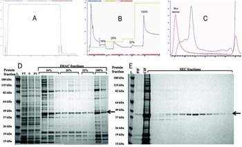
The cell pellet collected following expression was resuspended in 200 mL lysis buffer (20 mm sodium phosphate, 500 mm NaCl, 20 mm imidazole, 1% (v/v) Triton X-100, 2 mm MgCl2, 100 U Benzonase, complete protease inhibitor cocktail (Roche), pH 7·1) and sonicated on ice. The lysed cell pellet was further incubated for 1 h on an orbital wheel at room temperature to degrade released DNA. The supernatant was collected by centrifugation, filtered through a 0·45 μm filter and loaded onto a prepacked 1 mL HisTrap excel IMAC column (GE Healthcare). The column was washed with 20 column volumes wash buffer (20 mm sodium phosphate, 500 mm NaCl, 20 mm imidazole, pH 7·1) (Fig. 1A) prior to a multi-step elution, whereby increasing concentrations of elution buffer (20 mm sodium phosphate, 500 mm NaCl, 500 mm imidazole) (16, 26, 32 and 100% buffer) were used to elute the rSjB10 protein (Fig. 1B). Each concentration was held until a baseline absorbance was reached. Purification steps were performed on the AKTA Explorer FPLC system (GE Healthcare) at a flow rate of 1 mL min−1 at 4 °C. Eluted protein fractions were dialysed against phosphate buffered saline (pH 7·4) and loaded onto a S75 SEC column with the AKTA Explorer FPLC system at 0·5 mL min−1 at 4 °C. Collected protein fractions were analysed by SDS-PAGE (Fig. 1D and E) and the yield of protein determined by a standard Bradford assay (Bradford, Reference Bradford1976). The fractions were pooled and rSjB10 was stored at −20 °C until needed.
Purification of rSjB10 expressed in Sf9 cells by IMAC affinity chromatography followed by size exclusion chromatography. (A) An IMAC column was loaded with filtered protein sample and the column washed to remove unbound material. (B) Multi-step elution was carried out with increasing concentration of elution buffer (16, 26, 32 and 100%). The peaks indicate eluted protein fractions. (C) IMAC purified elution samples were pooled, concentrated and loaded onto the SEC column for further purification. The blue peak corresponds to the rSjB10 fraction. (D) Following protein expression, the soluble cell lysate was filtered and loaded onto the IMAC column. Unbound proteins were washed off and bound rSjB10 eluted with increasing concentrations of the IMAC elution buffer. (E) Pooled IMAC eluted fractions were concentrated and loaded onto the SEC column and purified fractions collected. Black arrows show the purified rSjB10. Representative samples collected from the eluted fractions were analysed by SDS-PAGE. L = protein ladder, FT = IMAC flow through, S = soluble IMAC load, FS = filtered soluble IMAC load, PIF = pooled IMAC fractions and CIF = concentrated IMAC fractions.

Western blot analysis of native SjB10
Production of rabbit anti-SjB10 serum was carried out at the Antibody Production Services (South Australian Health and Medical Research Institute, Australia). Briefly, a rabbit was immunized 3 times with 250 μg rSjB10 at 3-week intervals with complete Freund's adjuvant (FA) for the first immunization and incomplete FA for subsequent immunizations. The polyclonal antiserum was collected 7 days after the last immunization and stored at −80 °C until required. Western blotting was performed according to the LI-COR Biosciences (Lincoln, USA) protocol. Briefly, rSjB10 and S. japonicum SWAP were electrophoresed on a 4–12% (w/v) NuPAGE gel (Invitrogen). After separation, electrophoretic transfer of protein from the polyacrylamide gel to Immuno-Blot LF PVDF (Bio-Rad) membrane was achieved using a XCell II Blot Module (Invitrogen) at 25 V for 2 h. Membranes were blocked with Odyssey Blocking buffer (OBT, LI-COR Biosciences) overnight at 4 °C, then incubated with the primary antibodies (anti-rSjB10 serum and normal rabbit serum as control) for 1 h at room temperature (1:1000 dilution of rabbit serum in 0·2% (v/v) Tween-20 in OBT). The membranes were washed with PBST (PBS with 0·1% (v/v) Tween-20) and then incubated with secondary antibody (IRDye-labelled goat anti-rabbit IgG (H+L) at 1:20 000 dilution, LICOR Biosciences) in 0·02% (w/v) SDS in OBT for 1 h at room temperature. After further washes with PBST, immunoreactivity was visualized using a LICOR Odyssey Infrared Imager.
Immunolocalization of SjB10
Freshly collected adult worms and eggs were washed in PBS, fixed in 4·5% (v/v) paraformaldehyde, washed in PBS, dehydrated, embedded in paraffin wax and cut into 5 μm thick sections. Sections were air-dried, dewaxed through ascending grades of alcohol and then rehydrated into Tris buffered saline (TBS) (pH 7·6). After three washes in water, sections were subjected to heat antigen retrieval by immersion into 1% (v/v) Revealt-A solution (Histotechnology, QIMR Berghofer, Australia), autoclaving at 85 °C for 15 min and then at 80 °C for 5 min, blocking in 1% (w/v) foetal bovine serum albumin (BSA) in PBS, and incubating with a 1:100 dilution of rabbit anti-SjB10 antiserum or with pre-immune serum at room temperature for 1·5 h. After three washes, the sections were incubated for 1·5 h with secondary antibody (Alexa fluor 594 goat anti-rabbit IgG (H+L) antibody, Life Technologies) at a dilution of 1:2000, washed and mounted with Prolong gold anti-fade reagent with DAPI (Invitrogen). Sections were examined using a digital EVOS XL fluorescent microscope.
Serine protease inhibition assays
The inhibitory potential of rSjB10 was tested in vitro against a panel of serine proteases. All protease inhibition assays were performed in 96-well half-area black microplates (Corning). Assays involving trypsin and chymotrypsin were carried out as described (Mika et al. Reference Mika, Reynolds, Mohlin, Willis, Swe, Pickering, Halilovic, Wijeyewickrema, Pike, Blom, Kemp and Fischer2012) with slight modifications. Briefly, the assays were performed in standard assay buffer (100 mm Tris-HCl, pH 8·2 10 mm CaCl2 and 0·05% (v/v) PEG 8000) with the two proteases and their substrates (N α -Benzoyl-L-arginine-7-amido-4-methylcoumarin hydrochloride and N-Succinyl-Ala-Ala-Pro-Phe-7-amido-4-methylcoumarin) (Sigma Aldrich) at 37 °C. Increasing concentrations of rSjB10 and the proteases were pre-incubated at 37 °C for 10 min followed by addition of their respective substrates to start the enzyme reaction. Fluorescence was measured continuously at 30 s intervals at excitation and emission wavelengths of 370 and 460 nm, respectively. The blank control and whole activity control did not include trypsin/chymotrypsin or rSjB10, respectively. All reactions were carried out in four replicates.
Pancreatic elastase enzyme assays were carried out using the EnzChek Elastase Assay Kit (Molecular Probes). Briefly, elastase at a concentration of 0·5 UmL−1 was incubated for 15 min at room temperature with varying concentration of rSjB10. The proteolytic reaction was started by adding the substrate (DQ elastin) to a final volume of 0·1 mL. The reaction was carried out at room temperature (22–23 °C) for a total of 2 h with the fluorescence measured in a fluorescence multi-well plate reader set for excitation at 485 nm and emission detection at 530 nm. All values were corrected for background fluorescence and were expressed relative to the fluorescence obtained in the absence of inhibitor. Three replicates were carried out for each reaction.
Experimental challenge infection of rodents, measurement of serum anti-SjB10 antibodies
The laboratory rat is a resistant host for experimental infections with S. japonicum while the mouse is a susceptible host (He et al. Reference He, Salafsky and Ramaswamy2001; Jiang et al. Reference Jiang, Hong, Peng, Fu, Feng, Liu, Shi and Lin2010; Peng et al. Reference Peng, Gobert, Hong, Jiang, Han, McManus, Wang, Liu, Fu, Shi and Lin2011). Two infection time-course experiments were completed with outbred Wistar rats (7 weeks old) and Swiss outbred mice (8–12 weeks old) (Animal Resource Centre, Australia). Rats (N = 3) and mice (N = 5) were infected percutaneously with 200 and 60 S. japonicum cercariae, respectively, using the cover slip method (Dupre et al. Reference Dupre, Kremer, Wolowczuk, Riveau, Capron and Locht2001). Blood samples were collected from mice prior to infection and 6 weeks post-challenge while in the rat time-course experiment, blood samples were collected after a primary and a secondary cercarial challenge (as above) based on the observation that immune resistance in rats re-challenged with schistosomes was shown to be highest between 4–8 weeks after re-infection (Knopf et al. Reference Knopf, Nutman and Reasoner1977; Usawattanakul et al. Reference Usawattanakul, Kamijo and Kojima1982). Blood was collected via the tail vein (rats), the tail tip (mice) or from cardiac puncture at necropsy. Sera were obtained from clotted blood by centrifugation and stored at −80 °C until required.
The levels of anti-SjB10 antibodies in the sera of individual mice and rats were determined by standard ELISA. Briefly, microplates were coated overnight at 4 °C with 0·5 μg mL−1 of rSjB10. Plates were washed once with PBST and blocked with 5% (w/v) skim milk in PBST. After three washes with water and PBST, plates were incubated with rodent sera in blocking buffer for a further 1·5 h at 37 °C. With mouse sera, the plates were washed and incubated with second antibody (goat anti-mouse IgG conjugated to HRP, Sigma Aldrich) and subsequently developed with substrate solution (SIGMAFast OPD tablets). The endpoint antibody titre was defined as the dilution with an OD reading 2 times above the background level. The protocol for ELISA using the rat sera was essentially the same except for the secondary antibody used (goat anti-rat IgG conjugated to biotin, Sigma Aldrich) and an additional incubation step with HRP conjugated to streptavidin (BD Biosciences) was carried out before developing the plates with the substrate solution. SWAP was used as a positive control antigen source and sera collected from mice and rats prior to cercarial challenge were used as negative controls.
Statistical analysis
Data were expressed as mean±standard error of the mean. Changes in real-time PCR data and antibody levels were assessed by one-way ANOVA with post hoc Bonferroni testing (P≤0·05). All analyses were performed using the GraphPad Prism version 6.02 (GraphPad Software).
RESULTS
Cloning and characteristics of SjB10
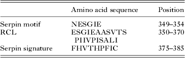
The cloned full-length cDNA (1212 nucleotides) of SjB10 contained a complete open reading frame (ORF) encoding a polypeptide of 403 amino acids with a predicted molecular mass of 45·7 kDa and a pI of 5·73, with the analysis of the deduced SjB10 peptide by ScanProsite (www.expasy.ch/tools/scanprosite/) indicating the presence of two conserved motifs, namely a serpin motif and a RCL (Table 2), located near the C-terminal. A serpin signature sequence is also present at positions 375–385 of the putative amino acid sequence (Table 2).
Characteristic serpin sequences in SjB10
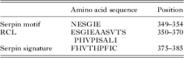
Six N-glycosylation sites (81NISE84, 104NTTL107, 134NQSI137, 152NQTN155, 211NFSV214 and 349NESG352) were identified on SjB10 but no glycosaminoglycan attachment site was found. The SjB10 polypeptide contains eight protein kinase C phosphorylation sites, a microbodies C-terminal targeting signal and a leucine zipper pattern. No transmembrane domain or signal peptide was found in the peptide sequence suggesting SjB10 is an intracellular protein. Analysis of the deduced amino sequence indicated the cleavage site of the RCL is at position 366I–S367.
Multiple sequence alignment of the deduced SjB10 protein sequence showed between 21–65% homology with known full-length parasitic helminth serpins from GenBank with the highest sequence homology being to Schistosoma haematobium serpin (accession number AAA19730.3) (Fig. 2). Analysis of the secondary structure using SWISS-MODEL (http://swissmodel.expasy.org/) and Phyre2 (Kelley and Sternberg, Reference Kelley and Sternberg2009) showed that SjB10 contains nine α-helices and 15 β-strands (Fig. 2). Analysis of the SjB10 RCL by visualization showed that the hinge region contained the consensus sequence typical of the RCL hinge region, a characteristic common to all inhibitory serpins ‘P17 [E]-P16 [E/K/R]-P15 [G]-P14 [T/S]-P13 [X]-P12−9 [AGS]-P8−1 [X]-P1′–4′’ (Hopkins et al. Reference Hopkins, Carrell and Stone1993; Irving et al. Reference Irving, Pike, Lesk and Whisstock2000), with residue variations only at positions P16 and P14 (Fig. 3).
Structure-based sequence alignment of SjB10 with human α1-antitrypsin and full-length serpins from other parasitic helminths. The alignment was constructed by comparison with human α1-antitrypsin (PDB 1HP7) as a modelling template using Phyre2. Secondary structures were assigned based on the 1HP7 tertiary structure; bold lines are α-helices and broken lines are β-strands. Helices are labelled from ‘hA’ to ‘hI’ and β-strands that constitute β-sheet A–C are labelled as ‘sA’, ‘sB’ and ‘sC’ respectively. The highly and lowly conserved residues are labelled in black and grey, respectively. A1AT (Human α1-antitrypsin), Sha (Schistosoma haematobium), Emu (Echinococcus multilocularis), SjB6 (S. japonicum), Sm1 and Sm2 (S. mansoni), Csi (Clonorchis sinensis), Pwe (Paragonimus westermani), Hco (Haemonchus contortus), Tvi (Trichostrongylus vitrinus), Asu (Ascaris suum), Bma1 and Bma2 (Brugia malayi).

Sequence alignment of the putative reactive centre loop (RCL) of SjB10 with that of human α1-antitrypsin and other parasitic helminth serpins. The residues in the RCL are numbered according to the nomenclature developed by Schechter and Berger (Reference Schechter and Berger1967), where residues on the amino-terminal side of the scissile bond (P1–P1′) are noted as unprimed and those on the carboxyl-terminal side are primed. Residues are marked grey in accordance with the consensus sequence of RCL. The boxed residues representing the scissile-bond and bold-faced residues were the predicted P1. A1AT (Human α1-antitrypsin), Sha (Schistosoma haematobium), Emu (Echinococcus multilocularis), SjB6 (S. japonicum), Sm1 and Sm2 (S. mansoni), Csi (Clonorchis sinensis), Pwe (Paragonimus westermani), Hco (Haemonchus contortus), Tvi (Trichostrongylus vitrinus), Asu (Ascaris suum), Bma1 and Bma2 (Brugia malayi).

Structure-based alignment analysis of SjB10
Alignment of the predicted tertiary structure of SjB10 showed that SjB10 is structurally similar to other known helminth serpins with the highest similarity again to S. haematobium serpin (accession number AAA19730) (Table 1). The root mean squared values ranged from 0 to 2. The root mean squared value indicates the degree of similarity between the three-dimensional structures of two proteins by measuring the root-mean-square distance between equivalent atom pairs from both proteins (Carugo and Pongor, Reference Carugo and Pongor2001).
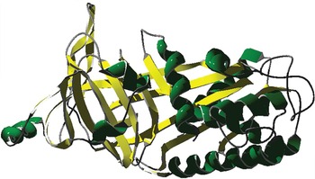
Predicted tertiary structure of SjB10. The figure shows the serpin SjB10 in its native conformation with the RCL being surface accessible by target protease. The green colour represents the α-helices while the yellow colour represents the β-sheets and loops are coloured grey.

Phylogenetic analysis
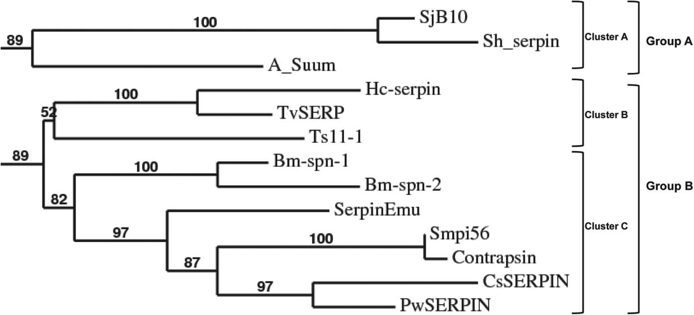
Phylogenetic analysis of the amino acid sequence of SjB10 with other helminth parasite serpins indicated two major groups – Group A and Group B. The former comprises serpins formed by cluster A while the latter comprises serpins formed by clusters B and C. The three phylogenetic clusters were supported by the aLRT statistical test (Anisimova and Gascuel, Reference Anisimova and Gascuel2006) showing branch support values of 90% for cluster A, 52% for cluster B and 83% for cluster C (Fig. 5). SjB10 grouped with Sh_serpin in cluster A, sharing 65% sequence identity; the two schistosome proteins are distantly related to the other parasitic helminth serpins with less than 30% sequence identity.
Phylogenetic tree of the amino acid sequences of SjB10 and serpins from other parasitic helminths. The alignment of retrieved sequences was created using MUSCLE. PhyML was used for tree building, TreeDyn for tree drawing and Gblocks for automatic curation. All sequences (accession numbers are shown in Table 1) were retrieved from GenBank.

Gene expression profile of SjB10
Stage-specific expression of the SjB10 transcript was examined by real-time PCR using cDNAs reverse transcribed from RNAs isolated from several developmental stages of S. japonicum and primers designed to specifically amplify the SjB10 gene. Expression was detected in the adult male, schistosomulum, egg and the cercaria with the cercarial stage showing at least 4-fold increase in expression compared with the other stages with the increase being highly significant (P≤0·0001) (Fig. 6). There was no evidence of SjB10 expression in either the female worm or the miracidium. The NADH-UR gene used as the internal control showed constant expression across all the life-cycle stages examined (data not shown).
Gene expression of SjB10 in different S. japonicum life-cycle stages. SjB10 expression was at least 4-fold higher in cercariae than in the other life-cycle stages. Error bars represent mean real-time PCR expression±standard error of mean (s.e.m.) of three biological replicates. **** P≤0·0001, *** P≤0·001.

Expression and purification of recombinant SjB10
The recombinant SjB10 protein was expressed using the flashBAC baculovirus expression system within Sf9 cells. In the first step of purification, IMAC affinity column was used to partially purify rSjB10. Further purification by size exclusion chromatography resulted in a single protein band of approximately 47 kDa as shown on a 4–12% NuPAGE gel (Fig. 1E).
Biochemical characterization of rSjB10
To investigate the potential inhibitory properties of SjB10, we tested its ability to inhibit a small panel of mammalian serine proteases, each having different substrate specificity. rSjB10 inhibited the enzymatic activity of porcine pancreatic elastase (PE) in a dose-dependent manner, showing complete inhibition of PE at a concentration of 2·5 nm, but the serpin exhibited limited or no inhibitory activity against trypsin, chymotrypsin or neutrophil cathepsin G (Fig. 7).
Concentration-dependent inhibition profile of rSjB10 against different serine proteases. (A) Inhibitory effect of rSjB10 against trypsin. (B) Inhibitory effect of rSjB10 against chymotrypsin. (C) Inhibitory effect of rSjB10 against pancreatic elastase. Increasing concentrations of rSjB10 were incubated with each serine protease (0·5 nm) over time and the inhibitory effect of the serpin determined. The assay utilizing 0 nm SjB10 represents 100% enzyme activity against which all subsequent assays were compared. All values were corrected for background fluorescence and three independent assays were performed in triplicate with each protease enzyme. Error bars represent the mean±s.e.m.

Western blot analysis of SjB10
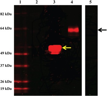
The reactivity of antibodies in rabbit anti-serum, raised against purified rSjB10, against native S. japonicum SWAP was tested by Western blotting. As shown in Fig. 8 (lane 3), a highly specific positive signal against a band in SWAP with a molecular mass of 64 kDa was detected. There was also a positive signal against a band of the expected molecular mass (47 kDa) (Fig. 8, lane 2) with the electrophoresed rSjB10 sample, thereby confirming the specificity of the rabbit antiserum. No specific signal was detectable when the SWAP sample was incubated with pre-immune control rabbit serum (Fig. 8, lane 4).
Western blot analysis of SjB10. Lane 1, pre-stained molecular-sized protein ladder; Lane 2, no sample; Lanes 3 and 4, rSjB10 and SWAP probed with rabbit anti-SjB10 antibodies, respectively; Lane 5, SWAP probed with pre-immune rabbit serum. The positive signals at 49 kDa (lane 3) and 64 kDa (lane 4) show the binding of rabbit anti-SjB10 antibodies to rSjB10 (yellow arrow) and native SjB10 (black arrow).

Immunolocalization of SjB10
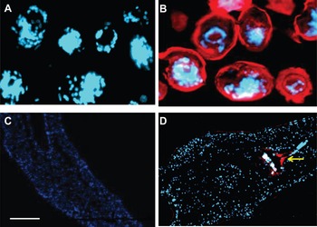
Native SjB10 was localized by immunohistochemistry using the rabbit anti-rSjB10 serum on sections of adult worms and eggs. Intense immune-staining was localized to the anterior gut (Li et al. Reference Li, de Castro-Borges, Parker-Manuel, Vance, Demarco, Neves, Evans and Wilson2013) of the adult worm and the cellular layer beneath the egg shell (Fig. 9) with signal localized within the extra-embryonic envelopes (von Lichtenberg's layer) (Neill et al. Reference Neill, Smith, Doughty and Kemp1988), the outer envelope and the cellular inner envelope of the egg (Jones et al. Reference Jones, Bong, Green, Holmes, Duke, Loukas and McManus2008).
Immunolocalization of SjB10 in the adult worm and eggs of S. japonicum using rabbit anti-SjB10 antibodies. Longitudinal sections of mature eggs (B) and an adult worm (D) showing localization of SjB10 to the extra-embryonic envelopes and the worm foregut, respectively. No significant staining was observed with pre-immune rabbit serum at the same dilution (A and C). White bar (panel C) indicate the scale bar (200 μm) for all panels while the blue stains indicate DAPI staining of the nuclei.

IgG antibody responses to rSjB10 in sera of rodents experimentally challenged with S. japonicum
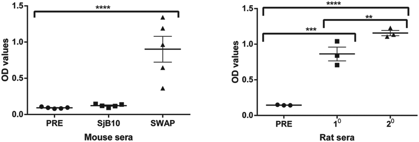
There was a strong serum IgG antibody response against rSjB10 in rats 6 weeks post-infection with a primary challenge of S. japonicum and the response was highly significant (P<0·0001) compared with rat serum obtained prior to challenge (Fig. 10). The total IgG antibody levels were even higher (P<0·001) in sera collected 6 weeks after a secondary challenge (Fig. 10). In contrast, there was no measurable IgG antibody response to rSjB10 in the sera of mice 6 weeks post-challenge (Fig. 10). There was a consistently strong IgG antibody response against SWAP in both the experimentally challenged mice and rats (Fig. 10).
Humoral immune profile of experimentally challenged rodents against rSjB10 determined by ELISA. Mice (n = 5) and rats (n = 3) were experimentally challenged with S. japonicum cercariae and blood samples for sera were collected 6 weeks post-infection with additional bloods for sera collected from the rats 6 weeks post-secondary challenge. Control blood samples for sera were also collected from the mice and rats prior to the primary cercarial challenge. Each data point represents an average of three ELISA replicates. There was a strong serum IgG antibody response against rSjB10 in rats 6 weeks post-infection with a primary challenge of S. japonicum and the response was highly significant (P<0·0001) compared with rat serum obtained prior to challenge (right panel). The total IgG antibody levels were even higher (P<0·001) in sera collected 6 weeks after a secondary challenge (Right panel). In contrast, there was no measurable IgG antibody response to rSjB10 in the sera of mice 6 weeks post-challenge (left panel). There was a consistently strong IgG antibody response against SWAP in both the experimentally challenged mice and rats (right and left panels). Error bars represent mean+s.e.m. 1o = primary challenge, 2o = secondary challenge, PRE = pre-immune sera. Statistically significant difference (two-way ANOVA: **** = P<0·0001, *** = P<0·001, ** = P<0·001).

DISCUSSION
It has been suggested that parasite-derived protease inhibitors, such as serpins, play essential roles in the survival of parasites in their hosts through host immune evasion by interfering with hosts’ immunomodulatory signals, as well as protecting the parasites from the deleterious effect of exogenous host proteases (Martzen et al. Reference Martzen, Geise, Hogan and Peanasky1985; Shepherd et al. Reference Shepherd, Aitken and McManus1991; Ghendler et al. Reference Ghendler, Arnon and Fishelson1994; Huang et al. Reference Huang, Haas, Biesterfeldt, Mankawsky, Blanton and Lee1999; Zang et al. Reference Zang, Yazdanbakhsh, Jiang, Kanost and Maizels1999; Knox, Reference Knox2007). In addition, serpins also participate in the maintenance of cellular homeostasis through regulation of endogenous proteases (Merckelbach and Ruppel, Reference Merckelbach and Ruppel2007; Hwang et al. Reference Hwang, Lee, Na, Lee, Cho and Kim2009). To date, although a number of serpins have been isolated and/or have been produced recombinantly from a range of parasitic helminths (Molehin et al. Reference Molehin, Gobert and McManus2012), there is relatively limited information on the identification and characterization of schistosome serpins. Here we have described the isolation and characterization of an intracellular serpin (SjB10) from S. japonicum; to our knowledge, the first serpin to be functionally characterized from this schistosome species.
Although the predicted amino acid sequence of SjB10 has low overall identity with serpins from other parasitic helminths (21–65% homology), SjB10 was identified as a typical serpin due to the presence of characteristic features such as the serpin motif, RCL and serpin signature (Irving et al. Reference Irving, Pike, Lesk and Whisstock2000; Gettins, Reference Gettins2002b ; van Gent et al. Reference van Gent, Sharp, Morgan and Kalsheker2003). Additionally, SjB10 is composed of 403 amino acid residues, with a predicted molecular mass of 47 kDa and a native molecular mass of 64 kDa, which is consistent with other members of the serpin superfamily (Patston, Reference Patston2000; van Gent et al. Reference van Gent, Sharp, Morgan and Kalsheker2003). Sequence analysis demonstrated that SjB10 did not contain an N-terminal signal peptide, C-terminal extension or transmembrane domain, suggesting that SjB10 is cytosolic. In addition, secondary and tertiary structure prediction analysis showed that SjB10 contains nine α-helices and 15 β-strands, features consistent with known serpins (Irving et al. Reference Irving, Pike, Lesk and Whisstock2000; Silverman et al. Reference Silverman, Bird, Carrell, Church, Coughlin, Gettins, Irving, Lomas, Luke, Moyer, Pemberton, Remold-O'Donnell, Salvesen, Travis and Whisstock2001; Gettins, Reference Gettins2002b ; Huntington, Reference Huntington2006). The RCLs of native inhibitory serpins are always exposed and accessible to target proteases (van Gent et al. Reference van Gent, Sharp, Morgan and Kalsheker2003) and this was clearly the case with the predicted tertiary structure of SjB10 (Fig. 4). These observations of the predicted tertiary structure of SjB10 further strengthen the argument that SjB10 belongs to the serine protease inhibitor superfamily.
The residues and structures involved in the inhibitory activity of serpin families have been identified and the conservation patterns are clearly correlated to the serpin inhibitory mechanism (Irving et al. Reference Irving, Pike, Lesk and Whisstock2000). The amino sequence within the RCL of a serpin determines not only its specificity but also whether the serpin is inhibitory or non-inhibitory (Gettins, Reference Gettins2002a , b) and this is based on the consensus 20/21 residue peptide ‘P17 [E]-P16 [E/K/R]-P15 [G]-P14 [T/S]-P13 [X]-P12−9 [AGS]-P8−1 [X]-P1′ – 4′’ (Hopkins et al. Reference Hopkins, Carrell and Stone1993; Irving et al. Reference Irving, Pike, Lesk and Whisstock2000). For inhibitory serpins, P15 is usually glycine, P14 threonine or serine, and positions P12–P9 are usually occupied by short side-chain residues such as alanine, glycine or serine. These residues allow efficient insertion of the RCL into the A β-sheet, a process critical to protease inhibition and subsequent conformational change within the serpin molecule (Irving et al. Reference Irving, Pike, Lesk and Whisstock2000). On the contrary, the corresponding regions of non-inhibitory serpins do not conform to this consensus sequence (Irving et al. Reference Irving, Pike, Lesk and Whisstock2000). Analysis of SjB10 showed high conservation among primary, secondary and tertiary structures of known inhibitory serpins, suggesting that SjB10 is a functional inhibitory serpin. In addition to these conserved consensus sequences, further analysis of the hinge region of SjB10 showed that it is occupied predominantly by hydrophobic amino acid residues which are thought to be advantageous for the construction of the skeleton conformation needed for the inhibitory activity of serpins (McCarthy and Worrall, Reference McCarthy and Worrall1997). Multiple structure alignment of the predicted tertiary structure of SjB10 with other known serpins showed that it is structurally similar to helminth serpins, particularly a S. haematobium serpin (Sh_serpin) with a root mean squared value of 0·26. This finding is supported by phylogenetic analysis showing SjB10 clustered closely with Sh_serpin (Fig. 5).
Rabbit anti-rSjB10 serum bound a single band (64 kDa) in a native soluble worm antigen preparation of S. japonicum. The molecular mass of native SjB10 was larger than that of the recombinantly derived protein (47 kDa) but this is not surprising as analysis of the SjB10 sequence indicated the presence of multiple N-glycosylation sites indicating that the protein undergoes post-translational modification including glycosylation as is the case with many other known serpins (Huntington, Reference Huntington2006; Law et al. Reference Law, Zhang, McGowan, Buckle, Silverman, Wong, Rosado, Langendorf, Pike, Bird and Whisstock2006). The importance of post-translational modifications such as N-glycosylation and the functionality of serpins was highlighted by the study of McCoy et al. (McCoy Reference McCoy, Pei, Skinner, Abrahams and Carrell2003) who demonstrated that changes in glycosylation patterns of the human serpin antithrombin affected its activity and affinity for heparin.
Gene expression analysis provided some insight into the biological function of SjB10 in S. japonicum. The results of qPCR indicated that SjB10 was expressed in the adult male, schistosomulum, egg and, particularly, the cercarial stage. Schistosome cercariae are known to utilize proteases, especially elastase in the case of Schistosoma mansoni, to digest host skin during invasion (McManus and Loukas, Reference McManus and Loukas2008; Ingram et al. Reference Ingram, Knudsen, Lim, Hansell, Sakanari and McKerrow2011) and we speculate that SjB10 might serve to protect these larvae from the deleterious effect of the proteases in case of leakage from its secretory vesicles in like fashion to the function of human intracellular serpin B9 which is found in the cytoplasm of cytotoxic T cell lymphocytes (CTL). Serpin B9 is thought to protect these CTLs against endogenous proteases leaking from their own secretory vesicles (Hirst et al. Reference Hirst, Buzza, Bird, Warren, Cameron, Zhang, Ashton-Rickardt and Bird2003; Zhang et al. Reference Zhang, Park, Wang, Shah, Liu, Murmann, Wang, Peter and Ashton-Rickardt2006). Our findings from the protease inhibition assays lend some support to this argument in that of the several proteases tested, SjB10 inhibited only PE in a dose-dependent manner in vitro.
SjB10 has a hydrophobic residue at position P1 (Fig. 3) with high specificity to its target protease (elastase). The specificity of similar hydrophobic residues for elastase was reported by Li et al. (Reference Li, Wang, Canagarajah, Jiang, Kanost and Goldsmith1999) who showed that when the original P1 residue in serpin-1B from the moth Manduca sexta was changed by site-directed mutagenesis (A343K), the inhibitory activity of serpin-IB against elastase was lost whereupon the serpin inhibited trypsin, plasmin and thrombin instead (Li et al. Reference Li, Wang, Canagarajah, Jiang, Kanost and Goldsmith1999). The Ixodes ricinus immunosuppressor (Iris) secreted in tick saliva is another specific elastase inhibitor with a methionine present at the P1 position (Leboulle et al. Reference Leboulle, Crippa, Decrem, Mejri, Brossard, Bollen and Godfroid2002). The mutation M340R (methionine→arginine) completely changed both the substrate specificity and affinity of Iris) so that, whereas it did not inhibit elastase, it was a powerful inhibitor of factor Xa and thrombin (Prevot et al. Reference Prevot, Adam, Boudjeltia, Brossard, Lins, Cauchie, Brasseur, Vanhaeverbeek, Vanhamme and Godfroid2006).
The relatively high gene expression of SjB10 in cercariae suggests, due to the dynamic nature of this life-cycle stage, that SjB10 may function in other important role(s) in addition to the endogenous regulation of serine proteases. SjB10 could be released during host skin penetration as a defence mechanism against host proteases implying an extracellular role, a phenomenon not uncommon with other intracellular serpins. Several studies have demonstrated extracellular roles for known intracellular serpins in which the serpins were released through a pathway independent of the endoplasmic reticulum. Examples include a tick serpin gene, Iris (Leboulle et al. Reference Leboulle, Crippa, Decrem, Mejri, Brossard, Bollen and Godfroid2002), human plasminogen-activator inhibitor 2 (Ritchie and Booth, Reference Ritchie and Booth1998) and Hc-serpin from Haemonchus contortus (Yi et al. Reference Yi, Xu, Yan and Li2010). There is also a possibility that SjB10 is released into the host tissue during cercarial penetration of the host skin as part of the released cytoplasmic content. In S. mansoni cercariae, during penetration the entire content of the acetabular gland is secreted including the cytoplasmic contents. However, no serpin was identified from the proteomic analysis of S. mansoni secretions (Knudsen et al. Reference Knudsen, Medzihradszky, Lim, Hansell and McKerrow2005). The argument for an extracellular role for SjB10 in the host-parasite interaction was further strengthened by the fact that rSjB10 was recognized by sera from rats experimentally infected with S. japonicum (Fig. 10), suggesting its release from the parasite and the induction of an immune response. This finding was supported by our Western blot analysis in which sera from rats experimentally infected with S. japonicum showed a high level of immunoreactivity against rSjB10 (data not shown). Surprisingly, in contrast to the rats, the sera from experimentally infected mice did not show any measurable reactivity against rSjB10. It is known that rats actively eliminate S. japonicum parasites upon infection, and it is possible to envisage the release of many internal cytoplasmic components of the parasite during the killing process and hence the possible exposure of SjB10 to the rat's immune system. This is however not the case in mice as experimental infection with S. japonicum leads to parasite establishment and disease progression. However, the mechanism by which SjB10 is shed into the host tissue is unknown and whether the immune response against SjB10 is protective requires further investigation.
Immunohistochemistry revealed that SjB10 was localized to the anterior gut of the adult worm and von Lichtenberg's layer of the egg (Jones et al. Reference Jones, Bong, Green, Holmes, Duke, Loukas and McManus2008), suggesting a possible role for SjB10 in protecting the schistosome from digestion by host proteases. This is supported by the proteomic analysis of S. mansoni vomitus carried out by Hall et al. (Reference Hall, Braschi, Truscott, Mathieson, Cesari and Wilson2011) in seeking insight into the function of the parasite gut which revealed the presence of protease inhibitors such as serpin (GenBank accession number Smp_090080) and α2 macroglobulin protease inhibitor.
The localization of SjB10 in the egg suggests another novel role in schistosome biology. The S. japonicum egg shell has an electron opaque matrix with two layers interrupted by preformed pores (Neill et al. Reference Neill, Smith, Doughty and Kemp1988; Ashton et al. Reference Ashton, Harrop, Shah and Wilson2001; Jones et al. Reference Jones, Bong, Green, Holmes, Duke, Loukas and McManus2008). Schistosome eggs must traverse tissues of the intestine or bladder to escape the host and further the life cycle and this is facilitated by the secretion of immune-reactive molecules from the outer envelope surrounding the differentiating miracidium (Neill et al. Reference Neill, Smith, Doughty and Kemp1988; Ashton et al. Reference Ashton, Harrop, Shah and Wilson2001; Jang-Lee et al. Reference Jang-Lee, Curwen, Ashton, Tissot, Mathieson, Panico, Dell, Wilson and Haslam2007). As the preformed pores on the egg shell allow for the release of secreted antigens into the surrounding host tissue, host molecules such as immune and/or digestive proteases could also gain access into the egg through these pores. The localization of SjB10 within this von Lichtenberg's layer indicates a possible protective role for this protein against potential proteolytic attack by host digestive and immune proteases present within the intestine as the eggs traverse this tissue. This argument is supported by studies that have localized proven immunomodulatory molecules such as IPSE/alpha-1 to the von Lichtenberg's layer of S. mansoni eggs (Schramm et al. Reference Schramm, Gronow, Knobloch, Wippersteg, Grevelding, Galle, Fuller, Stanley, Chiodini, Haas and Doenhoff2006; Wuhrer et al. Reference Wuhrer, Balog, Catalina, Jones, Schramm, Haas, Doenhoff, Dunne, Deelder and Hokke2006).
In summary, the full-length cDNA encoding an intracellular serpin (SjB10) of S. japonicum was cloned and expressed, and some of its characteristics determined, thereby providing some important insights into the biological functions of this protein. In vivo studies such as gene knock-down analysis and transgenesis experiments are now required to further our understanding of the biology of this and other schistosome serpins and to determine their possible roles in the host-parasite interaction.
ACKNOWLEDGEMENTS
The authors wish to thank Ms Mary Duke (QIMR Berghofer) for performing mouse perfusions and also the Protein Expression Facility, UQ for assistance with Baculovirus expression systems. We also wish to thank Histotechnology Department, QIMR Berghofer for their assistance with the immunohistochemistry studies.
FINANCIAL SUPPORT
This research was funded by The University of Queensland International Research Tuition Award (UQIRTA) to AJM and NHMRC grants to DPM. DPM is a NHMRC Senior Principal Research Fellow and Senior Scientist at QIMR Berghofer.